The sadly familiar toothache is actually not even a toothache at all - strictly speaking, there is nothing to hurt in the tooth itself, since it consists of hard tissues and enamel minerals. This is why caries
, and often
pulpitis
can go unnoticed for so long - until the destruction becomes serious, the tooth simply does not give any pain signals. The pain we experience actually comes from the nerve, or more precisely, from the pulp. So, when we say that a tooth hurts, it actually hurts the nerve in the tooth.
Pulp
- this is a bundle of neurovascular fibers, soft tissue that serves as a layer between the crown of the tooth and its root. The nerves located in it react to hot and cold, and nutrients and minerals enter the tooth through the vessels. However, when the destruction of the enamel affects the pulp, the nerves become inflamed and the tooth begins to hurt. This is pulpitis - inflammation of the pulp.
In case of pulpitis, it is too late to save the pulp and nerve, and therefore they are removed - this is a common procedure that completely anesthetizes the tooth and prevents the infection from spreading further to neighboring teeth. However, a tooth deprived of pulp becomes “dead”: it is deprived of nutrition, its tissues become thinner, and the color of the enamel darkens. This is unfortunately a side effect of nerve removal.
Number of roots in human teeth
There can only be one dental crown, but the number of roots a tooth has depends on its location and purpose. The number of roots is also influenced by hereditary factors. It is possible to determine how many roots a tooth has only with the help of an x-ray. The inner part of the tooth (root) makes up about 70% of the entire tooth.
Factors influencing the number of roots:
- Location of the tooth.
- The purpose of the tooth, its functionality (chewing or frontal).
- Genetic predisposition.
- Patient's age and race. In the European race, the number of dental roots is very different from the Negroid and Mongoloid races.
Teeth of ancient people
The dentofacial apparatus of prehistoric and modern humans differs significantly. Ancient people had more than 36 teeth, protruding fangs and a massive jaw. This was explained by the need to chew rough food and raw meat. With the addition of thermally processed foods to the diet, the dentition began to change. The canines were the first to transform, becoming aligned with the bite line. Then the jaw arch narrowed, the interdental spaces disappeared, and the teeth themselves decreased in size. Currently, 32 teeth in humans are the norm, but third molars are considered to be an atavism.
Interesting fact!
The teeth of ancient man cannot be called aesthetic, but they were healthy. According to scientists, cavemen never suffered from caries and other oral diseases.
Teeth numbering system
Dentists have developed a system in which all teeth have their own serial number. The numbering system for the teeth of the lower and upper rows will not allow you to “get confused” in the teeth.
The first number is the incisors - the frontal teeth of the upper and lower rows. There are two teeth on each side (left and right): No. 1 - central, No. 2 - lateral, behind which there are fangs, numbered No. 3. The small molars have numbers 4 and 5.
All of the listed teeth have one cone-shaped root.
Teeth No. 6, 7, 8 - large molars have three roots, and tooth No. 6 of the lower row has one root, with the exception of tooth No. 8, which can have 3 or even 4 roots.
Why do we need to know the structural features of roots?
X-ray diagnostics and computed tomography in particular allow us to carefully study the root system and at the same time identify the narrowest and most intricate branches. And if the length of the canal is determined instrumentally, then the exact number of internal passages will be clearly visible on the x-ray image. Information regarding their cross-country ability is also extremely important:
- curvature up to 25 degrees – tool processing available,
- within the range from 25 to 50 degrees – the cavity is considered impassable,
- from 50 degrees - it is impossible to carry out instrumental treatment, but if the corner is in close proximity to the mouth, a specialist can try to improve patency.
An X-ray examination allows you to examine the canals and roots of the teeth.
Due to severe narrowing or even overgrowth as a result of an advanced pathological process, the doctor may not find the canal at all. Among other reasons for this situation, experts in the field of endodontics identify age-related changes and previous medical errors.
The complex anatomy of teeth makes endodontic treatment a complex and risky undertaking. But with the advent of modern technologies in the arsenal of specialists, the likelihood of medical errors has noticeably decreased. We are talking about treatment under a microscope, when during work the doctor has a clear picture of what is happening in real time before his eyes. The use of optics can significantly reduce the risk of poor-quality filling or root perforation, which means that treatment can be carried out efficiently and without complications
- Dmitrienko CB, Krayushkin A.I. Anatomy of human teeth, 2003.
How many canals are there in teeth?
The number of tooth canals does not always coincide with the number of roots. The number of channels can only be determined using x-rays. The upper incisors usually have two or three canals. Some teeth have only one canal, which branches into two parts.
Number of canals in teeth:
- Upper Lower four – 1, less often 2 channels;
- Upper second – 1, less often 2 and even 3 channels;
- Bottom five – one channel;
- Upper first molar 3, 4 canals;
- Lower first molar – 3, less often 2 canals;
- Upper and lower seven – 3, 4 channels.
What is a human tooth made of?
The visible part or crown is covered with a layer of enamel - the strongest tissue in the human body. Beneath it is dentin, and then the walls of the pulp chamber. The pulp (nerve) is a neurovascular bundle that smoothly passes into the root canals. At the very end of the root there is a small apical opening - nerve endings, vessels and capillaries pass through it.
The photo shows the structure of the tooth
Thus, if for some reason the pulp becomes inflamed, the pathological process spreads very quickly through the canals. Essentially, these are communicating vessels, so if one of the passages is missed during treatment and filling, the inflammatory process will continue to develop and ultimately lead to the need to completely remove the diseased element.
Treatment of three-channel pulpitis in pregnant women
The immune system of a pregnant woman is weakened due to changes in hormonal levels. Together with changes in food preferences, this negatively affects the condition of the expectant mother’s teeth. The most favorable period for treating pulpitis of a three-canal tooth is the second and third trimester of pregnancy. In the presence of acute pain, treatment is carried out in the first trimester. Features of the treatment of pulpitis in pregnant women:
- Treatment is carried out without the use of anesthesia. If this is not possible, then drugs that do not contain adrenaline are used.
- Consultation with a gynecologist accompanying pregnancy is required.
- When carrying out therapeutic treatment, drugs are used that do not harm the health of the fetus and do not have a negative impact on its development.
- A pregnant woman needs to inform her doctor about her situation.
To avoid complications and the development of more serious diseases, you should contact your dentist if pain occurs. Delayed treatment may result in tooth loss.
Promotions of the DentBerg clinic
5% discount for following on Instagram
Subscribe to the DentBerg clinic page @dentberg_db on Instagram and receive a 5% discount on dental services.
More details
the promotion is valid until 02/28/2022
10% discount on a Muscovite social card
Receive a 10% discount on all clinic services using a Muscovite social card.
More details
the promotion is valid until 02/28/2022
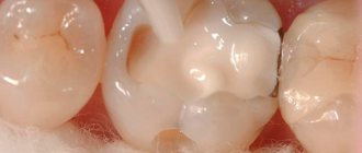
Treatment of caries “All inclusive”
Treatment of caries with placement of a heliocomposite filling at the DentBerg clinic at a fixed price.
6,500 5,500 rub. More details
the promotion is valid until 02/28/2022
10% discount on dental treatment for pensioners
Get a 10% discount on dental treatment at the DentBerg clinic.
More details
the promotion is valid until 02/28/2022
What is the cost of treatment?
How much does root canal treatment cost? The cost largely depends on the method used and the quality of the materials. When using laser and microscopic technology, nanocomposites and other advanced developments, the cost of tooth treatment with canal filling increases significantly. Approximate prices for root canal treatment in Moscow with gutta-percha filling and composite filling are presented in the table below.
| View | Price |
| Single channel tooth | 9,500 – 12,5000 rubles |
| Double channel tooth | 11,000 – 14,500 rubles |
| Three-channel tooth | 13,500 – 17,000 rubles |
The cost of root canal treatment should not be the determining factor in choosing a clinic. Contact only dentistry with a good reputation and experienced specialists who use modern equipment and the latest techniques in their work. Remember - the future fate of your teeth and the aesthetics of your smile depend on the quality of canal treatment!
Treatment under a microscope
It is possible to treat pulpitis under a microscope. This is a specialized dental instrument that allows the doctor to perform manipulations with high precision. The channels are visually enlarged. The doctor clearly sees the foci of inflammation, which allows them to be completely eliminated and avoid relapse.
The use of a microscope is necessary if the patient has small canals that are inaccessible to the human eye. This makes it possible to clean and seal the tooth efficiently.
Possible complications
Not all cases go smoothly. Let's consider the main problems that arise after the procedure.
- Perforation.
The phenomenon is the formation of holes between the dental canals and surrounding tissues. Treatment of perforation consists of medicinal treatment and filling. - Cheek swelling.
The reason why the cheek is swollen after root canal treatment is believed to be the impregnation of the periodontal tissues and mucous membranes with an anesthetic drug, which themselves are quite loose and easily absorb liquid. - Instrument fracture.
The probes for passing through the channels are very thin. If they break during medical procedures, the fragments are removed with special devices. Modern dental instruments made of nickel-titanium alloys are less susceptible to wear and break less often. - Adverse reactions to medications.
The range and severity of side effects of modern anesthetics are minimal. Before treatment, the doctor must collect an allergic history - information about drug intolerance - and the likelihood of a full-blown allergic reaction is practically reduced to zero. Adverse reactions of moderate and minor degrees are mostly short-term, can be easily corrected or can be overcome by changing the drug. - Other complications.
Situations such as swallowing particles of fillings, tooth dust, and small instruments now practically do not occur thanks to the use of a rubber dam - a latex plate that separates the tooth or teeth being treated from the oral cavity.
Complex cases
Double-canal teeth can be arranged non-standardly, have excessively narrow or curved canals. It also often happens that treatment has already been carried out, but the results were unsatisfactory. All this leads to serious problems.
If a broken pin is stuck in the tooth canal, or if the intervention was unsuccessful, then most clinics will recommend removal. However, we are ready to correct the situation. Our dentists have experience in dealing with the consequences of colleagues’ mistakes. Moreover, assistance is provided as delicately as possible. And the tooth is saved!
The Dentberg Clinic is a fusion of experienced professionals and advanced equipment. This allows us to successfully provide assistance in a variety of situations. Contact us!
What is pulpitis of a three-canal tooth?
Pulpitis is an inflammation of the connective tissue located inside the tooth. The development of pulpitis occurs as a result of infection entering the tooth. Most often this occurs through a carious cavity.
In a three-canal tooth, the pulp is located simultaneously in three branches. When inflamed, the pulp swells. This is accompanied by severe pain. The pain may be paroxysmal and throbbing. In some cases, pain intensifies at night. The tooth is affected by temperature changes, sweet or sour foods.
If pain occurs, you should consult a specialist. Delaying the treatment process can lead to aggravation of the situation and development of the disease into a chronic form. This will lead to gradual tissue destruction and tooth loss. Taking painkillers will reduce pain, but will not eliminate the problem.
Features of treatment of individual groups of teeth
The treatment tactics for incisors, canines, premolars, molars of the upper and lower jaws are different. Each tooth carries a different load. One part of the row is visible when you smile, while the other remains invisible to others. Let's look at the features of treatment for each area of the oral cavity.
Treatment of wisdom teeth
Eights are the outermost molars in the row. They erupt later than others, practically do not participate in chewing food, and regularly cause trouble. 1, 16, 17, 32 teeth can erupt incorrectly, displacing the entire row, injuring the gums, and causing the formation of a soft tissue hood. This increases the risk of plaque accumulation and tooth decay.
| Features of wisdom teeth treatment | |
| Question | Dentist recommendation |
| Do I need to remove wisdom teeth? | In most cases, this is necessary and recommended. |
| Is it possible to treat caries on the 8th tooth? | If they do not disturb the structure of the dentition or cause discomfort, then after consultation with a doctor you can do this. |
In most cases, eights must be removed. If they grow at an angle, disrupt the bite, displace other molars, cause discomfort, or have underdeveloped enamel, it is recommended to get rid of them as quickly as possible. Extraction of teeth 1, 16, 17, 32 does not affect the chewing function or appearance of the patient.
Is caries of eights curable? If they have a normal structure, do not cause discomfort, and are involved in chewing, superficial caries can be treated using the traditional method - drilling, installing a filling. The procedure is performed at the request of the patient. If a person refuses conservative therapy, the extreme molars are removed.
If there is significant damage to the visible part of the figure eights, their removal is indicated. Artificial crowns are not installed on them due to their location and anatomical features. For acute pain, the only way to solve the problem is extraction. The figure eights are located in the far corners of the oral cavity, which makes depulpation and endodontic treatment impossible. Lack of visibility and access to instruments prevents nerve removal and canal filling.
Extreme molars are unreliable and often destroyed, so they are not recommended for use as a support for bridges. If eights are lost, prosthetics are not performed; there is no such need or possibility.
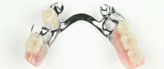
Treatment of molars
2, 3, 14, 15, 18, 19, 30, 31 teeth are molars that bear the main load when chewing. They are large, with a wide crown, have several roots, so they are firmly held in the jaw. They are not visible when you smile, but the defeat or absence of one of them causes severe discomfort to the patient. When treating this group, they pay attention to strength and strive for complete restoration of function.
| Features of treatment of molars | |
| Question | Dentist recommendation |
| What treatment methods are there if a molar is severely damaged? | In case of severe destruction of molars, ceramic inlays are most often used. |
| Which crowns are best for molar prosthetics? | More often, patients are recommended to install metal-ceramic crowns in the molar area. |
| What to do if you lose molars? | The best option in this case is implantation; patients also consider removable dentures. |
The main method of treating caries of sixes and sevens is drilling and filling. If the process does not affect the canal with the neurovascular bundle, the doctor drills out the affected areas and closes the defect with a filling made of composite materials. If the affected area is large, it is advisable to use ceramic inlays. They are made from impressions for each patient and close the cavity formed after treatment. The use of inlays allows you to increase the service life of the molar and ensures its resistance to chewing loads.
If the visible part of the six or seven is significantly destroyed, but the root is preserved, restoration is carried out using a crown. The best material for prosthetics of 2, 3, 14, 15, 18, 19, 30, 31 teeth is metal ceramics. Such designs are very durable and allow you to chew even hard food. A thin strip of metal that is visible between the metal-ceramic crown and the gum is not a disadvantage when restoring molars. It is completely invisible because these teeth are not located in the smile area. You can choose crowns made of zirconium dioxide, but they will cost more than metal-ceramics. Ceramic structures are not installed on chewing teeth.
The loss of molars affects the condition of the gastrointestinal tract and leads to displacement of the entire dentition, so prosthetics are necessary. To replace 6-k bridges, bridges are often used. If nearby units can serve as support, this is the fastest, least expensive, painless option for restoring mastication. For multiple defects, removable clasp and plate prostheses are used.
The optimal way to replace molars is implantation. The condition of adjacent teeth does not matter. With the help of titanium structures, it is possible to restore both one and several units. In most cases, root-shaped prostheses are used for prosthetics. They are able to withstand heavy loads. Such implants can serve as a support for artificial crowns and bridges.
Treatment of premolars
Teeth 4, 5, 12, 13, 20, 21, 28, 29 are premolars. They also perform the function of chewing, but are not as large and massive as molars. Some premolars are located in the smile zone, so when treating them, not only strength and functionality are important, but also beauty and naturalness.
| Features of treatment of premolars | |
| Question | Dentist recommendation |
| What to do if premolars are destroyed? | In case of significant destruction, ceramic inlays or crowns are used. The best material for premolar restoration is zirconium dioxide. |
| What to do if there are no premolars? | In this case, there are several treatment options - bridge structures, butterfly prosthesis or implantation of a missing tooth. |
The most common lesion of fours and fives is caries. Destruction occurs especially often in patients with deep fissures and narrow interdental spaces.
Due to the location of small molars, caries on them can be noticed in the initial stages. Sometimes even patients themselves pay attention to the problem and seek dental help. If a carious lesion is detected at the spot stage, modern clinics carry out treatment without drilling using the ICON system. Careful removal of damaged enamel using an etching gel allows you to save the tooth, and sealing the cavity with a special compound stops further destruction. For deeper lesions, destroyed tissue is removed using a drill or laser. Small defects are restored with durable photopolymer materials. You can choose the shade of the filling that perfectly matches the color of the enamel. In case of significant destruction, leading dentists recommend using ceramic inlays, which are made in a dental laboratory using individual impressions.
The destroyed outer part of the premolar is an indication for the installation of crowns. If only the root remains, a stump tab is installed to securely secure the prosthesis. The choice of crown material depends on the financial capabilities of the patient. The best choice is zirconium dioxide. Such designs are aesthetic and resistant to chewing loads. Their only drawback is their high price. Metal ceramics are available to everyone. It is strong and durable, but the strip of metal under the gum can be very noticeable for some. Ceramics are not suitable for fours and fives. This material is too fragile; with intensive chewing, chips will quickly appear on it.
The absence of premolars in a person is immediately noticeable to others. When they are lost, most turn to dentistry for prosthetics. If you want to quickly restore chewing and restore an attractive smile, installing an inexpensive bridge will help. The design consists of several interconnected crowns made of metal ceramics or zirconium dioxide. The outer crowns are placed on the supporting teeth, and the middle ones cover the defect in the dentition. The main disadvantages of this type of prosthetics are the need to grind healthy teeth and uneven distribution of load on the jaw.
If installing a bridge is not possible, the prosthetist will suggest a removable clasp prosthesis. Typically, the design is used when several teeth are missing. This method saves the budget and has virtually no contraindications. Lightweight butterfly prostheses are used to temporarily replace the defect. They last no more than six months, but this is enough for patients who are awaiting the manufacture of a permanent prosthesis or implantation.
The best method of replacing a lost premolar is implantation. If there are no contraindications and your budget allows, it is better to choose this modern method. A titanium rod is inserted into the jaw bone tissue and becomes a support for an artificial crown. To install such a prosthesis, you do not need to grind down the adjacent teeth. In addition, the load during chewing is distributed physiologically. Externally, an artificial premolar is difficult to distinguish from your own. Even a dentist will not always do this at first glance.
When treating 4- and 5-k, it is important to maintain a balance between practicality and aesthetics. For this reason, this group is the most difficult for dentists.
Treatment of fangs
There are four fangs in the oral cavity. These are 6, 11, 22, 27 teeth according to the universal numbering system. They have a different shape from other teeth, so during restoration it is necessary to have experience working with this group.
| Features of canine treatment | |
| Question | Dentist recommendation |
| What treatment options are there for lost fangs? | In this case, implantation of the lost canine is recommended. |
| How can you correct abnormally growing fangs? | After consultation, the patient is offered options for either orthodontic treatment or the installation of veneers or crowns. |
The fangs do not bear the chewing load. They help you bite off small pieces of food. In the process of evolution, the role of fangs in humans was lost; we do not use them to hold food or tear it into fragments. That is why the treatment of triplets is aimed at restoring their natural appearance.
In case of carious lesions, the preparation of fangs is carried out carefully, trying to preserve healthy tissue as much as possible. This will allow for high-quality restoration. With superficial caries, it is possible to preserve the volume of hard tissue necessary for high-quality filling, but deep lesions serve as an indication for installing a crown. To maintain a natural smile, it is recommended to use structures made of ceramics or zirconium dioxide.
If 6, 11, 22, 27 teeth are lost, implantation is recommended. Manufacturers offer titanium prostheses of a special configuration, taking into account the anatomical features of the jaw. Such prostheses are already classic, they have a special thread that allows you to securely fasten the structure in the narrow alveolar process.
A popular dental service is canine shape correction. Some patients have teeth that are too long, pointed, or significantly narrower than the incisors, which makes the smile less attractive. The best way to give the fangs the ideal shape is to use ceramic onlays made from individual impressions, as well as modeling with composite materials.