Usually, the healing of the hole after tooth extraction occurs painlessly and does not cause any sensations in the patient. It would be good if everyone knew how this process works and how to speed it up.
Proper oral care after tooth extraction and following all the dentist’s recommendations will help speed up the healing of the hole and avoid complications.
Stages of socket healing by day
- The bleeding that occurs after tooth extraction stops after a few minutes , and a blood clot is formed that protects the wound from the penetration of microbes. Under the clot, damaged tissue is being restored, so it is so important to maintain its integrity until the hole heals completely.
- Day 1 – the processes of epithelization (healing) of the wound surface begin.
- Day 3 - signs of epithelization become noticeable, healing begins from the edges of the wound and moves towards the center.
- Day 4 - granulation tissue begins to form - a type of connective tissue that is formed in the body only during wound healing by secondary intention, when there is a strong defect in the skin.
- Day 7 - granulation tissue replaces part of the blood clot, it remains only in the center of the hole. Osteoid (bone) beams begin to appear, indicating bone formation. At the same time, epithelial tissue grows significantly from the edges of the gums.
- 14th -18th day - epithelization of the wound is completely completed, its surface is tightened and heals completely. By this time, the hole is completely filled with granulation tissue, and osteoid tissue is actively developing.
- After a month, a significant part of the hole is filled with new bone tissue. By the end of the second, sometimes third month, this process is completely completed.
- 4th month - bone beams in the newly formed osteoid tissue calcify, it matures: it becomes spongy and does not differ from the rest of the bone. At the same time, the edges of the socket dissolve, the alveolar edge in the area of the extracted tooth becomes thinner and lower than it was before surgery.
Variants of manifestation of dry socket syndrome
Dry socket can manifest itself in different ways. It is very important to notice the pathology in time and consult a dentist.
Options for the manifestation of a dry socket:
- the serous nature of the inflammatory process - the cavity is open, and there are food debris and blood clots in it, the removal area hurts all the time;
- chronic inflammation with the release of pus, granules form along the entire perimeter of the hole, the gums swell;
- purulent-necrotic inflammation - inflamed, very swollen gums, an opening with a gray, dirty coating and dead tissue.
Suppuration of the socket
The last variant of the syndrome is considered the most dangerous. The pain is such that it radiates even to the ear and temple, and the person’s temperature almost immediately rises. Therapy in this case should only be surgical.
Pus in the hole
Healing of the socket with gum inflammation
If tooth extraction was carried out against the background of inflammation, or the inflammatory process in the gums developed later, epithelization of the wound begins on the 10th -14th day, bone beams appear only by the 15th day. A significant part of the socket is filled with young osteoid tissue only by the end of the second month.
After a complex tooth extraction, when the gums rupture and the walls of the socket are traumatized, the edges of the gums cannot come together for a long time and the epithelization process slows down. Wound healing can only be completed after 1-1.5 months. In this case, the development of new bone tissue is delayed.
Socket bleeding
Socket bleeding most often appears immediately after surgery, although sometimes several days pass between tooth extraction and the onset of bleeding.
Bleeding from the socket of an extracted tooth can occur for several reasons:
- The patient actively disturbs the socket of the extracted tooth - he feels it with his tongue or hands, inaccurately brushes his teeth or aggressively treats the sore spot with an irrigator;
- Physical damage in the socket area - gum injury, fracture of the alveoli, and so on;
- Concomitant diseases of the patient: hypertension, leukemia, etc.
Treatment for alveolar bleeding depends on the cause and source of bleeding.
Healing of the hole
If the gums bleed in the area of the socket, then the problem may be complications after surgery, and the wound will need to be stitched.
If blood comes from the vessels directly in the walls of the socket of the extracted tooth, then it is first cooled, after which the vessels are compressed and a tampon with a hemostatic agent is placed in the socket for 4-5 days.
If local remedies do not give the desired effect, the dentist prescribes the patient to take general hemostatic agents.
How to speed up the healing of a hole
To speed up the healing of the hole after tooth extraction, you need to follow these rules:
- Do not touch the blood clot on the socket with your tongue, much less with your hands or a toothpick, so as not to damage it. The presence of a clot is a guarantee of speedy healing of the wound.
- For three hours after visiting the dentist, you should not eat or rinse your mouth. Compliance with this point will also help maintain the integrity of the blood clot.
- · Drinks and food should not be too hot or cold for several days after removal. The food chosen is soft, without coarse inclusions, to prevent injury to the gums.
- You should not engage in heavy physical work for several days to avoid opening the wound and resuming bleeding.
- Since tobacco smoke and alcohol irritate the mucous membrane and inhibit the healing of the hole, you need to stop smoking and drinking alcohol for a while.
- To prevent infection from entering the wound and reduce inflammation after a traumatic tooth extraction, antiseptic baths are used. For them, solutions of chlorhexidine or furatsilin, infusions of sage, chamomile, and eucalyptus are used. The solutions are taken into the mouth, held for several minutes and carefully spat out.
- On the third day after removal, you can carry out antiseptic rinses with the same agents.
- Taking anti-inflammatory drugs (nimesulide, ibuprofen) will help not only eliminate pain, but also relieve the inflammatory process, which also helps accelerate socket regeneration.
- If the doctor has prescribed antibiotics, you should not refuse them, since eliminating the microbial infection shortens the healing time.
- When brushing your teeth, be careful not to touch the wound with the brush.
- To speed up healing, you can use Solcoseryl dental paste. It enhances intracellular energy exchange, due to which cell regeneration and restoration of damaged tissues are accelerated. Solcoseryl also creates conditions for the growth of granulation tissue. The paste is applied to a previously dried surface, then moistened with water. Before using the medicine, it would be more correct to ask your dentist about when to start using Solcoseryl after tooth extraction and how many times a day to apply it to the gums.
What happens to the gums during tooth extraction
To completely remove a tooth from its socket, surgeons have to traumatize the soft tissue of the gums. Before the procedure, the patient is given an anesthetic, since the extraction causes significant pain.
After the onset of anesthesia, the doctor bends the gum away from the tooth and wraps the neck of the element with special forceps. After this, the problematic unit is removed from the hole using rotational and rocking movements.
What happens to the gums after tooth extraction? The blood vessels that fed the pulp rupture, leading to excessive bleeding. Damaged nerve endings signal injury with severe sharp pains that appear immediately after the anesthetic wears off. The symptom worsens when talking and eating. During extraction, the ligaments that fix the root in the jaw alveolus are also damaged.
Surgical intervention is acutely perceived by the body, so the area of the hole begins to be intensively supplied with blood. An inflammatory process develops in the damaged area. This is necessary so that most of the leukocytes can penetrate through the blood vessels, thereby preventing infectious complications.
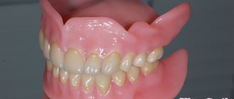
What to do when the hole has healed?
After the hole has healed and filled with full bone tissue, it is necessary to take care of restoring the dentition. The absence of even one tooth can lead to displacement of the remaining teeth, changes in bite, diction, aesthetic problems, and resorption (atrophy) of bone tissue at the site of the extracted tooth. A tooth can be restored using implantation, when an artificial root is implanted into the bone and a crown is installed on it. This method of prosthetics is by far the most comfortable and effective for the patient.
If bone tissue atrophy has already occurred, bone tissue augmentation is performed before installing the implant. The procedure allows you to perform high-quality prosthetics and restore the functionality and aesthetics of your teeth.
How to remove a tooth
There are two extraction methods used in dentistry: simple and complex. Their choice depends on which teeth are being removed - premolars and molars with tangled branched roots are removed using a complex method. It is very difficult to pull out such elements entirely due to the fact that the tooth socket is penetrated by retaining ligaments and alveolar processes. Errors during the procedure or insufficient experience of the specialist lead to serious complications. Therefore, even despite the acute condition, always find out in advance where you can have a tooth removed from a good doctor with positive recommendations.
Factors complicating the operation:
- complete destruction of the coronal part;
- high fragility;
- acute inflammatory diseases;
- Unerupted or misaligned wisdom teeth.
The technology of the procedure depends on which teeth are removed. In some cases, tissue incision and suturing are performed.
Is it possible to put a crown on a granuloma on the root of a tooth?
Dental prosthetics imposes special requirements on the doctor and the patient. If we are talking about installing a crown on a tooth of which no more than 1/3 remains, that is, only the root, then it must be carefully prepared: the canals are sealed, the infection is eliminated. A granuloma under a tooth is an indicator that an inflammatory process is occurring in the tooth, and before dental prosthetics it is necessary to get rid of it, or sooner or later the granuloma will still have to be treated, since it can become inflamed under the crown. So why not do this right away, before installing a denture?
Treatment of dental granuloma with antibiotics and physiotherapy
Dentists often suggest treating dental granuloma with antibiotics. As a rule, this is only a temporary solution to the problem, since it does not eliminate the source of inflammation, and after some time the disease will return. Dentists successfully use physiotherapeutic treatment methods. For example, depophoresis works great for curved or complex tooth canals. Its action consists in the effect of a weak electric current on a suspension with copper hydroxide, which relieves inflammation, penetrating into all corners of the tooth.
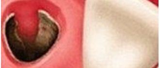
How to detect pathology
If you examine the hole itself, you can find a yellowish or slightly greenish coating. It is possible that there will be a rotting clot inside this cavity. There are situations when even exposed bone tissue is noticeable. At the same time, a repulsive putrid odor comes from the person’s mouth.
- Ear hurts after wisdom tooth removal
Greenish coating
The process of decay and the accompanying inflammatory process lead to intoxication (poisoning) of the patient’s body.
Therefore, the symptoms of dry socket syndrome can also be considered:
- weakness, lethargy, fatigue;
- deterioration of health;
- enlarged lymph nodes in the neck and, most likely, under the jaw;
- increase in body temperature.
Temperature rises, lymph nodes enlarge
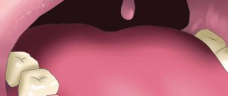
A dry socket also leads to damage to the gums, and the cheek may become swollen. After about two days, pain will appear, the pain can spread even across the face, and also radiate to the ear. If this happens after extraction of a molar tooth in the lower jaw, the patient may well experience difficulty eating.
Cheek swelling
Severe pain may radiate to the ear
Cyst and granuloma of the tooth
Previously, it was generally accepted that if a neoplasm on the root of a tooth measures up to five millimeters, it is a granuloma, and if it is more than ten millimeters, then it is a dental cyst. Some dentists also distinguish a certain transitional stage - cystogranuloma, but practice shows that there are granulomas reaching even twelve millimeters, so an accurate diagnosis cannot be made only by the size of the formation and its shape. In addition to X-rays, histological examination of tissues is necessary.
The question often arises of how a granuloma differs from a dental cyst. The fact is that a cyst is a bladder filled with fluid or pus, with a shell formed by connective tissue and lined on the inside with endometrium. Secretory fluid is produced by the membrane, and due to this the cyst increases in size. Granuloma grows due to the growth of tissues containing infected cells.
Associated symptoms
Dry socket may be accompanied by symptoms such as:
- High temperature accompanies acute and purulent inflammation, a dangerous symptom that requires immediate attention from a dentist.
- Swelling of the cheek indicates the accumulation of pus, the development of a dangerous purulent form of the disease.
- Severe pain is an obligatory companion of alveolitis and increases with exposure of the bone and purulent inflammation.
Causes
Causes of dry socket may include:
- blood clotting disorder;
- incorrectly selected anesthesia;
- severe tissue injury during extraction;
- active rinsing of the mouth in the first days;
- smoking immediately after the procedure;
- a tooth fragment remaining in the socket;
- poor tissue washing, which leads to infection.
Important! If at the time of tooth extraction there are foci of infection in the oral cavity, this will become a factor of alveolitis. Before extraction, professional hygiene must be carried out and inflammatory processes are eliminated.