Dental implants are one of the most widely used methods to replace missing teeth. To do this, you will visit your dentist, and after an examination, your dentist may inform you that a bone graft is required before the implant can be placed. And this is often scary for patients, especially when they are not familiar with bone grafts for dental implants. But don't worry because the dental bone grafting procedure is simple and painless. If you are reading this, you are probably a candidate for bone graft surgery and want to learn more about it. You have come to the right place because this article will explain everything you need to know about bone grafting, including the technique, different types of grafts, surgery cost, success rate, risks and side effects.
What is a Dental Bone Graft?
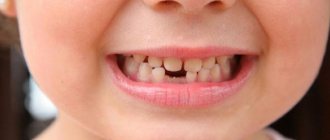
Part of the jawbone may degenerate over time due to the loss of one or more adult teeth. In this case, you may need a bone graft before getting a dental implant to increase the amount of bone in the affected jaw area. Overall, dental bone graft surgery is a painless and simple procedure in which bone taken from any part of the body is fused to the existing jaw bone. These bone grafts are taken from the patient's bones, such as the hip, thigh, or back of the jaw. It may also be artificial. You may ask, “Are dental bone grafts necessary?”
In answer to this question, we must state that this surgery is necessary if the patient does not have enough bone in the jaw to support the implant. Factors such as gum disease, facial injury or trauma, and developmental problems can also damage part of the jawbone, in which case bone grafting is necessary.
Also visit our bone graft products: Monib Bone Graft
Types of grafts for bone grafting
Natural bone material for bone grafting in modern medicine is not used as often as it seems. Innovative technologies for the production of artificial transplants make it possible to avoid many of the disadvantages and risks of using natural materials.
There are four types of grafts for bone grafting:
- Autogenous material. Transplants that are taken from different parts of the jaw, chin or hard palate from the patient himself. It takes root the fastest and most successfully, since it does not have antigens to which the human immune system will react. However, the installation of such grafts requires additional intervention. During the recovery period, multiple surgical approaches will need to be monitored and cared for.
- Allogeneic material. Natural bone material that is taken from another person. As a rule, corpses are used, that is, the use of such transplants requires special laws that allow the removal of any body parts from deceased people. Allogeneic material takes root less well, but is also absolutely safe, as it undergoes a number of treatments and quality controls.
- Xenogeneic transplant. Materials of animal origin are enjoying great success in modern technologies. They are made from bovine tissue and carefully modified into a human-fit structural design or shavings. By filling the space with such material, the doctor increases the likelihood of increased regeneration of the patient’s own bone.
- Synthetic material. Artificial structures are widely used for several reasons. The main one is the low cost of the material. In addition, the patient can have 100% no doubt about the safety of a synthetically created graft.
The patient is involved in the selection of material for bone grafting, since the financial side of the issue is not in last place. The dentist is obliged to explain to the person in detail all the features of natural and synthetic transplants, as well as recommend the most optimal options.
How does Dental Bone Graft Work?
There are several methods for performing bone graft surgery. However, the general process involves the dentist making a small incision in the jaw and inserting a bone graft into the jaw. So how do these bone grafts or powders work? Bone matrix is found in the skeletal system. It is a hard material that helps strengthen bones. The matrix is formed and maintained by the living bone cells within it. Doctors connect bone grafts containing matrix and living cells to the jaw. The cells inside the new bone can then attach to the old bone. As a result, the living bone cells in the bone grafts form a matrix and heal the damaged bone. As a result, your jawbone will eventually become strong enough to support the implant.
Methods for installing bone blocks
There are two main techniques used in dental practice for grafting bone blocks. They have their advantages and disadvantages.
Veneer technique
– a thick block of bone is tightly fixed to the jawbone with several screws. The fusion of biomaterials is quite slow, since it is difficult for blood vessels to break through the thickness of the graft. After 6-8 months, a fairly dense bone with a small number of blood vessels is formed. The ability to regenerate such bone is low, which does not guarantee successful implantation in the future.
With the veneer technique, complications quite often arise due to the fact that hard bone plates cut through the gum. As a result, the bone block is partially or completely resorbed, and the traumatic surgery to increase bone mass has to be repeated.
Laminate technology
– a thin plate of bone is fixed parallel to the narrow jaw ridge. In this way, the inner and outer bone walls are formed, and powdered bone substitute is poured into the space between them. It is easier for blood vessels to break through the soft, porous “filler”, and the formation of bone tissue occurs faster. The implant takes root better in such “living” bone.
The prognosis when using the laminate technique is more favorable. But this technique is much more complicated, and only a doctor with special skills and extensive experience can perform all the manipulations with a predictably good result.
When transplanting a bone block, in many cases plastic surgery of the soft tissues of the oral cavity is also required. The need for this operation arises because the stretched gum above the graft becomes thinner, and sometimes the bone block is even exposed. This is fraught with infection in the affected area. Elimination of such defects leads to the fact that the process of tooth restoration is delayed.
Types of Dental Bone Grafts
Another concern for patients is the type of dental bone graft. Because the quality and material of the grafts, as well as the procedure, are important to them. Dental bone grafts are divided into four types based on their origin and components:
Allografts
Allografts are obtained from the bones of another person, usually a corpse. Patients generally do not prefer to use these types of grafts, but dentists use them widely today. Allografts themselves come in various forms, such as the socket graft and the lateral ridge sparing graft . The main goal of socket grafting is to avoid alveolar bone atrophy. A lateral ridge preservation graft is used to expand the jawbone to accommodate a dental implant.
Autografts
These types of grafts contain bones from your own body, such as your hip or jaw. Block bone graft is a well-known example of an autograft. Dentists use this type of graft when there are significant defects in the jawbone.
Xenografts
These grafts are made from bones from other species, such as cows, pigs or coral. Dentists often use these grafts to treat jaw bone defects.
Alloplasts
The newest type of dental bone graft is completely artificial and made from compounds such as calcium phosphate or calcium phosphosilicate.
Best Candidate for Dental Bone Implantation
Is it possible to install a dental implant without a bone graft? Good question and the answer is yes. Dental implants do not always require bone graft surgery. This is possible when you go to the dentist immediately after losing a tooth. However, if you wait a few months, the jawbone in the place of the missing tooth will begin to deteriorate. In these conditions, you will need a bone graft before getting a dental implant. Dental bone grafts are not just for implants. This surgery is necessary in circumstances such as gum disease, trauma and trauma to the jawbone, and developmental abnormalities. As a result, choosing an experienced dentist and using high-quality bone grafts allows the operation to be performed without complications.
When is the transplant performed?
The collection and replanting of bone blocks is a rather traumatic intervention. Therefore, experienced implantologists prefer to restore teeth using implants of a special design, without resorting to bone grafting. But in certain clinical situations, bone block transplantation is an inevitable procedure.
For example, it is performed when it is impossible to split the bone ridge - due to the fact that the ridge is uniformly narrow throughout its entire height or has a curved shape, for example, in the form of a moon or an hourglass. The extremely low height of the gingival bone is also an indication for its augmentation using bone blocks.
Dental Bone Graft Rejection
Another concern for patients is the failure of dental bone grafts. You may ask, is this possible? Although dental bone implants have a success rate of approximately 99.3%, failure can occur. But what causes this failure? There are several factors involved, but the leading cause of bone graft failure is bone graft surgery performed by an inexperienced surgeon. But this can happen to professional surgeons too. Bone graft rejection or failure can occur for the following reasons:
- If the bone grafting material is contaminated with bacteria, the procedure will fail;
- Infection of surgical instruments can lead to the transfer of bacteria to the surgical site and graft failure.;
- Failure is possible if the patient does not properly follow the postoperative care prescribed by the surgeon,
- If you do not maintain proper oral hygiene after surgery, you may experience problems.
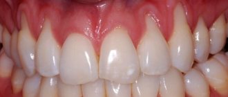
Symptoms of Single Dental Graft Rejection
Bone graft rejection can occur in early and late stages, each with its own symptoms. You may experience early problems within three to four months after surgery. In this case, the following symptoms may occur and you should visit your dentist immediately:
- Swelling of the dental bone graft: Swelling in the surgical area and part of the face is common a few days after surgery. However, if the swelling lasts more than a few weeks or gets worse, it is a sign that your body is rejecting the bone graft.
- Acute pain: A few days after surgery, pain and swelling are normal because bone implant pain is one of the most common side effects of this surgery. However, if the pain persists and worsens over time, it may indicate that the bone graft has failed.
- Large amounts of leakage: As with other surgeries, some drainage from the treated area is expected. However, if the leak persists and a large amount of drainage occurs, this is one of the most important symptoms of graft failure and you should see a doctor immediately.
- Gum disease: If you have a persistent gum infection for three or four months after surgery, this may indicate a problem with your treatment.
These were signs of early bone graft failure, but sometimes this failure can occur 6-12 months after surgery. Symptoms of late stage failure vary, and you should pay attention to whether it is due to graft rejection or another medical condition. These symptoms include the following:
- A bacterial infection in the mouth that affects the gums and other teeth;
- If your oral health worsens despite serious and regular care;
- You notice that you are clenching your teeth much harder than usual;
- If you notice that your gum tissue is receding or that the amount of bone around the surgical site is decreasing over time, contact your dentist;
- You may feel pressure on the implant when you eat or chew
- You suffer from neck or head pain.
As a result, constant vigilance for many years is recommended after any surgery. If you are aware of these symptoms and see your doctor promptly, you can prevent many oral diseases that may be bothering you. By contacting a specialist as soon as possible, your surgeon will be able to offer a suitable solution to the problem. However, even a small delay can have irreversible offsetting consequences.
Introduction
Elimination of jaw defects plays one of the leading roles in craniomaxillofacial surgery. Bone grafts are used to fill bone deformities and defects in the craniofacial skeleton. Bone tissue is the only tissue in the body capable of reparative osteogenesis without scar formation and organotypic regeneration, subject to a number of conditions. Free bone grafts are bone blocks obtained from donor areas. In maxillofacial surgery they are usually divided into intra- and extraoral [7, 8]. Extraoral zones include both parietal bone and fibula grafts. It should be recognized that such a division is somewhat arbitrary, since in terms of their structure and properties all types of grafts, including intraoral ones, have a sufficient number of differences among themselves, which makes such a division convenient only when planning surgical treatment, but not from the point of view of predicting its effectiveness [9 , 11, 12].
The method of autotransplantation of bone blocks is widely used in such areas as traumatology and orthopedics, maxillofacial surgery, reconstructive surgery, oncology, neurosurgery, etc. In literature sources it appears as the “gold standard” in the treatment of many diseases due to the possibility of obtaining a predictable clinical result [3, 17]. Most publications on the high effectiveness of bone block transplantation are based on clinical and aesthetic results. The few works containing the results of clinical and histological studies are, as a rule, descriptive in nature [4, 19]. The main mechanism for integrating the transplanted block is considered to be the osteoconductive properties of cancellous bone, which maintains viability. As for the cortical plate, its role has not been fully determined [2, 14]. However, clinical data indicate signs of dystrophic changes in the transplanted free bone beams, which is manifested in a decrease in mineralization over time [15]. Since replacement of extensive bone tissue defects with free grafts is associated with a risk of complications, the next generation of autografts became vascularized grafts, or grafts with a vascular pedicle. They are part of an organ with a preserved vascular network, take root in the bed and undergo integration and restructuring under load much faster than non-vascularized ones, which is explained by the same structural features of bone tissue [18]. The few scientific publications containing the results of histological studies do not shed light on the mechanisms of graft engraftment, regeneration, or their future fate [6, 10]. A comparative analysis of vascularized and nonvascularized grafts using radiation and morphological research methods showed that a graft on a vascular pedicle retains the ability to retain the mineral component and reconstruct the graft according to the type of cortical bone in the anastomotic area [5, 13, 16].
Thus, at present, favorable conditions have developed for the dynamic study of reparative osteogenesis during autologous bone tissue transplantation.
Risks of Bone Tissue Implantation
Bone graft for dental implants is a simple and generally low-risk procedure. However, patients are always concerned about the risks and side effects of this surgery and are willing to learn more about it. Generally, surgeries may have some complications, some mild and some very severe. The severity of side effects depends on various factors such as the experience of the surgeon, the use of quality materials and the physiology of the patient. Although bone graft surgery is a low-risk procedure, you may experience some of the following side effects, which are normal:
- Light bleeding from the gums that may last for several days;
- Swelling of the gums and parts of the face, which recovers after a few days;
- Difficulty eating, speaking and chewing,
- Pian
These are common side effects that all patients experience three to four days after surgery. However, in rare cases, serious complications may occur during bone implantation, which can have dangerous consequences for the patient. Possible complications include:
- Severe bleeding and infection in the surgical area;
- Inflammation, swelling and excruciating discomfort;
- Damage to nerves of parts of the face or gums during surgery;
- An adverse reaction to anesthesia that may occur during or after surgery;
- Your body may reject the grafted bone;
But remember that these complications are rare and may occur in less than 1% of patients. Additionally, by choosing your surgeon carefully and taking the necessary precautions after surgery, you can greatly reduce the likelihood of serious side effects.
Recovery Time After Bone Graft Surgery
The recovery period after bone graft surgery can range from two weeks to three months, depending on factors such as the type of surgery and the patient's physical strength. However, the bone graft will take three months to heal. Some patients resume their normal activities within two weeks after surgery, while others may require 6-12 months to recover. Post-operative care is essential, and if you do it carefully, you will have a shorter recovery period. Here are some steps of bone graft healing that will help you recover quickly:
- You will have light bleeding 12 to 24 hours after surgery. To stop this, place a gauze pad on the surgical area and bite down lightly; keep it in this position for an hour.
- Swelling around the mouth, chin, eyes and some parts of the face is normal. Use an ice pack to reduce swelling for 48 hours at 30-minute intervals.
- Complete medications prescribed by your doctor, such as pain relievers and antibiotics.
- Avoid vigorous activity for two weeks after surgery as this may cause bleeding.
- Smoking should be avoided for one month before surgery and during the healing period as smoking delays the healing process and the development of new bone tissue.
- For the first few days after surgery, eat a soft, moderate-temperature diet to avoid damaging the surgical site.
results
Histological structure of bone regenerates after transplantation of mandibular branch blocks
Histological examination of bone trephine biopsy samples from the area of transplantation with a free graft revealed that the bone tissue consists of a well-defined cortical plate and cancellous bone. The intertrabecular space is filled with reticular stroma of the bone marrow without foci of hematopoiesis and with areas of coarse fibrous unformed connective tissue. The bone mass of the cortical bone had a lamellar structure with well-defined Haversian canals. Part of the trabeculae consisted of a heterogeneous bone matrix—the internal structure of the trabeculae was represented by a bone matrix with empty lacunae with slits and stratified areas of bone substance (see Fig. 1, a). On the surface of the beams there was bone tissue with osteocytes, sometimes immature. The inner surface of the Haversian canals and the outer surface of part of the trabeculae showed signs of smooth resorption - disintegration of the bone substance with a change in the tinctorial properties of the dye. The interbeam space was filled with unformed loose fibrous connective tissue of the regenerative type. In turn, the trabeculae of the cancellous bone of the graft with preserved viability had a mature structure; in places, osteoid formation and proliferating osteoblasts were detected on the surface. Osteoclastic activity was evident by the presence of erosive surfaces on the surface of the bone beams. In contrast to the cortical plate, trabeculae carried osteoclasts in moderate quantities. Haversian canals were often without vessels. In some cases, the entire column of trephine biopsy specimen was represented by necrotic bone.
During the morphometric study, attention was drawn to the presence of two variants of bone structures in the following proportions: necrotic bone (BDV/CV=9.76±1.04%) and viable, including newly formed bone tissue (BTV/CV=45.07± 3.88%). The tissue of the intertrabecular space was represented by yellow bone marrow (MaV/CV=40.08±5.84%) and unformed dense fibrous connective tissue (Fb.V/CV=3.67±0.57%).
Osteogenesis processes occurred both on the surface of the graft, which retained its viability, and, to a greater extent, on the surface of bone structures with signs of necrosis. The phenomena of osteoresorption took place according to the mechanism of smooth and osteoclastic resorption, and on the surface of necrotic areas of transplantation, osteoclasia prevailed (Ra.Oc = 15.6 ± 1.5%). Assessment of bone balance showed a predominant contribution to the regenerative osteogenesis of the graft from newly formed bone tissue with preserved viability (BB-BT=5.6±1.8) compared to necrotic areas (BB-BD=2.54±0.75) at this time observations.
According to multislice computed tomography (MSCT), performed 4 months after transplantation of a free bone block of the mandibular ramus, when examining transverse and axial sections, a moderate decrease in the bone density of the graft compared to the cortical plate of the alveolar process of the mandibular ramus was noteworthy. There was no pronounced fusion of the receptive bed with the graft (see Fig. 1, b).
Histological structure of bone regenerates after transplantation of iliac bone blocks on a vascular pedicle
Histological examination of trephine biopsy specimens obtained from a bone graft, 12 and 24 months after autotransplantation of a fragment of the iliac bone, determined that the bone tissue of the column at the initial stage consisted of a cortical plate and underlying cancellous bone. The cortical plate was of moderate thickness, the trabeculae of the cancellous bone were large and wide. The intertrabecular space was filled with red bone marrow, sometimes with signs of replacement by yellow bone marrow. The cortical plate often had large Haversian canals that were lined with endosteum. In them, reticular stroma of the bone marrow was observed, turning into yellow bone marrow. The trabeculae of the cancellous bone were also covered with endosteum, had signs of osteoclastic resorption with the formation of characteristic lacunar grooves, and osteoblastic activity was moderate (Fig. 2, a).
Rice. 2. Disorganization of the bone matrix of the transplanted bone block of the lower jaw branch 4 months after surgery. Papanicolaou staining. ×400 (a). Decrease in bone mineral density of the graft according to MSCT data after 4 months (b). After 24 months, the intertrabecular space was only partially filled with red bone marrow. The trabeculae of the graft were thickened due to the layering of newly formed bone tissue. Signs of osteoclasia occurred both on the bone substance of the graft and on the surface of the newly formed bone.
A morphometric study also observed the presence of two variants of bone structures: necrotic bone (BDV/CV=5.7±1.5%) and viable bone, including in newly formed bone tissue (BTV/CV=48±3.75%). The tissue of the intertrabecular space was represented by yellow bone marrow (MaV/CV=38.8±7.4%). Assessment of bone balance (BB) demonstrated a predominant contribution to regenerative osteogenesis of newly formed bone tissue (BB=44.2±5.8) compared to the graft and its necrotic areas at this observation period. The phenomena of osteoresorption were determined by the mechanism of both smooth and osteoclastic resorption (Ra.Oc=36%). The processes of osteogenesis and osteoclasia on the surface of the graft and necrotic bone resulted in the formation of a lamellar bone matrix around them.
In an MSCT study of transverse and axial sections of the lower jaw after plastic surgery with a bone block on a vascular pedicle from a branch of the iliac crest, 12 months after transplantation, the formation of an artificial lower jaw was observed through integration and restructuring of the bone graft with initial signs of the formation of the cortical plate and well-defined spongy substance. The bone density of the artificial jaw was comparable to the contralateral half (see Fig. 3, b).
Rice. 3. Bone regenerate 12 months after transplantation of the iliac bone block on a vascular pedicle. Masson staining. ×200 (a). MSCT of the transplant area: after 6 months, the density of bone tissue in the graft is comparable to the skeletal bones not involved in the process (b).
Histological structure of bone regenerates after transplantation of free parietal bone blocks with bone chips
According to a histological study of trephine biopsy specimens obtained from patients after osteoplasty with autologous parietal bone grafts, the bone tissue was represented by a loose cortical plate, under which a layer of spongy bone was located. The cortical plate is narrow, surrounded by Volkmann canals. The bony trabeculae of the cancellous bone were narrow in places and of normal thickness in others. Bone tissue of trabeculae with small areas of incompletely mineralized bone tissue (Fig. 4, a).
Rice. 4. Trephine biopsy from bone regenerate 12 months after transplantation of a parietal bone block in combination with bone chips from the parietal bone. Papanicolaou staining. ×200 (a). Formation of bone structures of the artificial jaw from the graft according to MSCT data 12 months after surgery (b). Osteoblasts of a round or cubic shape, with a basophilic nucleus and nucleolus, surrounded by osteoid or lying freely on the surface, were found on the surface of the trabeculae. For the most part, the trabeculae were focally lined with flattened cells, the precursors of resting osteoblasts. In some places, osteoclastic activity was recorded, accompanied by smooth resorption of the surface of the bone beam. The interbeam space was filled with reticular soft fibrous connective tissue and was moderately vascularized. The reticular stroma did not contain adipose and blood tissue. Interbar space in the cortical layer with some fibrosis.
A morphometric study revealed the presence of two variants of bone structures in the following proportions in trephine biopsy specimens 6 months after surgery: necrotic bone (BDV/CV=5.7±1.5%) and viable, including newly formed bone tissue (BTV/CV =48±3.75%). The tissue of the intertrabecular space was represented by yellow bone marrow (MaV/CV=38.8±7.4%). After 12 months, the necrotic part of the graft was determined by a few islands of bone tissue located in the center of the bone structures. It was not possible to identify the graft tissue. The relative bone volume (BV/CV) was 72.4±5.4%. The reticular stroma did not contain hematopoietic elements. Osteoresorption 6 months after transplantation was carried out according to the mechanism of osteoclastic resorption (Ra.Oc=14±3.8%). After 12 months, a decrease in resorption activity was observed (Ra.Oc=12±5%). Assessment of bone balance (BB) showed that a greater contribution to regenerative osteogenesis was made by newly formed bone tissue (BB=44.5±8.6) than by the graft and its necrotic areas. Reparative osteogenesis remained active after 12 months (BB=25.7±7.6).
MSCT results 6 months after transplantation of bone blocks of the parietal bone and bone chips: when studying transverse and axial sections of the lower jaw, it was recorded that the structure of the lower jaw at the transplant site anatomically corresponds to the bone structure on the contralateral side, the cortical plate and spongy substance are pronounced. Bone density was comparable to the opposite side (see Fig. 4, b).
Histological structure of bone regenerates after transplantation of vascularized fibular bone blocks
Histological examination of trephine biopsy samples obtained from a fibular bone graft 6, 12 and 24 months after transplantation revealed that the trephine column mainly consisted of a dense mass of bone tissue, which could correspond to the cortical plate of the tubular bone. The Haversian canals were almond-shaped and tended to widen over time. In the early stages, only vessels and reticular stroma of the bone marrow were observed in the lumen of the Haversian canals, which over time was supplemented with newly formed bone tissue; by 24 months, the lumens were partially filled with newly formed bone tissue. The process of graft resorption began as a smooth resorption through swelling and disintegration of the matrix, which changed its tectorial properties with polychrome stains. Osteoclasts in the early stages were rare. Osteoblastic activity is minimal and is represented by single osteoblasts. 12 months after transplantation, the Haversian canals were widened, often irregularly shaped, and branched, resembling cancellous bone. In the area of physiological bone restructuring, single osteoclasts were found. Osteoblastic activity was minimal and was represented by a few osteoblasts in the areas of reversion (Fig. 5a).
Rice. 5. Trephine biopsy from bone regenerate 12 months after transplantation of a fibular bone block on a vascular pedicle. Papanicolaou staining. ×200 (a). Moderate decrease in bone mineral density of the graft according to MSCT data 12 months after transplantation. After 24 months, reticular bone marrow stroma and vessels containing formed elements, as well as bone structures resembling spongy bone tissue, were detected in the lumen of the Haversian canals. In some cases, accumulations of hematopoietic tissue cells were encountered. The trabeculae were unevenly distributed in space, their thickness varied significantly both in the thickness of the column and in its length. The inner surface of the Haversian canal with endosteal lining extending to the bone structures within the canals often showed signs of osteoclastic resorption. Single osteoclasts were detected in the area of physiological bone restructuring. Osteoblastic activity was minimal and was represented by a few osteoblasts in the areas of reversion on the surface of both the graft and the newly formed bone inside the Haversian canals. The bone substance contained osteocytes with a basophilic nucleus; some of the osteocyte lacunae did not have cells, especially at a distance from the blood vessels. The bone plates in the bone substance predominantly retained a characteristic pattern corresponding to the lamellar bone tissue of the cortical plate of the tubular bone - osteonic structures with circular plates and a few intercalated plates (Fig. 6, a).
Rice. 6. Trephine biopsy from bone regenerate 24 months after transplantation of a fibular bone block on a vascular pedicle. Papanicolaou staining. ×200 (a). Formation of cortical plates and spongy substance in the graft according to MSCT data 24 months after transplantation (b).
Thus, the microscopic picture in dynamics corresponded to a surviving graft, partially necrotic, with signs of bone tissue restructuring at the beginning according to the smooth type, and later 1 year according to the osteoclastic type, as well as signs of reparative osteogenesis inside the lumens of the Haversian canals with the formation of unorganized bone substance and sometimes with foci of hematopoiesis.
The morphometric study also determined two types of bone structures in the following proportions in samples 6 months after transplantation: partially necrotic bone (BDV/CV=42±7.5%) and viable, including newly formed bone tissue (BTV/CV=22 ±9.75%). The tissue of the intertrabecular space was represented by yellow bone marrow (MaV/CV=33.4±8.2%). 12 months after transplantation, the relative volume fraction of bone tissue decreased mainly due to resorption of necrotic bone (BDV/CV=18±4.1%, BTV/CV=27±3.7%). 24 months after transplantation, the relative volume fraction of bone tissue tended to increase mainly due to graft resorption and bone tissue formation (BDV/CV=23.7±4.4%, BTV/CV=37±5.9%).
The dynamics of reparative osteogenesis in the surviving graft consisted of a six-month period of relative stagnation (BB=2.25±0.6, Ra.Oc=5.7%±0.3%), characterized by some moderate surge in regeneration by 12 months against the background of pronounced osteoresorption (BB=3±0.3, Ra. Oc=7.2±0.6%) and subsequent slowdown of osteogenesis by 24 months against the background of ongoing resorption (BB=1.5±0.03, Ra. Oc=9, 4%±0.8%).
A CT study of transverse and axial sections of the lower jaw of patients after plastic surgery with fibular grafts revealed that the structure of the graft changed starting from 12 months (see Fig. 5, b). If during this period the structure of the graft completely repeated the fibula in terms of anatomical structure and bone density, then by 24 months there was a thinning of the cortical plate, an increase in the size of the artificial jaw, an expansion of the size of the graft with a change in the ratio of the cortical plate and cancellous bone in favor of the latter. Bone density was comparable to the opposite side (see Fig. 6, b).
How Much Does a Dental Bone Graft Cost?
The cost of dental bone graft surgery varies depending on various factors, including the patient's geographic location, the type of graft used, and other medical procedures. The average cost is between $200 and $1,300 per graft. This cost is for artificial bone grafts only. Thus, patients who require an autologous bone graft will have to pay more because an additional procedure will be performed in these circumstances. Overall, the cost of dental bone grafts is very reasonable and therefore we advise patients not to delay undergoing this procedure.
How to prepare for a bone transplant?
It is worth discussing in detail with your doctor how to prepare for bone graft surgery.
- You should tell your doctor about all the medications you take, including over-the-counter drugs such as aspirin. You should ask if you should stop taking any medications early, such as blood thinners.
- If the patient smokes, you should try to quit smoking before the procedure to speed up healing.
- You should also tell your doctor about any changes in your general health, such as a recent fever.
Additional tests may be required before the procedure, such as x-rays, computed tomography, or magnetic resonance imaging (MRI).
Additional measures may need to be taken beforehand, depending on the reason for the bone graft. For example, if a patient cannot stand on his legs after surgery, his living conditions may have to be changed.
You cannot eat or drink after midnight before the procedure.
summary
A dental bone graft is a simple procedure used to replace missing jawbone. In this method, surgeons use different types of bone grafts according to the patient's needs and the severity of the problem. When is bone graft surgery necessary? If you know how dental implants are made, you will understand the importance of bone graft surgery. There must be enough bone in the jaw to hold the implant posts in place so that the tooth is securely anchored. Therefore, the use of bone grafts is essential for bone growth in the target area. If you are also a candidate for bone graft surgery and have questions about a specific topic, please write to us so we can guide you.
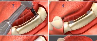
What happens during a bone transplant - stages of the operation
The details of a specific bone graft surgery will vary greatly, depending on the reason for the procedure being planned. You should ask your doctor about the details of your specific surgery.
An orthopedic surgeon will perform the procedure with the help of a team of medical professionals.
You can expect the following steps:
- The patient will receive anesthesia so that they will not feel pain or discomfort during the procedure. The anesthesiologist will closely monitor all vital signs - such as heart rate and blood pressure - during surgery.
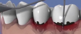
- After treating the affected area, the operating surgeon will make an incision through the skin and muscle surrounding the bone that will receive the bone graft.
- In some cases, the surgeon will also make another incision to collect the bone graft. This may be material from your own femur, leg bone or ribs. Using special instruments, your surgeon will remove a small portion of the bone.
- The surgeon will then place a bone graft between two pieces of bone that are meant to grow together. In some cases, your doctor may secure the bone graft with special screws and make any other necessary bone repairs.
- The layers of skin and muscle around the donor bone will be surgically closed - and, if necessary, around the site where the affected bone was harvested.