Each person goes through the stages of the eruption of the first teeth, the development of milk teeth and their subsequent replacement by permanent ones. Despite their similar appearance and function, temporary and permanent teeth have differences, which we will talk about, at the same time we will consider the timing of the appearance of the main teeth, possible problems with them in the process of their development.
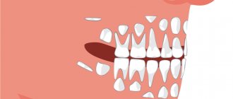
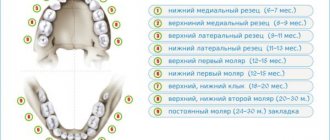
The photo shows a diagram of the structure of human teeth
Content:
- Is there a difference? 2.1. Dairy 2.2. Indigenous
- How to tell the difference?
- Is there always a change?
- Are there baby wisdom teeth?
- Can milk root remain in the gums?
- When does radiography come to the rescue?
Children's milk and permanent teeth raise many questions from their parents.
The most attentive mothers and fathers try to clearly monitor the change in temporary bite. It happens that they do not understand whether the baby’s milk units remain in the baby’s mouth or are no longer there. How to determine whether a molar or baby tooth is? To do this you need to have certain information.
Daily doctor's advice on caring for baby teeth:
- Start brushing your baby's teeth only after they have erupted; use a special brush for babies.
- When your child turns one year old, buy a brush with soft bristles and toothpaste without fluoride; it will not harm the child if he swallows it.
- Brush your baby's teeth 2 times a day (morning and evening)
- Don't put off going to the dentist. They should be regular, even if there are no signs of concern.
- Include more fresh greens, fruits and vegetables in your diet.
Is there a difference?
It happens that, trying to determine which children have milk teeth, mothers make a significant wave of their hand and say that there are no differences between the new and old units. In fact, they are.
Dairy
They take their places for closer to two to three years. The very first ones erupt when the baby is 6-12 months old. There are only twenty primary teeth (while there are 32 primary teeth). Therefore, they are arranged in a row quite freely. As the jaw grows, the spaces between them increase significantly. This can be seen with the naked eye.
At the age of 5-6 years, temporary teeth begin to loosen and fall out. This process is completed by the age of 10-12 years.
Indigenous
They appear at five or six years old or a little later. First, molars grow , which become “sixes”. After this, the incisors begin to wobble. They are pushed out by a constant shift.
Children with permanent dentition usually have 28 teeth. The four terminal units (which are popularly called “wise”) usually appear after 20 years. For many, it grows at 30-40 years old. And this is the norm.
How to tell the difference?
It is possible to determine which “generation” a tooth belongs to by its:
- Size and shape. Molars are larger, because they are designed for the jaw of an adult. Their edges and boundaries are clear and lumpy. Dairy ones have rounded, smooth edges.
- Blossom. Children's units are white with a bluish tint, adults are yellowish-gray. This is due to the fact that the latter has a higher percentage of mineralizing components. Over the years, they also turn white, but at first they don’t look very presentable.
- Position in the oral cavity. The molars face towards the lips and cheeks. They appear to be slightly tilted. Dairy plants grow evenly and straight.
If we talk about the differences in more detail :
- Six and seven are always radical. You need to count from the middle line of the row (located between the central incisors) towards the ear. In the milk row there are only fives, since there are ten teeth at the bottom and at the top.
- If you have questions regarding the fours and fives, then maximum attention should be paid to their form. Primary teeth have wide crowns and chewing cusps (usually four). If a change has already occurred, then there will be fewer tubercles and the crown will be narrow.
- Temporary fangs are smaller than permanent ones. By the time they fall out, their sharp tips become dull. Instead, units with clearly pointed apexes grow.
- When assessing the nature of the incisors, you need to look at the size. In dairy ones, the width is about 4 mm, the height is 5-6. Permanent ones are usually larger. During the period of change of bite, children's incisors have smooth, even edges, while adults' incisors are lumpy.
Is there always a change?
Not a single child's tooth should be preserved - they all fall out. Instead of each temporary one, a permanent one is cut through. But it also occurs in dental practice that the rudiment of the root unit is missing. This can be seen in the X-ray of the jaws.
In this case, the life of the baby tooth is extended to the maximum - after all, nothing will grow in its place. When an empty space appears in the dentition, the issue of prosthetics is discussed.
To eliminate partial congenital edentia, you can implant an implant or install a dental bridge. In the first case, there is no need to file adjacent teeth. The doctor, under local anesthesia, installs an artificial titanium root into the jaw tissue. When it takes root, it fixes the dental crown.
In the case of a bridge, it is necessary to depulpate and prepare the neighbors . This is not very healthy, since their service life after such manipulations is significantly reduced. Therefore, young patients who do not have separate molars are recommended by dentists to resort to implantation.
The structure of human teeth
Teeth are not only intended for mechanical processing of food, but are also necessary for the formation of speech, breathing, and influence facial features. To navigate what dentists advise, how to take care of your teeth, and what the risks of disease are, it is useful to know how they work.
Anatomical structure
3 parts that make up a tooth:
- Crown. The visible part of the tooth used for chewing. The outside is covered with durable enamel, which protects it from bacteria, chemicals contained in food, water, and saliva. The surfaces have their own names: Facial (vestibular) - in contact with the lip or cheek.
- Lingual (lingual) – the opposite of the facial, involved in the formation of speech.
- Occlusion – the upper surface in contact with the tooth of the opposing jaw.
- Contact (approximal) – contacts with adjacent teeth.
Milk teeth, having a largely similar structure, also have differences in anatomy:
- They are noticeably smaller in height than permanent ones.
- The crown is much wider than the root.
- Enamel is thinner and more fragile.
- The roots are more round.
- The wear of baby teeth, as well as their spontaneous loss, is a normal physiological process.
Histological structure
The structure has several layers:
- Enamel is the most durable fabric. When a tooth just erupts, a cuticle is located on it, which is gradually, under the influence of saliva, replaced by a pellicle.
- Dentin is a highly mineralized tissue that resembles bone, but has better mechanical strength. Instead of enamel, the root part of the dentin is covered with cement.
- The pulp, the central part of the tooth, is a soft connective tissue containing a large number of blood vessels. Caries and inflammatory processes “owe” pain to the pulp with its large number of nerve endings.
Milk teeth are distinguished by dentin with a lesser degree of mineralization, which weakens their protection against caries. The volume of pulp occupies most of the tooth, and small protective layers (enamel and dentin) provide less protection against the penetration of bacteria and the development of inflammatory processes.
Types of teeth
There are 4 groups:
- Incisors. 4 chisel-shaped cutters. The largest are a pair of upper central incisors, and the situation is opposite from below - the lateral incisors are slightly larger than the central ones.
- Fangs. 2 on the upper and the same number on the lower jaw. Their length is longer than the others, the front wall is convex.
- Premolars. There are 8 in total, prismatic in shape, the upper surface with two tubercles (buccal and lingual). Premolars have 2 roots. The second premolar has a larger buccal surface. There are no primary premolars.
- Molars. The first molar (molar) is the largest tooth in the upper jaw. The chewing surface has four tubercles, 3 roots. The cubic-shaped second molar is smaller, and the buccal tubercles are larger than the lingual ones. The third (“wisdom tooth”) is in many ways similar to the second, but not everyone has it.
Can milk root remain in the gums?
If you carefully examine fallen children's teeth, you will not be able to see any semblance of roots. Some mothers unknowingly begin to panic - it seems to them that a significant part of the unit remains in the deep tissues of the gums.
There is no need to worry - this is how it should be. The absence of roots is the result of their gradual resorption. This process starts long before the day of loss. That is why during a natural change (when a tooth falls out without outside help), the child does not experience pain.
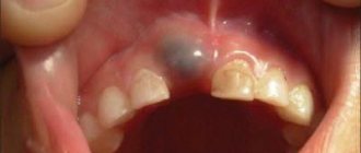
Injuries
An accident or incident, such as a fight, can cause a tooth injury. And it doesn’t matter whether a small part has broken off, or the tooth has cracked, as they say, “to the point of bleeding” - the help of a doctor is definitely needed. In some cases, lost dental tissue is replenished. If a tooth is broken into pieces, it will most likely need to be completely removed and a prosthesis replaced every year. And the answer is simple - the dental tissues have not yet fully matured, the body is growing. And it is necessary to take full care of your teeth at such an early age. In case of extension, the operation is performed by introducing composite materials that replace enamel and dentin.
When does radiography come to the rescue?
Very rarely, but still situations occur in dental practice when even the doctor doubts which tooth he has to work with - a child’s or a molar. In this case, the capabilities of radiography are used. In the photographs you can see:
- length and structural features of the roots;
- presence/absence of radical primordia;
- the location of the unerupted unit and the direction of growth of its incisal edge.
If you need to find out exactly what the situation is with a change in bite in a child, contact your dentist. He will tell you the number of units that should fall out in the near future, and tell you whether it is worth interfering with this process.
Possible problems
Despite the fact that changing teeth is a natural physiological process, some children and their parents may encounter a number of problems that require contacting a pediatric dentist.
No molars
The absence of permanent units can be caused by congenital edentia - the complete or partial absence of tooth buds.
Another reason for the absence of molars is previous inflammatory diseases - periostitis or periodontitis, resulting from progressive caries. Inflammatory diseases of the periosteum and periodontal tissues have an extremely negative effect on the condition of the tooth buds and can lead to their death.
Important! It is absolutely necessary to treat baby teeth for caries. You should not assume that the problem will go away on its own with the change of teeth. The progression of the disease can negatively affect the health of the tooth buds.
Molar tooth hurts
The enamel of newly emerging permanent teeth is still poorly formed. The low level of its mineralization makes teeth vulnerable to cariogenic microflora. This can lead to the development of caries and cause pain.
Due to poorly formed enamel, tooth sensitivity to external irritants (cold, hot, sour, sweet) may increase, which is also accompanied by painful sensations.
Important! Normally, permanent teeth do not hurt. If pain occurs, you should contact your pediatric dentist. The specialist will determine the cause of the pain, carry out the necessary treatment, fluoridation or remineralization of tooth enamel.
Molars grow crooked
The incorrect position of permanent teeth can be caused by two reasons - the growth of the permanent unit outpaces the process of loss of baby teeth or they were removed ahead of schedule, which led to incorrect formation of the rudiments of permanent teeth.
In this case, there is only one way out - orthodontic treatment of malocclusion.
Important! A malocclusion must be corrected. The sooner you contact a dentist, the more successful the treatment will be. The child will be prescribed to wear removable or fixed orthodontic appliances that will help straighten the permanent teeth and bite.
Injuries
Due to their activity and lack of experience, children can accidentally injure a newly emerging permanent tooth. Due to mechanical damage, cracks and chips may appear on it. The damage looks unattractive. Caring for such teeth is complicated, since food debris can get stuck in the cracks, which will certainly lead to the development of caries.
Important! If a child accidentally injures a permanent tooth, it is necessary to seek help from a dentist. The specialist will assess the complexity and depth of the damage and will build up the missing volume of tooth tissue with composite materials.
Tooth loss
Loss of healthy permanent teeth can only occur as a result of severe trauma to the jaw, for example, during a child’s fall or fight. A diseased molar may fall out on its own. In this case, you will also need to consult a specialist. Most likely, the child will undergo temporary prosthetics for the lost unit, which will not disrupt the formation of a correct permanent bite.
The tooth is loose
Looseness of a permanent tooth is an alarming symptom indicating a pathology of the dentofacial apparatus or the presence of inflammation. Consultation with a specialist is required!