When visiting a dentist, you have probably heard them more than once say such phrases as, for example, “lower left five” or “16th tooth” or “2nd premolar.” Experienced patients know that this is the numbering of the teeth, which is necessary for the doctor to describe the condition of the oral cavity when filling out the card. There are several systems used in dental practice for this purpose.
The dentofacial system and the universal principle of numbering
Each dental unit in the lower and upper rows performs its own functions. They are determined by its position and structure. For example, the purpose of incisors is cutting, canines are tearing off, holding, premolars are preparing, grinding, molars are grinding, chewing.
The structure of the dentofacial rows in the oral cavity:
- four incisors;
- two fangs;
- four premolars;
- six molars.
“Pairedness” refers to the symmetrical arrangement of the same number of teeth on the left and right. Its middle is located between pairs of central incisors - two are assigned to the left side, two to the right. Taking into account the location, teeth numbers are obtained in dentistry:
- two incisors - numbers 1 and 2 (counting from the center);
- fang - number 3;
- two premolars - numbers 4 and 5;
- three molars - numbers 6, 7, 8.
The calculation formula is valid for the left and right sides of the row. To prevent confusion during treatment and to make the records in the medical record clear to other dentists, doctors use the concept of jaw segments. They indicate the upper or lower location, the side of growth. The image below shows how the teeth are arranged by number in adults on the upper/lower jaw.
Segments:
- the top right is the first (10 is added in the records, that is, the 6th molar on the top right will be 16, the central incisor will be 11);
- left above - second (add 20, that is, the 6th molar on the left above will be 26);
- lower left - third (add 30);
- lower right - fourth (add 40).
With full rows, there will be 48 teeth in the pattern. Therefore, there is no need to panic if the doctor at the clinic sends the patient to take an X-ray of 48 units. We are talking about wisdom tooth treatment here. In standardized schemes, counting is done clockwise.
Price
- Primary appointment (examination, consultation) with a dentist (special offer) 100001
For free
Promotion
- Dental prosthetics using an implant with a temporary crown using the laboratory method (PMMA) 152006
7 500 ₽
- Simple tooth extraction 160001
1 900 ₽
- SGS implant + turnkey crown
24 900 ₽
Promotion
- Osstem implant + turnkey zirconium crown
29 900 ₽
Promotion
- Astra Tech implant + turnkey zirconium crown
59 900 ₽
Promotion
- Nobel implant + turnkey zirconium crown
59 900 ₽
Promotion
- Straumann implant + turnkey zirconium crown
59 900 ₽
Promotion
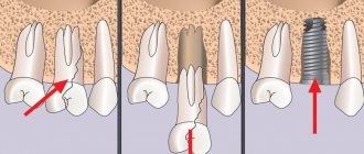
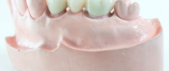
Implantation of the seventh tooth involves inserting an implant into the jaw and fixing an artificial crown on it. The method ensures complete restoration of the functions of the seven, but is associated with some risks. This is a surgical operation, with preparation and a rehabilitation period of 3-4 months. To replace the 7th lower or upper tooth, a classic two-stage treatment protocol is suitable, the implementation of which may require bone tissue augmentation. If the protocol is followed correctly, the patient will receive a tooth that matches the natural one.
Children's dental chart
The given counting system cannot be used to count baby teeth. It is valid only for permanent ones. To avoid confusion in the future, a different jaw segmentation scheme is used, but the serial numbers remain the same. That is, the incisors are still assigned the numbers 1 and 2, and the clicks are assigned the number 3. Let’s find out which tooth numbers dentistry uses in children’s counting schemes.
Segments:
- top right - first (marked as 50 in entries);
- top left - second (marked 60 in records);
- bottom left - third (marked 70 in records);
- the bottom right is the fourth (marked as 80 in the records).
Numbers are assigned clockwise. But the children's counting scheme can cause difficulties due to the incompleteness of the dentition.
Is it possible to remove a cyst while saving the tooth?
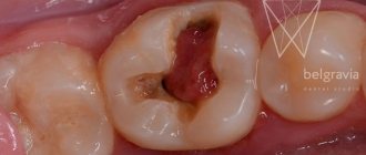
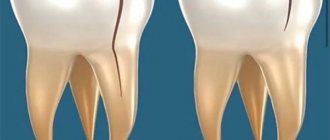
A cyst is a formation that appears at the root of a tooth and is a capsule filled with yellowish exudate. It may not manifest itself; it is often discovered by chance, when clarifying another diagnosis.
Such formation is fraught with displacement of teeth and destruction of periodontal tissue. Therefore it must be deleted. And our clinics have all the conditions to save a tooth by removing the capsule.
Dental-sparing cystectomy
– excision of the cyst, the secondary appearance of a formation in this place is excluded. It is performed on the lower and upper jaws under anesthesia, which relieves pain and reduces blood circulation in the operating area. Sedation allows you to undergo surgery to remove a cyst or granuloma of a tooth without fear or worry. You are conscious, will not lose self-control and can comply with the doctor’s requests, but you will not feel any discomfort during the manipulations.
It is also practiced to remove a cyst through a tooth, when the dental canals can be opened. Exudate is removed from the internal cavity of the cyst, and the medicine is injected into the root canal. Laser removal of a dental cyst is performed in the same way. But the beam simultaneously disinfects the cavity and channels, and cauterizes the capillaries. The operation is practically bloodless.
Other counting systems
It’s easier to represent the location of teeth by numbers - the diagram is called the 2-digit Viola system. It has been operating since 1971. Its advantage is the convenience of calculation without the need to map the series. But in international dental practice, other systems are also used: Haderup, Palmer-Zsigmondy (used by orthodontists and surgeons), alphanumeric (in the USA). Each of their schemes has its own system for counting teeth in children and adults.
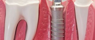
Number of roots and canals in human teeth
Most of the oral cavity is occupied by organs whose main function is to chew and grind food into smaller pieces.
This promotes its complete digestion and better absorption of nutrients. A tooth is an organ that has a characteristic shape and consists of several parts. In dentistry, the outer visible part is called the crown, and the inner part is called the root. The element connecting the crown and root is the neck. An interesting fact is that, unlike a crown, a tooth can have more than one root. How many roots a tooth has, as a rule, depends on the location and purpose of the organ. In addition, its structure and number of roots are influenced by hereditary factors. The situation can only be definitively clarified with the help of an x-ray.
The article provides detailed information about how many roots there are in the frontal, lateral chewing teeth, as well as the number eight, or the so-called wisdom tooth. In addition, you can find out what the purpose of the tooth root is, why the chewing units need nerves. The dental advice provided in the following material will help prevent the development of dental diseases.
Channels in baby teeth
There are as many nerves in baby teeth as there are in molars—one. In addition, temporary units are similar to permanent ones in the structure of the root system. That is, a milk tooth such as the upper six or second molar has a canal system similar to its molar brother, the second premolar.
Nerve endings perform standard functions:
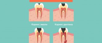
- signal about developing caries;
- responsible for the growth and development of teeth;
- control the flow of water and nutrients to dentin and enamel.
The root canals of baby teeth are also treated and filled, but the tactics of their treatment depend on how long ago they erupted. Under the temporary units, permanent ones are formed, so treatment should be aimed at preserving them. Milk teeth can only be removed if the permanent teeth are ready to emerge.
Read also: How long does a tooth grow after milk
The roots of permanent incisors, canines and molars do not form immediately, but over the course of about 3 years. Treatment of permanent teeth with unformed roots also differs from the standard one. The canals in the teeth of patients four, five, six years old (depending on the rate of formation of the dentoalveolar apparatus) are filled with a special paste with calcium and fluoride, which helps close the roots.
Process and stages of channel healing
The root canal healing process has a clear sequence of steps:
- First you need to make a correct diagnosis of the disease (disturbances in normal functioning, performance)
. In complex cases, the dentist sends the patient for an x-ray. - After this, the doctor carries out the necessary preliminary oral procedures for healing.
- Healing canals is a rather painful procedure, so the tooth must be numbed by injecting anesthesia (usually into the gums near the diseased tooth).
- After this, complete asepsis of all instruments is carried out and the unhealthy tooth is separated from the others using a special rubber film (caffedram).
- Next, the unhealthy tooth is opened with the help of a drill, providing maximum access to the unhealthy canals, after which their initial cleaning is carried out, the infected pulp is removed, and at the same time the canals are treated with special pharmaceutical products.
- Afterwards I dry them and seal them with special materials.
Wisdom tooth
(*7*)An eight or a wisdom tooth is quite unique and does not fall under standards and statistics. The upper one can have a completely different structure with channels from 1 straight to 5. The lower eight is most often three-channel, but often when opened during healing, additional branches can be discovered.
Apart from the rest, a wisdom tooth differs from the rest in that its canals are quite rarely of the correct shape, often very curved and with a narrow passage, which makes their healing and filling very difficult.
Wrong worldview
Because a tooth consists of roots and a pre-coronal part, from time to time there is a misconception that there are as many canals in teeth as there are roots . This is far from true, because the canals quite often branch off and bifurcate near the pulp. Moreover, several channels can run parallel to each other in one root. There are also cases of their bifurcation at the apex, which is why it turns out that one root has two apexes and this, naturally, complicates the work of doctors when filling similar teeth.
Taking into account all the individual characteristics of the teeth, dentists need to be very careful when treating (the process of alleviating, removing or eliminating symptoms and diseases)
and filling, so as not to miss any branch. Indeed, from time to time, without an x-ray, it is very difficult, even during an autopsy, to identify how many canals are in the teeth.
Prevention after healing
After the procedure for healing the root system you should avoid putting stress on the treated tooth for some time; moreover, you should not eat food earlier than two hours after therapy (therapy is a process to relieve or eliminate the symptoms and manifestations of the disease)
, otherwise the filling is not completely hardened It might just fall out. But the same thing can happen if the doctor uses low-quality drugs or performs incorrect treatment (for example, the canals were over-dried or not dried before filling).
Also, after filling, for some time (up to several days) the tooth may feel pain (an unpleasant sensory and emotional experience associated with actual or potential tissue damage or described in terms of such damage) when pressed, or simply ache, cause discomfort, or have excessive sensitivity. This is usually a common condition if pain (physical or emotional suffering, painful or unpleasant sensation)
powerful, you can take painkillers.
If pain (physical or emotional suffering, painful or unpleasant sensation)
does not go away after a certain time, this may also be an indicator of disgusting healing (insufficient cleaning of infection
(The term means various types of interaction of foreign microorganisms with the human body)
or infected pulp, leaky filling, introduction of low-quality pharmaceuticals or materials).
From time to time, there are cases of allergic reactions , which are also accompanied by incessant pain, and from time to time itching and rashes appear on the body. It may be caused by a reaction to a pharmaceutical product or the material that was used for the filling. In this case, it must be changed to something else that will not cause allergies.
In all these situations, it is imperative to consult a doctor as soon as possible for a re-examination and dental prophylaxis in order to identify the cause of the defects.
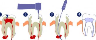
Root canal treatment
The treatment plan for dental canals consists of several stages:
- First, access to the problem area is freed: using a special dental instrument, the filling or the area of the crown damaged by caries is removed.
- Then the contents of the pulp are removed, and the canals are cleaned mechanically using antiseptic drugs.
- After this, the root is prepared for filling. At this stage, the dentist can form the correct conical shape of the canal passage.
- Then the canals are carefully sealed. If baby teeth are treated, the dentist uses a special filling paste, which gradually dissolves as the root dissolves.
- After this, a filling is placed on the crown.
This treatment regimen is standard and does not depend on exactly how many canals there are in the diseased tooth. The main thing is that all dental canals are cleaned, treated with an antiseptic and carefully closed. If treated incorrectly, it may be necessary to remove the tooth and visit an oral surgeon.
Teeth can be single-channel, two-channel, three-channel and even eight-channel. If one of the ducts becomes inflamed, it is necessary to clean and seal not only it, but also all other canals, since the infection could penetrate into them.
Tips from dentists for disease prevention
The development of dental pathologies can be prevented only by following the advice of qualified doctors and observing the rules of oral hygiene.
So, for prevention purposes, dentists recommend:
- do not abuse the rules of hygiene, brush your teeth only in the evening and in the morning. More frequent exposure to tooth enamel contributes to its wear;
- hygiene procedures should be carried out half an hour after eating;
- use rinses to destroy germs remaining in the mouth after brushing;
- Cleaning should be carried out for at least 3 minutes, performing circular movements.
The main rule is that if the first signs of the disease are detected, you should immediately contact your dentist. This will help prevent further development of pathology and preserve teeth.