3D CT – what is this procedure and what is it for?
3D imaging of the jaw is one of the main dental procedures used in diagnosis. The data obtained allows us to assess the condition of a specific maxillofacial area and plan further treatment.
This diagnostic tool is universal; it is used by both therapists and orthopedists, periodontists and implantologists. Using a 3D image, the therapist assesses the condition of the roots and canals of the tooth, and clarifies the localization of the inflammatory process.
An orthopedist can examine the anatomical structure of the temporomandibular joints, an implantologist can examine the volume and density of bone tissue in the area of the upcoming operation, as well as the parameters of the maxillary sinuses (maxillary sinuses).
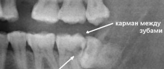
Sometimes a large part of the tooth remains, as they say, “behind the scenes”, and the dentist simply cannot see it. Then computed tomography comes to the rescue, which allows you to examine the hidden area and prevent unpleasant consequences for the patient.
Until recently, the “gold standard” for instrumental diagnostics was a panoramic image. Today there is a more advanced method in which the maxillofacial area is scanned using computer technology.
Safety of X-ray diagnostics
The standard of safe radiation exposure is regulated by the federal law “On Radiation Safety of the Population”. Article 9 of this law “State regulation in the field of radiation safety” states that the average effective dose for the population per year is 0.001 sievert (1 millisievert). In this case, the dose received during one cone-beam computed tomography is 0.09 millisieverts. Accordingly, at least 10 CT scans of teeth can be safely performed per year.
For comparison, during air travel the radiation dose is on average 0.08 millisieverts. The dose of so-called natural radiation is 2.4 mSv. It consists of the effects of cosmic and solar rays, the influence of atmospheric radioactive atoms (radon), radiation from soil and building materials, and food radionuclides. Thus, performing a dental CT scan is absolutely safe for your health.
Unique visualization method
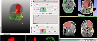
A computed tomograph produces a high-quality three-dimensional image of an individual tooth, maxillary sinuses, one or both jaws. Unlike standard panoramic images, a 3D tomogram allows the doctor to see the desired anatomical structures in a virtual section, from any angle.
During the procedure, the doctor can enlarge, rotate and study the maxillofacial area of interest at the required angle, which is unrealistic with conventional radiography.
Computed tomography is an integral and primary stage of examination before implantation. It allows you not only to assess the condition of bone tissue, but also to measure its height, width and density. Moreover, three-dimensional beams help to choose the optimal method for installing implants through preliminary virtual surgery.
3D CT is a multi-purpose and indispensable diagnostic tool that makes it possible to avoid many medical errors and complications. Thanks to this examination, the quality of treatment increases significantly and eliminates unnecessary traumatic operations.
What does a CT scan of the jaw show when diagnosing serious diseases?
As a diagnostic test, CT is also suitable for identifying fairly serious diseases. High-precision three-dimensional scanning allows you to identify pathology in the early stages and begin timely treatment.
Jaw cancer
It is a bulky neoplasm that can be found in epithelial tissues. It is not common, but according to statistics, jaw cancer is diagnosed in about 0.8% of the total number of people with malignant tumors. The disease can affect both the lower and upper jaw (here it is more common). The most common forms of pathology are osteogenic sarcoma and carcinoma.
Detection requires differential diagnosis using MRI and CT, as well as a pathomorphological examination of the material taken during biopsy. CT results show data that allows us to determine not only the nature of the tissue manifestation of the tumor, but also the boundaries of bone structures. Most importantly, tomography can reveal signs of infiltrative osteolysis.
Osteomyelitis of the jaw
It is expressed as an infectious-purulent inflammation that affects the jaw bones. It can be chronic and acute. Quite often, traumatic osteomyelitis can be diagnosed, which is a consequence of complications after injuries (for example, fractures) of the lower jaw. If ignored, the pathology can lead to the development of osteonecrosis.
Computed tomography is a priority technique for timely diagnosis of the disease. CT scan of the jaw shows:
- volume of affected bone tissue;
- the condition of the soft tissues surrounding the affected bones;
- form of the disease - limited, widespread or diffuse;
- degree of subperiosteal spread.
As a result, the attending dentist is able to determine what phase the disease is in - exacerbation or remission.
The Smile Factor network of clinics invites residents and guests of St. Petersburg for dental diagnostics using innovative equipment with minimal radiation exposure.
Indications
Computed tomography is prescribed to identify:
- hidden carious lesions;
- defects in the structure of the jaw and dentition;
- fully or partially unerupted teeth;
- dystopic dental units with incorrect location or direction of growth;
- supernumerary teeth;
- damage to the dentition due to jaw fractures and other injuries;
- pathologies of the temporomandibular joint TMJ;
- tumors, cysts and other neoplasms in the jaws;
- condition of periodontal and periodontal tissues in case of gum disease, inflammation in the root area;
- number of roots, canals of teeth;
- cracks in the roots of teeth;
- features of the structure of bone tissue before jaw surgery (installation of implants, bone augmentation).
A 3D photo must be taken before dental implantation. The fact is that the jaw bone is clearly visible on a regular x-ray, but it does not allow assessing the soft tissues. On a three-dimensional tomogram, you can see in detail not only the bone, but also the nerve of the lower jaw, as well as blood vessels.
A 3D tomogram is much more informative than a panoramic image or targeted photographs of all teeth.
Is special training needed?
The preparatory stage includes an initial consultation with a dentist, who conducts a survey to identify possible contraindications. Further preparation depends on what type of research needs to be carried out:
- Normal (without contrast injection). Such a CT scan will not require additional preparation, except for the general requirements: it is recommended to remove all metal objects from the head - earrings, hairpins, etc. if present, remove the hearing aid and removable dentures; warn the radiologist if there are non-removable orthopedic structures in the mouth (this will allow the specialist to adjust the device in a special way).
The lack of serious preparation before taking a CT scan of two jaws or teeth allows diagnostic studies to be carried out immediately after receiving an order from a doctor.
CT scan before implantation
Diagnostics using a computed tomograph before installing implants allows, first of all, to determine whether implantation is necessary at all. The image will give a complete picture, and the doctor will see where the teeth are missing, whether there are problem units, and whether they can be cured.
The 3D tomogram will show:
- hidden carious cavities;
- unerupted and “extra” teeth, which may interfere with the installation of artificial pins;
- properties of roots, canals - curved, narrow and long canals require a special approach, which should be taken into account before implantation;
- bone dimensions in height and width, on the basis of which the type and size of the implant is selected;
- condition of bone beams, partitions, voids in the jaw bone;
- the presence of inflammatory processes in the root area - cysts, granulomas, abscesses where implants are planned to be installed. All this needs to be treated or removed before surgery;
- inflammation in the paranasal sinuses and lacrimal ducts, which can become a temporary obstacle to implantation;
- density, size, inclination of the alveolar process, thickness of the cortical bone layer, taking into account which the optimal type of artificial pin is selected;
- the physiological structure of the maxillary sinuses, the mandibular canal to determine the angle of inclination of the implant rod;
- defects and anomalies in the structure of the dentofacial apparatus;
- quality of installation, strength of fixation of implants after surgery for their implantation;
- severity and nature of traumatic injuries in fractures.
Based on the results obtained, a virtual operation to install the rods is performed. The appropriate size of the titanium pin is selected, its inclination and the point of implantation are determined, bypassing the anatomical structures. Thus, the final outcome of implantation is modeled.
Next, the tomographic data is loaded into the computer, and the program creates a three-dimensional model. The patient’s personal surgical template is printed on a 3D printer - an overlay with guide holes for inserting rods.
During implantation, the template is placed tightly on the gums, and the placement of the pins is carried out with extreme precision.
How long does it take to do a CT scan?
The duration of tomography depends on the type of diagnostic equipment used. Before doing a CT scan of the jaw and teeth, it is better to ask what kind of tomograph is used in the clinic. Modern devices allow you to carry out the necessary manipulations as quickly as possible with minimal radiation. If you are offered a procedure that will last more than 15 minutes, then you should refuse. Only innovative equipment allows you to carry out the required research in just a few minutes, guaranteeing highly detailed images.
On average, everything is done in no more than 10 minutes, taking into account the instruction time and the preparatory stage. Please note that 3Shape is also used for 3D diagnostics, but this intraoral scanner is not suitable for all types of diagnostic studies. The attending physician will be able to determine which scan is required for you after collecting a general clinical picture and based on the reason why you came to the dentist.
Multispiral or cone beam?
A multispiral tomograph performs layer-by-layer scanning of an object along a spiral trajectory caused by the continuous movement of the table and the X-ray tube relative to each other.
Most often, MSCT - multislice computed tomography - is used in maxillofacial surgery for facial injuries and pathologies of the temporomandibular joint.
In dental practice, especially when planning implantation, this method is not widely used due to insufficient data accuracy. Since the patient lies down during the examination, the jaw connection is distorted.
In addition, the radiation level of MSCT can reach 1000 μSv, which is unacceptable, since implantological treatment involves more than one procedure over several months.
Cone beam CBCT is a more modern, accurate and safe method compared to MSCT. Its radiation exposure is less, about 25-50 μSv, which makes it possible to carry out the procedure several times a year.
Contraindications for examination
Research is not recommended if:
- you are overweight;
- you have an exacerbation of pain;
- restlessness is brought to the point of hyperkinesis (uncontrolled movements);
- the period of bearing a child is underway;
- there is an allergy to iodine;
- there is renal failure.
How often can a CT scan of the jaw be done?
Computed tomography is based on the already familiar X-ray radiation. It would seem that in this case it should be carried out quite rarely, for fear of tangible consequences for health. However, this is not true: the tomograph itself is built differently than an x-ray, which helps keep the radiation dose to a minimum. Therefore, you should not expect any irreversible consequences even with repeated use of this technique.
3D CT or panoramic image?
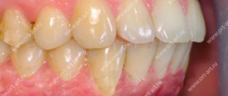
An orthopantomogram allows the doctor to assess the condition of the teeth, root canals, and soft tissues. With its help, hidden inflammations, abscesses and abnormally located teeth are revealed.
However, a panoramic photo does not give a 100% accurate picture; the error is about 20%. Even a slight shift causes the focal spot to shift, and the image is compressed or stretched.
Due to the difference in the refraction of X-rays by tissues of different densities, it is impossible to assess the properties of the cancellous bone layer, since it is simply not visible behind the denser periodontium.
A two-dimensional orthopantomogram is, in fact, an auxiliary technique that gives a general idea of the condition of the oral cavity and identifies mainly obvious pathologies. It does not show the configuration and structure of the alveolar process at the desired level.
The advantage of a three-dimensional 3D tomogram is that it produces not one flat photo, but several consecutive images from different angles.
The doctor sees and evaluates all necessary objects located at any depth, from all sides and at different angles.
Preparatory stage
CT scan of the upper and lower jaw according to the basic (native) protocol does not require special preparation. If a CT scan of the jaw with contrast is prescribed, the diagnosis is usually carried out with a break in food for 2 hours. If the patient suffers from renal failure, tests to determine the level of creatinine in the blood are preliminarily prescribed before contrast tomography. This way, the doctor can assess the possible risks of developing nephropathy after administration of a contrast agent. Before entering the CT room, it is better to change into comfortable clothes and remove all jewelry.
How to make a 3D tomogram
Usually the procedure is performed standing, the patient bites a small flat plate with his teeth and stands without moving for 15 to 30 seconds. The device makes several rotations around the head, managing to take about two hundred pictures in various projections.
In 10-15 minutes, the information is processed and transferred to electronic media.
We invite you to make a three-dimensional tomogram in our clinic using the latest generation dental tomograph. Sign up for the procedure online or by phone at a time convenient for you.
How is a CT scan of teeth and two jaws done?
After the preparatory stage described above, the procedure itself begins with a short instruction - the radiologist explains how and why it is necessary to remain still while the device is operating. After this, the CT scan proceeds as follows:
- if necessary, a contrast agent is introduced at this stage;
- The patient wears a vest with a lead layer, which will provide additional protection to the chest area from radiation;
- depending on the type of scanning equipment used, the patient is asked to stand or sit near the tomograph;
- the radiologist fixes the patient’s head with special supports provided in the tomograph (fixation is carried out in the frontal, temporal and chin parts), and the hands are asked to be placed comfortably on the handrails;
- the specialist starts the equipment and the sensor, which takes and transmits 3D images, begins to rotate around the head (in one revolution, the sensor takes 150-200 high-resolution 2D images, after which it converts them into a three-dimensional picture);
- the sensors take readings for no more than 30 seconds and to obtain the highest quality results and accurate images, it is recommended to remain motionless during this time;
- The radiologist releases the patient's head from fixation on the supports and this is where the procedure ends.
Next, it will take a few minutes for the device to process the scanned data and write it to disk. You may be interested and useful to know - in the article “CT of the jaw for dental implantation” we talk in detail about the features of tomographic studies before implanting artificial roots.
You might be interested in:
Dental diagnostics
Computed tomography of teeth and jaws
X-ray of teeth
3D image of teeth