- Pediatric orthodontics - correction of malocclusion in children
- Adult orthodontics - bite correction in adults
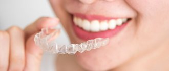
- Treatment with removable orthodontic appliances
- Correction of malocclusion without braces using mouthguards in adults.
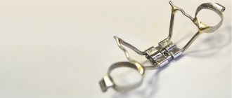
- What orthodontic devices do our orthodontists use to correct the bite?
- How is braces treatment performed?
- What types of braces are there?
- How and why to choose an orthodontist?
- Reminder to the brace bearer
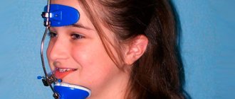
- What is orthognathy?
Do you know what awaits a patient who has braces installed? Let's find out!
When is orthodontic treatment with braces necessary?
A good reason to seek orthodontic treatment may be:
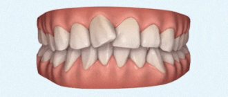
- Ugly smile, crooked teeth.
- Functional disorders of the dental system that worsen the patient’s quality of life: there is no ability to bite or chew well, teeth are destroyed and worn out.
- Problem with the jaw from overload of the temporomandibular joint (TMJ).
- Lack of space for prosthetics due to deformation of the dentition.
But it happens that the patient himself does not see the reasons for orthodontic treatment, and his dentist sends the patient to an orthodontist. Why?
Because his correct interdental contacts are impaired. The patient may not be aware of this; he is satisfied with the smile and is not bothered by anything. But the dentist sees the issue at the level of functioning of the dental system. If the teeth do not close properly, this means that the chewing load on the teeth is distributed unevenly. Sooner or later this will lead to illness. Timely orthodontic treatment is the only way to maintain healthy teeth for the rest of your life.
Numerous works by foreign and domestic authors emphasize that the thickness of the alveolar bone determines the boundaries of orthodontic tooth movement. Neglecting this fact can lead to undesirable side effects from periodontal tissues. Tooth movements that decentralize the teeth from the alveolar ridge beyond the cortical plate pose the greatest risk for the development of bone defects such as dehiscence and fenestration. The criticality of the process depends on the condition of the bone tissue of the alveolar process, the vestibulo-oral inclination of the teeth and the patient’s periodontal biotype. Historically, the term “periodontal biotype” was introduced by S. Ochsenbein in 1969. Based on criteria such as the height and width of tooth crowns, the thickness of the alveolar bone and the volume of gum tissue, as well as the size of the attached gum zone, two types of periodontium were classified - thin and thick. Loss of periodontal attachment and recession of the marginal gingiva during orthodontic treatment are adverse effects that are observed in patients with thin periodontal biotypes. The thin periodontal biotype is characterized by high and narrow tooth crowns, a small zone of attached gums, multiple dehiscences (slit-like defects of the alveolar bone with exposure of the root) and fenestrations (window-shaped defects). Thin, scalloped periodontium, according to research, is more common among the female population. A thick periodontal biotype is usually combined with short and wide tooth crowns and a pronounced and significant area of attached gum; The marginal bone contour is massive, the gums have a more pronounced fibrous layer. According to the literature, forced loads during orthodontic treatment of a patient with a thin biotype are one of the main iatrogenic factors leading to the formation of multiple recessions. Under the influence of biophysical laws, orthodontic movement of the anterior teeth when eliminating their crowding most often occurs by moving them into protrusion. In some cases, forced uncontrolled orthodontic load on the periodontium leads to virtually complete “progression” of the roots of the teeth from the alveolar process of the jaw. Extended diagnosis of this group of patients before orthodontic treatment and clinical vigilance can assist practitioners in making appropriate decisions about the degree of incisor movement in the vestibulo-oral direction. Accurate radiographic imaging is key to preventing periodontal complications in patients during orthodontic treatment. Cone beam computed tomography (CBCT) allows visualization of the level and thickness of the alveolar bone in the vestibulo-oral direction, which cannot be determined on OPTG and TRG in the lateral projection.
Cone beam computed tomography data are applicable to determine the morphology of the bone tissue of the alveolar process before starting orthodontic treatment. CBCT shows the individual possible amplitude of tooth movement depending on the thickness of the bone tissue in the vestibulo-oral direction and influences the choice of treatment tactics. This information may change the usual treatment plan by indicating a therapeutic limit for tooth movement.
Purpose of the study
— determination of the risk group of patients whose orthodontic treatment planning requires the use of CBCT.
Material and methods.
To solve this problem, 20 female patients aged 23 to 30 years old with a diagnosis of “crowding of the lower dentition in the frontal region” were selected for the study. In each patient, during the clinical examination, signs of a thin periodontal biotype were identified: high and narrow tooth crowns, a small area of attached gum (less than 1 mm), as well as the absence of clinical manifestations of gum recession. A diagnostic plan for orthodontic treatment has been drawn up. To determine the limit of tooth movement in the vestibulo-oral direction, patients were sent to CBCT of the lower dentition. The resulting images were processed using computer software. Hard tissue thickness was measured 2 mm inferior to the alveolar ridge and perpendicular to the inner cortical plate of the tooth using a cross-sectional slice along the midline of the selected tooth.
Results.
After studying CT images of cross-sectional sections of the frontal group of teeth of the lower dentition in patients with a thin periodontal biotype before the start of orthodontic treatment, it was revealed that the thickness of the bone tissue of the vestibular surface of the alveolar process in the area of the lower incisors ranged from 0.01 to 0.8 mm . Studies of the lingual surface of the alveolar process in the area of the lower incisors showed bone thickness from 1.3 mm to 1.9. In 5 patients, 3D bone reconstruction of the jaw showed dehiscence in the area of the central incisors of the lower dentition. On the axial section of the alveolar process of the lower jaw at the level of the alveolar ridge in 15 patients, a decentralized displacement of the incisors to the vestibular side can be traced.
Conclusion.
Patients with crowded dentition and signs of a thin periodontal biotype require a thorough clinical and radiological examination before starting orthodontic treatment. CBCT in the studied group of patients is a mandatory method of examination, as it reveals clinically unexpressed but already existing bone defects, and also shows the individual possible amplitude of tooth movement. Thus, it becomes obvious that leveling the crowding of the dentition by moving the frontal group of teeth in the vestibular direction in patients with a thin periodontal biotype is extremely undesirable. Bringing teeth into protrusion will increase the likelihood of bone defects and multiple recessions. For this group of patients, in order to limit the movement of teeth in the vestibular direction, we recommend the use of segmental arches, fixed appliances with a self-ligation system, rational grinding of the proximal surfaces of the lower incisors, and the use of light class III traction at the beginning of treatment on round arches. It would also be optimal to choose equipment with individual torque.
How much does orthodontic treatment with braces cost?
The main part of the cost of orthodontic treatment consists of the cost of the orthodontic apparatus on which the treatment is performed and the cost of the orthodontist’s work.
You also need to include indirect costs here:
- Cost of initial orthodontic consultation.
- Cost of diagnostics.
- The cost of preparation for orthodontic treatment (sanitation of the oral cavity, professional hygiene, tooth extraction, if your treatment plan includes it.
- The cost of the necessary additional orthodontic equipment (springs, miniscrews, orthodontic rods - elastics, etc.)
- The cost of regular professional hygiene, which you will have to undergo every 4-6 months throughout the entire orthodontic treatment.
- Cost of special orthodontic daily hygiene products.
The cost of orthodontic treatment indicated on the website and in other public sources is always approximate, indicative for the patient. You can accurately calculate it specifically for your case after diagnosis and drawing up your treatment plan.
There are 2 main payment schemes for orthodontic treatment:
- Payment for each visit.
In this case, the patient pays for his orthodontic apparatus and for each separately provided service, according to the price list. - Payment in installments in equal payments.
With this payment scheme, after the treatment plan is approved, the patient makes an advance payment, which is usually equal to the cost of the orthodontic appliance with installation, and begins treatment.
The remaining amount is divided equally by the number of predicted months of treatment, and the patient pays a monthly payment. If the treatment is still ongoing, but its cost has already been paid, the patient continues treatment until the desired result is obtained without additional payments.
Patients of our clinic "GALA DENT" on Prosvet pay for orthodontic treatment according to the second payment scheme - equal monthly payments
, because we consider this form of payment more convenient and fair.
Read more about prices for dental services and payments in the section Payment system and prices.
Coordination of treatment tactics
The last stage of preparation is the approval of the correction scheme by the Patient. He should be familiarized with the condition of the dentofacial apparatus, the degree of pathology, and the consequences in the absence of therapy. The Patient should also be familiarized with correction options, advantages and disadvantages of the chosen methods. A mandatory step is cost calculation indicating prices for each step or product.
Before starting treatment, a contract and written consent to the actions taken are signed. If the Patient is under 16 years old, his parents do this for him. But the teenager’s opinion is still taken into account; if he categorically refuses surgical intervention, alternative methods of correcting the pathology are chosen.
Additionally, the dentist provides recommendations on the care of the oral cavity and orthodontic apparatus. Personal hygiene includes:
- daily cleaning with non-abrasive pastes;
- using an irrigator to remove food debris;
- changing the diet, excluding solid foods, nuts, seeds, excessively cold or hot foods.
A doctor’s visit plan is drawn up to monitor and correct the installed structure. If one of the elements is broken, you must immediately notify the orthodontist and visit the clinic to repair or replace the device.
Why is it important to choose the right orthodontist?
Orthodontic treatment is long-term, it will last from a year or more, so the patient cannot immediately determine whether it is being treated well or not. If something in the treatment goes wrong due to the doctor’s fault, you will find out about it months, or maybe years, later. In order not to waste time, money, nerves and health, it is important to immediately choose a good orthodontist with whom you will also enjoy communicating for a long time. If you want to know our recommendations on how to do this so as not to regret it, read the article: “How and why to choose an orthodontist?”
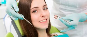
Diagnostics
A diagnostic examination of the patient begins with the collection of complaints and a visual examination of the oral cavity. The course of pregnancy in the mother of the child, details of feeding, when the eruption and appearance of molars began are clarified. Particular attention is paid to:
- Functionality of the jaw;
- Type of facial profile;
- The position of the dentition and their condition;
- Bite appearance;
- Condition of the oral mucosa;
- Age characteristics of the body.
Further diagnostics include:
- Creating models from plaster or creating it using 3D modeling. It makes it possible to determine the presence of occlusion and allows you to select the optimal prosthesis.
- OPTG. An x-ray provides information about the condition of the teeth and jaw. With its help, it is possible to determine the presence of carious changes, complete destruction and periodontal damage.
- TRG. Its use allows you to obtain an image of the skull and jaw in a lateral projection. This test evaluates the structure of the bones, their relationship, the location of the teeth and identifies the presence of developmental abnormalities.