Studies using a magnetic resonance imaging scanner are informative. A distinctive feature of MRI is a targeted examination of a selected area and the ability to detect pathology at the very beginning of development. But patients are prevented from undergoing this procedure by the presence of contraindications. Many people are particularly interested in whether it is possible to do MRI of the pituitary gland or other parts of the body with dental implants and metal crowns. More details later in the article.
Safety of MR Imaging in Patients with Dental Appliances and Devices
An MRI scanner generates the powerful magnetic fields needed to produce images. Metals interact with magnetic fields in different ways. As a result of this interaction, a number of effects arise that affect the results of the examination - dark spots or so-called artifacts appear on the images, which can complicate or make diagnosis impossible, metal products can heat up or move under the influence of a magnetic field. The degree of this influence depends on what metal was used for the manufacture of metal structures for dental purposes.
Artifacts
Most often in dentistry, inert metals are used - titanium, gold and its alloys, zirconium, nickel, zinc, chromium, copper, silver, iron (stainless steel) - which do not interact with living tissues. Depending on the ability of these materials to cause artifacts, a distinction is made between MRI-compatible and non-MRI metals.
MRI-compatible metals that cause no or minimal artifacts include zirconium (the metal from which dental crowns are made), silver, zinc, copper alloys, titanium and its alloys, gold and its alloys, nickel and its alloys. Stainless steel, alloys containing large amounts of chromium and cobalt are not compatible with MRI and cause artifacts that complicate diagnosis. You can find out detailed information about the composition of the materials used to make dental structures from your dentist.
Heating the metal
The magnetic fields generated by an MRI machine can cause heating of metal products, up to temperatures sufficient to cause a burn. However, this is only true for metal objects that are on the surface of the patient's body - which is why it is recommended to remove all metal objects, jewelry, piercings and jewelry before the examination. Metal crowns, dental pins, implants, and dentures are less susceptible to this effect - studies on the safety of MRI in the presence of metal in the mouth have found that the heating of such products during research does not exceed 2-3 degrees Celsius. Therefore, MRI of the head or internal organs is a safe procedure for the patient, even in the presence of dental structures.
Displacement of metal structures for dental purposes
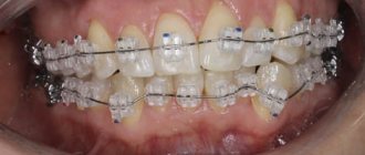
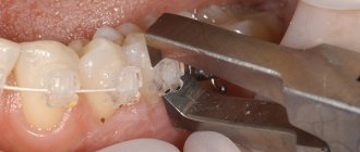
The magnetic fields generated during magnetic resonance imaging are capable of attracting metal objects made from magnetic metals, giving them significant acceleration. It is for this reason that before performing an MRI of the brain, spine or any other parts of the body, it is strictly forbidden to carry any metal objects on you. Otherwise, a piece of metal flying into the tomograph magnet can cause serious injury. In this case, is it possible to do an MRI with metal crowns on the teeth, with pins or braces? It is possible, since the mass of such products does not exceed several grams, and the structure itself is firmly fixed in the oral cavity. The exception is braces, dental crowns, bridges and dentures made of stainless steel. Removable structures must be removed before performing the procedure - this will not only make the examination safer, but will also improve the quality of the resulting images. If the structure is not removable, it is recommended to consult a dentist to check the strength of the fastening and prevent the product from moving.
Features of the procedure in the presence of dentures
Not long ago, metal implants were widely used, the main disadvantage of which is the distortion of diagnostic results. Metal structures reduce the quality of the image obtained during the examination. For example, if the spine is being examined, the presence of implants will not cause distortion of information. But if the jaw or brain is examined, this may affect the reliability of the information.
But dental implants do not directly harm health, since they are not exposed to a magnetic field. In addition, you should not worry that the pins may change their position after the procedure. Only those elements that are not tightly fixed in the bone, but are located in soft tissues, are subject to displacement. Therefore, an MRI of the head with pins in the teeth can be performed without any fear.
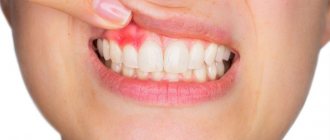
If you have metal teeth, the situation becomes somewhat more complicated, since metal products in some cases become an obstacle to a full diagnosis. The main disadvantage of metal dental crowns is the possibility of them heating up during the examination. And the larger the structure, the higher the chances of heating. Therefore, there are still contraindications for MRI with metal teeth. After all, the presence of metal prostheses can not only affect the reliability of the information received, but also harm the patient’s health. Therefore, in some cases they resort to removing the crowns or refuse to carry out the procedure.
Make an appointment now!
With which implants is MRI of the brain contraindicated?
Implants are not a contraindication for brain MRI, since they are mainly made of titanium. Titanium and its alloys are compatible with MRI and result in virtually no artifacts in images. However, if non-removable stainless steel dental products are installed in the oral cavity, artifacts can cover most of the cervical spine and head, making the diagnosis of diseases of the brain and neck uninformative. If this happens, such research will not be paid for. You can obtain more detailed information from the doctors of our medical center.
| MRI of the head and neck, which shows |
| Stroke on MRI of the brain |
| MRI of the pituitary gland with contrast, what does it show? |
| MRI of the brain with contrast, how is it done? |
| Decoding MRI of the brain |
| MRI of cerebral vessels, which shows |
The way out is metal-ceramics
The situation is completely different if the patient has metal-ceramic crowns installed. There are no contraindications for MRI with metal-ceramics, since metal alloys are used in the manufacture of such products. Their peculiarity lies in their inertness to the effects of electromagnetic waves emitted by the tomograph. In most cases, metal ceramics do not affect the general health in any way and do not distort diagnostic results. But MRI with metal-ceramic crowns can be hampered by fastening systems. Sometimes a metal-ceramic prosthesis has fasteners made of metal. Such a metal-ceramic design can degrade image quality and affect diagnostic information.
Magnetic resonance imaging with pins in teeth or other dental structures should be carried out only after consultation with specialists and an assessment of possible risks. In this case, only the latest equipment should be used, in which settings can be changed to improve image quality.
- Why do you need an MRI?
- What diseases can MRI detect?
- MRI of the head for children.
- MRI of the TMJ and temporal bones.
- MRI of cerebral vessels.
- What to choose for a head examination - MRI or EEG.
Is it possible to do an MRI if you have dental implants?
Many patients: “Can I do MRI with dental implants?” After all, today every third person has pins installed.
Indications for use
The reason for prescribing this procedure may be a suspicion of a tumor, disruption of vascular activity or blood supply to tissues. Most often, MRI is prescribed as the main examination of the brain, spine or spinal cord to diagnose diseases of the osteoarticular system and abdominal organs. The doctor can prescribe diagnostics for ligament injuries, gynecological diseases, kidney diseases, suspected tumors and metastases.
Features of the procedure
This procedure can be carried out both for the patient’s entire body and for a separate part. The patient lies down on the couch with the head fixed. The couch is then moved inside the MRI machine. The procedure takes from 30 to 90 minutes. The patient spends this time in a relaxed and motionless state. A special feature of the procedure is the loud noise of the device, so the patient is provided with either headphones or earplugs. Throughout the procedure, the patient has access to an emergency interrupt button. Before the procedure, all metal objects must be removed. In some cases, the procedure is performed under anesthesia. The images are usually ready immediately after the examination or the next day.
Should you tell your doctor that you have dental implants?
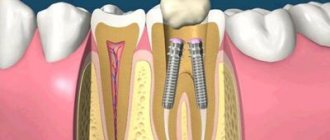
Dental replacement implants are made primarily from titanium. This is a paramagnetic material that does not resonate with a magnetic tomograph. But this does not mean that there are no restrictions on the examination.
To avoid having to undergo an MRI twice, notify your doctor about the implant so that he can change the machine settings
The diagnostician must be informed about artificial teeth for two reasons:
- Titanium is capable of distorting the picture. Yes, the metal does not heat up and does not move. But it can create minor interference. In order not to undergo an expensive procedure twice, it is better to immediately notify the doctor about the implant - he will change the settings of the device.
- The titanium pin is not the only element of the implant. In addition to the rod itself, there is also an abutment (gum former) and a crown. They are made from ceramics or various types of metal and then lined with porcelain.
Therefore, patients are always asked about the presence of implants at least twice: when giving a referral and immediately before diagnosis.
Features of MRI in human treatment
Magnetic resonance imaging is a relatively new method for diagnosing serious pathologies in the human body. Its essence lies in the effect of a special magnetic field on enzymes that are found in human tissues and mucous membranes. When conducting an examination, you must be extremely careful, since each organ is diagnosed using its own method.
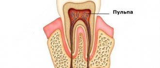
This is due to the fact that each tissue of the system has its own number of hydrogen atoms. It is they, under the influence of a magnetic source, that make it possible to obtain information about the presence or absence of a problem. The tomograph picks up the received signals, and the doctor deciphers them. During diagnostics, it is important to remove all possible interference, which can be even small metal and metal-ceramic elements.
Attention! The procedure itself can take quite a long time, the average diagnostic time is 40 minutes. It all depends on the tissues that need to be assessed. If the patient is afraid of closed spaces, the diagnosis is carried out in an open apparatus.