One of the most used methods of prosthetics in dentistry today is metal-ceramics; it is installed for many patients. However, in the case when a person, for one reason or another, needs to undergo a magnetic resonance imaging procedure, he will have a logical question: is it possible to do an MRI with metal-ceramic crowns or are they a contraindication for such a diagnosis?
Safety of MR Imaging in Patients with Dental Appliances and Devices
An MRI scanner generates the powerful magnetic fields needed to produce images. Metals interact with magnetic fields in different ways. As a result of this interaction, a number of effects arise that affect the results of the examination - dark spots or so-called artifacts appear on the images, which can complicate or make diagnosis impossible, metal products can heat up or move under the influence of a magnetic field. The degree of this influence depends on what metal was used for the manufacture of metal structures for dental purposes.
Artifacts
Most often in dentistry, inert metals are used - titanium, gold and its alloys, zirconium, nickel, zinc, chromium, copper, silver, iron (stainless steel) - which do not interact with living tissues. Depending on the ability of these materials to cause artifacts, a distinction is made between MRI-compatible and non-MRI metals.
MRI-compatible metals that cause no or minimal artifacts include zirconium (the metal from which dental crowns are made), silver, zinc, copper alloys, titanium and its alloys, gold and its alloys, nickel and its alloys. Stainless steel, alloys containing large amounts of chromium and cobalt are not compatible with MRI and cause artifacts that complicate diagnosis. You can find out detailed information about the composition of the materials used to make dental structures from your dentist.
Heating the metal
The magnetic fields generated by an MRI machine can cause heating of metal products, up to temperatures sufficient to cause a burn. However, this is only true for metal objects that are on the surface of the patient's body - which is why it is recommended to remove all metal objects, jewelry, piercings and jewelry before the examination. Metal crowns, dental pins, implants, and dentures are less susceptible to this effect - studies on the safety of MRI in the presence of metal in the mouth have found that the heating of such products during research does not exceed 2-3 degrees Celsius. Therefore, MRI of the head or internal organs is a safe procedure for the patient, even in the presence of dental structures.
Displacement of metal structures for dental purposes
The magnetic fields generated during magnetic resonance imaging are capable of attracting metal objects made from magnetic metals, giving them significant acceleration. It is for this reason that before performing an MRI of the brain, spine or any other parts of the body, it is strictly forbidden to carry any metal objects on you. Otherwise, a piece of metal flying into the tomograph magnet can cause serious injury. In this case, is it possible to do an MRI with metal crowns on the teeth, with pins or braces? It is possible, since the mass of such products does not exceed several grams, and the structure itself is firmly fixed in the oral cavity. The exception is braces, dental crowns, bridges and dentures made of stainless steel. Removable structures must be removed before performing the procedure - this will not only make the examination safer, but will also improve the quality of the resulting images. If the structure is not removable, it is recommended to consult a dentist to check the strength of the fastening and prevent the product from moving.
How does MRI work?
Let's figure out what a magnetic resonance imaging scanner is and how it works. This device is designed for targeted examination of tissues and blood vessels of the body. Modern devices with a power of 1.5-3 Tesla are capable of identifying problem areas smaller than 0.2 mm. The principle of operation is based on radio waves and the magnetization properties of the nuclei of hydrogen atoms, which are located everywhere in the body (and we remember that a person is 70% water).
The nuclei of hydrogen atoms “respond” to a powerful magnet (enter into resonance), and a special program records a picture and displays it on a computer. The diagnostic result consists of many virtual sections or sections of the desired organ - the brain or spinal cord, liver, vascular system, etc. And the radiologist makes a written report on the data obtained.
%akc54%
In general, there are two types of tomographs that are installed in specialized institutions - a cone-beam tomograph (CT) and an MRI machine itself. The first, even with implants, does not distort the image, but is also not suitable for studying small areas (for example, brain tumors). During an MRI, a magnet may cause some image distortion in the area of metal products in the body - but whether implants are included in this list will be discussed later.
On a note! Tomographs are also available in dentistry - they are less powerful and give a much worse picture (however, for the needs of the clinic this is enough). Neither the presence of implants nor the presence of metal crowns is a contraindication to this study. However, such devices are cone-beam and do not contain magnets.
With which implants is MRI of the brain contraindicated?
Implants are not a contraindication for brain MRI, since they are mainly made of titanium. Titanium and its alloys are compatible with MRI and result in virtually no artifacts in images. However, if non-removable stainless steel dental products are installed in the oral cavity, artifacts can cover most of the cervical spine and head, making the diagnosis of diseases of the brain and neck uninformative. If this happens, such research will not be paid for. You can obtain more detailed information from the doctors of our medical center.
| MRI of the head and neck, which shows |
| Stroke on MRI of the brain |
| MRI of the pituitary gland with contrast, what does it show? |
| MRI of the brain with contrast, how is it done? |
| Decoding MRI of the brain |
| MRI of cerebral vessels, which shows |
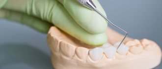
Is it possible to have access to tomography if I have crowns?
Are there any special features in MRI diagnostics for patients with dental crowns? This question also worries many. Some people worry that under the influence of magnets, metal, metal-ceramic crowns and pins can fly out of their places and damage something in the mouth. Experts note that metal crowns and pins have absolutely no effect on MRI results, but only if they are made of pure ceramics, zirconium dioxide or high-quality alloys. If the crown is made of metal-ceramics, then it is worth clarifying the metal content in the base - it is likely that you will not be allowed to conduct research.
How are crowns removed from teeth, indications?
In cases where the crown or bridge has moved during the MRI, they are removed and installed again or replaced with new prostheses. They do this in the following ways:
- sawing - the prosthesis is drilled out using drills with a cooling system; after removal, the structure is unsuitable for further use;
- ultrasound – ultrasonic waves destroy the cement, and the crown is easily removed from the stump;
- Koch apparatus - a special drill breaks the bond with gentle pushes, then the structure is lifted and removed;
- Coronaflex apparatus - compressed air is supplied under the edge of the prosthesis, this is the only method that eliminates damage to the structure and supporting teeth;
- special tools or sliding crown removers - used after destruction of the adhesive base using one of the above methods.
Orthopedic structures are also removed in case of resorption of cement, chips of ceramic lining, damage to the frame, inflammatory processes in the root canals, or the patient’s desire to replace the prosthesis with a better one.
It doesn't hurt to remove crowns
Is it possible to shoot on your own?
Sometimes the bridge or crown is so loose that the patient can tear it off himself. However, this should not be done, because... possible:
- damage to the prosthesis;
- fracture of the stump or root system of the tooth;
- traumatization of the mucous membrane or adjacent units.
If the denture becomes loose, the only correct option is to contact a dental clinic.
Does it hurt?
Removing dentures does not hurt at all, unless, of course, the patient rips them off himself. Because crowns and bridges are placed on already decayed teeth; in most units the pulp has been removed. Therefore, unpleasant sensations are excluded. There may only be a feeling of pressure on the jaw.
If the tooth under the crown is not pulpless or its roots are inflamed, dentures are not removed. The doctor administers anesthesia to eliminate pain.
How long does the procedure take?
The duration of prosthesis removal depends on the method. So, using a cut, the structure is removed in 10-15 minutes. But preliminary decementing of crowns using ultrasound, a Koch or Coronaflex apparatus can take several visits.
Artificial teeth are not an obstacle to magnetic resonance imaging. Modern designs are made from bioinert materials that do not harm the patient and do not affect the examination. If there is one of the items prohibited for examination in the mouth, it is removed and then fixed again or replaced with a new one.
Possible complications and precautions
MRI in the presence of electronic implants can seriously harm a person or even lead to his death. Performing the study on persons with coronary walls and clips on cerebral vessels can provoke massive bleeding, which will lead to death. Endoprostheses made from some alloys may move out of place or heat up during an MRI, causing burns.
MRI installation before the procedure.
People with certain types of implants are strictly prohibited from undergoing magnetic resonance imaging. But for patients with implants made of “dangerous” alloys, you can still try to perform the study. As a precaution, a button is placed in the person's hand. If he feels a strong burning sensation, he presses it and the study is stopped.
Fact! Metal prostheses tend to “fade”, making the image of nearby tissues unclear. Therefore, it is pointless to try to obtain an MRI image of the replaced joint or bone held together by fonts or plates.
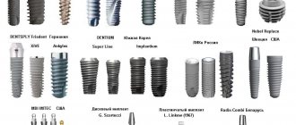
Manufacturing companies
Over the past 20 years, medicine has mainly used implants made from chromium-cobalt alloys (as we have already found out, these metals actively react to a magnetic field). Many models made from better materials have appeared on the market. They are better tolerated by patients and do not cause allergies or MRI problems.
Table 2.
| Company manufacturer | Characteristics and Application | Behavior of implants during MRI diagnostics |
| Biomet | Produces high-quality implants that take root well and do not cause allergic reactions. | Due to their small size and low magnetic susceptibility, they do not interfere with MRI. |
| Zimmer | It produces products not from titanium, but from tantalum. The implants have a porous coating and fuse perfectly with the bone tissue. | They do not cause unexpected complications during magnetic resonance imaging and do not distort the results of the study. |
| Johnson&Johnson | The company produces implants using all available standards and technologies. | Do not interact with magnetic field. When available, MRI is absolutely safe. |
| Smith&Nephew | Manufactures endoprostheses from an alloy containing zirconium and niobium. | Smith&Nephew implants are hypoallergenic and practically do not interact with the magnetic field. |
| Stryker | World-famous company of beta-titanium endoprostheses and fixators for internal osteosynthesis. | Owners of Stryker implants can undergo MRI without any worries. Additional precautions may only be necessary if you have several large prostheses. |
| Aesculap | Produces endoprostheses from titanium, zirconium ceramics, chrome-cobalt alloys. | Most implants can easily withstand magnetic resonance imaging. |
If you have a prosthesis from one of the companies listed in the table, you can do an MRI without the slightest fear. However, in any case, you should not undergo the study without first consulting a doctor.
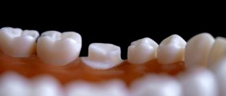
How long does a crown stay on a tooth?
The service life of entire crowns primarily depends on the material. So:
- metal ones last an average of 5 years;
- metal-ceramic – 8-10 years;
- ceramic and zirconium dioxide - 10-15 years and above.
The duration of wearing dentures is also affected by: how well the supporting tooth was treated and ground, the skill of the dental technician and the quality of oral care. The dentist is visited every six months to remove plaque from products and adjust them. And after 7-10 years, most crowns become unusable and are replaced with new ones.
But loose and broken dentures will not last long: from several days to a couple of weeks. Therefore, people contact the clinic immediately: the faster the doctor adjusts or replaces the bridge or crown, the greater the chance of saving the abutment tooth.
This is how a crown is sawed
Impact of implants on the quality of results
The MRI machine will not have a negative effect on dental structures, but the presence of metal may negatively affect the quality of the image. It will become fuzzy, blurry, and the doctor will not be able to determine the condition of the structures.
Distortion is observed during examination:
- thoracic region;
- brain, skull;
- shoulder joints.
Not long ago, improved devices with PET/MRI technology began to be introduced. The new model eliminates the influence of noise from metal objects on the image. Before carrying out the procedure, you need to clarify which tomograph is used in the office . If an old-style device is installed, the doctor will adjust it taking into account the foreign body in the body, which will help improve the image of the area being examined
How to find out if you can have an MRI
Remember that an MRI can be done with the permission of a specialist. Only he will determine whether you need this research and whether it will harm you. Perhaps the doctor will make a diagnosis without magnetic resonance imaging. Spinal spondylosis and deforming osteoarthritis of stages II-IV can be detected using conventional radiography.
Comparison of visual diagnostic methods. MRI is on the right.
Should you tell your doctor that you have dental implants?
Dental replacement implants are made primarily from titanium. This is a paramagnetic material that does not resonate with a magnetic tomograph. But this does not mean that there are no restrictions on the examination.
To avoid having to undergo an MRI twice, notify your doctor about the implant so that he can change the machine settings
The diagnostician must be informed about artificial teeth for two reasons:
- Titanium is capable of distorting the picture. Yes, the metal does not heat up and does not move. But it can create minor interference. In order not to undergo an expensive procedure twice, it is better to immediately notify the doctor about the implant - he will change the settings of the device.
- The titanium pin is not the only element of the implant. In addition to the rod itself, there is also an abutment (gum former) and a crown. They are made from ceramics or various types of metal and then lined with porcelain.
Therefore, patients are always asked about the presence of implants at least twice: when giving a referral and immediately before diagnosis.
For which examinations does crown material matter?
The ban on performing an MRI on a patient with metal-ceramics applies only to examination of the brain. If it is necessary to diagnose the condition of the musculoskeletal system, the genitourinary system; cardiovascular system, upper and lower extremities, the doctor will not ask questions about the material of the crowns. In this case it does not play a role.
Pulses of magnetic radiation will not be able to resonate with the smallest particles of molecules of the organ under study. Therefore, the results of the survey will be reliable.
Contraindications to the procedure
If prostheses, pins and plates are firmly connected to the bone tissue and cannot move, then implants of other locations can easily move under the influence of a magnet. Therefore, it is STRICTLY PROHIBITED to conduct magnetic resonance imaging if they are present.
Implants that cannot be used for MRI:
- artificial heart valves;
- stents and clips on vessels of any location;
- middle or inner ear implants;
- pacemakers;
- artificial lens;
- Illizarov apparatus;
- insulin pump;
- large metal implants.
Minimally invasive endoprosthetics in the Czech Republic: doctors, rehabilitation, terms and prices.
Find out more