Treatment of non-carious dental lesions is the elimination of congenital and acquired pathologies in the development of enamel and dentin, which are non-infectious dental diseases. The destruction of enamel and dentin that progresses with age worsens the appearance of teeth and creates problems with chewing food, which is why it is so important to visit the dentist in a timely manner, i.e. at least 2 times a year to start treatment at an early stage. This group of diseases includes hypoplasia and hyperplasia of enamel, as well as erosion, wedge-shaped defect, pigmentation, dental trauma, hyperesthesia, necrosis of hard tissues.
- Hypoplasia
- Hyperplasia
- Hyperesthesia
- Wedge-shaped defect
- Hard tissue necrosis
- Pigmentation
- Erosion
- Fluorosis
- Injuries
- Advantages of treatment at GrandMed
- On a note
Non-carious dental lesions are a large group of diseases of a non-infectious nature that lead to the destruction of hard dental tissues. The destruction of enamel and dentin, which rapidly progresses with age, worsens the appearance of teeth and creates problems with chewing food. Non-carious lesions are diagnosed during an examination of the oral cavity, and treatment, the effectiveness of which directly depends on the timeliness of contacting a specialist, is aimed at restoring the mineral composition of tissues and eliminating the aesthetic defect of the dentition.
Dental lesions are divided into two groups:
- Congenital or occurring before teeth eruption (enamel hypoplasia and hyperplasia).
- Acquired or occurring after teething (erosion, wedge-shaped defect, pigmentation, dental trauma, hyperesthesia, necrosis of hard tissues).
About half of people in the older age group (45–65 years) suffer from acquired diseases.
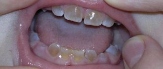
Dental hypoplasia
The pathology is manifested by underdevelopment or absence (aplasia) of tooth enamel; It can be congenital, but also develops after the birth of a child due to insufficient intake of minerals into the body. Hypoplasia itself refers to non-carious lesions of teeth, but thanks to it, excellent conditions are created in the oral cavity for the development of microbes, including those that cause caries.
Causes of hypoplasia:
- metabolic disease;
- mechanical damage to the jaw bone;
- dystrophy;
- diseases of the gastrointestinal tract;
- developmental pathologies;
- During pregnancy, the woman had: severe toxicosis, rubella, ARVI, toxoplasmosis, conflict of Rh factors, as well as premature birth and birth trauma.
Symptoms of hypoplasia
Pathology can be systemic (affects all teeth) and local (affects several teeth). In the early stages, underdevelopment of the enamel (too thin a layer or complete absence), deformation of the teeth and the appearance of grooves, yellow and white spots on their surface are noted. Painful sensations are noted during meals, and later, in the absence of treatment, the tissues are destroyed (erased) and an abnormal bite is formed.
Treatment of hypoplasia
Since underdevelopment of hard tissues and tooth decay is associated with a lack of minerals, when treating hypoplasia, it is necessary to remineralize the enamel with special preparations. To improve the appearance, teeth that have retained their normal shape are whitened, and if this is not enough, the defects are filled or prosthetics are performed. Congenital dental hypoplasia in children, in most cases, can be prevented; for this, during pregnancy, a woman’s diet must be balanced and contain sufficient amounts of vitamins D, A, C, group B, as well as calcium and fluorine. In addition, the expectant mother, in order to avoid non-carious lesions of the child’s teeth, should pay due attention to oral hygiene.
Treatment of caries in the cervical area
All video presentations
The tactics of treating cervical caries depends on many factors, but the main one is at what stage of development of the pathology treatment began.
Treatment in the early stages
In the early stages of the disease, conservative therapeutic methods are used. In particular, the treatment of caries at the stage of the appearance of a stain is reduced to saturating the surface layer of enamel with calcium. For this purpose, special remineralizing agents containing fluorine and highly active calcium are used (for example, Tiefenfluorid or ICON). Before applying such drugs, hard and soft pathological deposits are removed from the teeth.
Treatment of caries in later stages
In the later stages of the disease, treatment is reduced to filling the tooth.
The treatment program in such a situation includes the following steps:
- cleaning teeth from soft and hard pathological deposits using abrasive pastes and polishing brushes;
- determining the color of the enamel of a damaged tooth and selecting suitable filling materials;
- preparation of a carious defect, removal of all dental tissues damaged by caries;
- isolation of the dental cavity from saliva;
- treatment of the prepared carious cavity with an adhesive for reliable adhesion of the filling to the dental tissues;
- introducing filling material into the treated cavity;
- giving the tooth its physiological shape;
- grinding and polishing the filling using special burs and fine-grained discs.
Dental hyperplasia
With hyperplasia, teeth acquire an unaesthetic appearance, becoming covered with growths due to excessive formation of hard tissue. The so-called “pearl drops” reach 5 mm in diameter and consist of tooth enamel, dentin, and connective tissue. Depending on the location, there are root, coronal and cervical (main area of localization) formations.
Causes of dental hyperplasia:
- metabolic disease;
- congenital developmental pathologies;
- mechanical injuries of teeth and jaw bones;
- diseases of the gastrointestinal tract;
- disorders of intrauterine development of the fetus.
Symptoms of dental hyperplasia
Excessive formation and deposition of hard tissues on the surface of teeth is a cosmetic defect, clearly visible when the frontal group of teeth is affected; pain or discomfort is not typical for the pathology. Sometimes “drops” consisting of enamel form inside the dentin of the crown or root parts of the tooth and do not show themselves in any way.
Treatment of dental hyperplasia
If “drops” worsen the appearance of the teeth, then they are polished, thus leveling the surface and giving it an aesthetic appearance. The pathology does not require any special treatment, but since cervical growths often cause inflammation of the gums, they are removed with a diamond bur and treated with drugs containing fluoride.
Signs of cervical caries
Caries in the cervical zone, just like other varieties of this pathology, goes through 4 stages in its development:
- spot stage;
- superficial stage;
- average caries;
- deep carious lesion.
Stage 1 – spot stage
At the spot stage, the disease is asymptomatic, almost imperceptible to the sick person.
The main signs of the development of the pathological process at this stage are:
- loss of shine of tooth enamel in the affected area;
- the formation of a pigmented or white carious area with a smooth surface.
Stage 2 – superficial caries
With superficial caries, the clinical picture of the disease is complemented by the gradual destruction of enamel in the cervical area, while the carious spot loses its smoothness and becomes rough. The patient may complain of increased sensitivity of the tooth to temperature, mechanical and other influences.
Stages 3 and 4 – medium and deep caries
Further development of the disease is characterized by the appearance of carious cavities in place of the spots. With moderate caries, dentin and enamel are damaged, and with deep caries, all dental tissues are damaged.
Dental hyperesthesia
In most cases, hyperesthesia or increased sensitivity of teeth is not an independent disease, but a symptom of other non-carious lesions associated with changes in the structure and thinning of the enamel. The pathology is characterized by short-term acute or aching pain in one or more teeth that occurs when exposed to external irritants. Teeth with an intact pulp have increased sensitivity, both those that have lost part of their own tissues as a result of caries treatment, and those that maintain their integrity.
Causes of dental hyperesthesia:
- congenital structural features of enamel;
- thinning of enamel with age;
- exposure of the cervical part of the tooth due to receding soft tissue of the gums;
- violation of the integrity of tooth enamel by bacteria accumulating in the cervical area due to poor oral hygiene;
- mechanical damage to enamel, including hard toothbrushes and toothpastes containing coarse abrasives;
- enamel damage associated with exposure to chemically active substances that disrupt the acid-base balance (consumption of sweet and sour foods, as well as carbonated drinks);
- cracking of enamel when eating too hot or cold food;
- grinding and strong clenching of teeth (bruxism) lead to microtraumas of the enamel;
- unprofessional teeth whitening and abuse of such procedures;
- lack of calcium and phosphorus in the diet, or poor absorption of microelements due to diseases of the digestive system;
- general demineralization of the body, for example, in women during pregnancy or menopause.
Symptoms of dental hyperesthesia
Increased sensitivity is indicated by fairly severe pain that occurs as a response to any irritation. The tooth “aches” from hot and cold food, when consuming sour and sweet foods.
Treatment of dental abrasion and hyperesthesia
In case of increased tooth wear, orthodontic treatment (bite correction) is carried out, and if the patient has exposure of the cervical area due to pathological receding gums, then surgery is performed. In the case of a mild form, hypersensitivity is relieved by special toothpaste, application of fluoride-containing applications to areas with damaged enamel, as well as restoration of demineralized enamel with filling material. Prevention of hyperesthesia consists of maintaining oral hygiene (including regular professional cleanings) and a balanced diet, that is, sufficient intake of calcium, phosphorus and vitamins K, E, D. After sour and sweet foods, the mouth should be rinsed with water.
Carious lesions of teeth
Caries is one of the most common dental diseases, which is a consequence of the activity of pathogenic microorganisms. Bacteria, breaking down food debris (especially sugar), form certain types of acids, under the influence of which dental tissue is demineralized, softened and destroyed.
The rate of degeneration of dental material due to caries depends on the following factors:
- Oral hygiene. With careful care (especially before bed), food debris, plaque and pathogenic bacteria are destroyed, which helps slow the spread of carious lesions.
- Metabolic state. Depending on the health of the endocrine system, the destruction process may be slower or faster. This also includes eating disorders - with an excess of sweets in the diet, caries progresses much faster.
- Gastrointestinal health. This is due to the chemical processes of breakdown of simple carbohydrates that provoke caries.
- Presence of sleep disorders. Since the bacteria that cause tooth decay are anaerobic organisms (that is, they live without access to air), sleep disorders that cause the patient to sleep with the mouth open or snore significantly reduce the rate of bacterial growth. This group of factors is joined by an active-conversational rhythm of life, in which the patient has to talk a lot, as a result of which regular access of air in the oral cavity reduces the activity of microorganisms.
Symptoms of caries
The first manifestations of caries are white or dark spots on the tooth enamel. Most often this goes unnoticed, especially in the case of damage to distant teeth.
The next stage is increased sensitivity of the teeth to the effects of the external environment - a reaction to sweet foods, temperature changes.
In cases where caries has affected the surface of the tooth, destruction may be accompanied by sharpening of its edges, as a result of which the mucous membrane of the oral cavity next to the diseased tooth is often injured.
If caries is not treated at this stage, it will continue to destroy the tooth until it reaches the pulp, the area of soft tissue in which the dental nerve is located. This is accompanied by severe pain, with which, in most cases, the patient comes to the dentist. The sooner the patient consults a doctor, the more tooth tissue can subsequently be preserved.
Treatment of caries
Caries is treated by removing the affected tissue. To do this, the carious cavity of the tooth is opened and prepared using a drill, abrasives, laser or ultrasound. After this, the tooth is filled..
Filling allows you to restore the functionality of the tooth in case of mild to moderate destruction of dental tissues. In cases where the tooth has been almost completely destroyed, prosthetics are used.
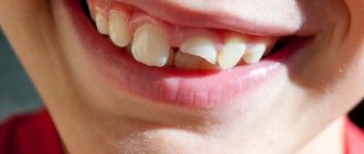
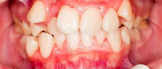
Wedge-shaped (abfraction) defect
Non-carious lesion of teeth, in which a defect is formed on the neck of the tooth that looks like a wedge. In the early stages, a wedge-shaped defect is difficult to diagnose, since the tooth retains its structure and color, so dentists, as a rule, detect deep lesions accompanied by darkening of the tissue and pain felt by the patient while eating. Often, a wedge-shaped defect develops against the background of periodontal tissue diseases (periodontitis and gingivitis) and is usually diagnosed in older and middle-aged people, mainly on canines and premolars (small molars).
Causes of wedge-shaped defect:
- deterioration of tooth nutrition due to age-related problems with blood supply;
- metabolic disorders, including due to thyroid diseases;
- improper distribution of the load on the teeth, for example, chewing on one side;
- bruxism or teeth grinding;
- improper brushing technique;
- brushing your teeth immediately after eating acidic foods;
- increased stomach acidity;
- gum diseases, including gingivitis and periodontitis.
Symptoms of a wedge-shaped defect
The disease is non-carious in nature, that is, it is not associated with pathogenic microflora, and therefore develops rather slowly. The enamel in the gingival zone is mainly affected: in the early stages, the strongest tissue of the human body becomes thinner with the formation of a pit, but remains smooth and retains color. Gradually, the enamel is completely destroyed, dentin is exposed, and the teeth react sharply to sour and sweet, hot and cold foods, “aching” pain also appears when brushing your teeth.
Treatment of wedge-shaped defect
In case of significant damage, they resort to filling the defect cavity with a material that has a certain degree of elasticity. If the tooth is not strengthened in this way, its supragingival part may break off. Another effective way to protect a tooth with a wedge-shaped defect is to perform microprosthetics and install a veneer on it. In case of a small depth of the defect, remineralization (saturation with calcium and fluoride) of the enamel is carried out in a dental clinic. It is recommended to maintain the mineral composition of teeth yourself using toothpaste and mouth rinse selected by a specialist. In addition, it is important to master the correct technique for brushing your teeth and avoid using hard-bristled brushes, as well as toothpastes with abrasives, that injure enamel and gums.
Necrosis of hard dental tissues
Necrosis or death of hard tissue is a lesion of enamel and dentin, manifested by the formation of multiple defects on the surface of the teeth. The disease is non-carious in nature, develops after teething and is equally common in men and women. Necrosis causes a change in cell structure, which manifests itself in the destruction of hard tissues and pulp vessels. For successful treatment, it is important for the dentist to establish the cause that caused the necrosis, so the patient must undergo a comprehensive examination, including an endocrinologist and gastroenterologist.
Causes of necrosis of enamel and dentin:
- hormonal imbalance;
- digestive disorders;
- work in hazardous industries and contact with toxic substances;
- exposure, including radiological treatment
Symptoms of necrosis of hard dental tissues
At the early stage of this disease, the enamel loses its shine and becomes covered with chalky spots, which gradually acquire a dark brown color. The matter is not limited to a change in color - the hard tissues under the spots soften, destruction of enamel and dentin occurs, which ends in the destruction of the supragingival part. The process of necrosis involves several teeth at once, which become sensitive to external irritants. The foci of the disease are located, as a rule, in the cervical part.
Treatment of hard tissue necrosis
The pathology is non-carious in nature and its symptoms resemble erosion and a wedge-shaped defect. In the early stages, calcium-containing preparations are applied to strengthen hard tissues and reduce sensitivity. In case of severe damage, orthopedic treatment cannot be avoided, during which the affected tissue is removed and the tooth is covered with a crown. At the same time, disease therapy and the elimination of negative factors that led to the development of necrosis must be carried out.
Clinic, diagnosis and differential diagnosis of wear of hard dental tissues
Only a third of practicing dentists in Europe report erosive or other tooth wear in their medical records [13]. The codes and criteria used in medical documentation do not allow recording of non-carious lesions.
The classic description of erosion in the literature is “a saucer-shaped defect with clear boundaries and a smooth bottom, located on the vestibular surface between the neck of the tooth and its equator” [14].
| Table No. 1. Variants of clinical manifestations of erosive lesions of teeth depending on the etiological factor | |||
| Clinical picture | Saucer-shaped defects in the cervical region of the anterior group of teeth of the upper jaw, unilateral or bilateral | Defects on the oral surface of the chewing group of teeth in the upper jaw | Defects on the oral surface of the anterior group of teeth or all teeth of the upper jaw |
| Cause of erosion | Diet, medications | Gastrointestinal diseases gastroesophageal reflux | Vomiting in eating disorders |
However, today the clinical manifestations of erosion are more varied and depend on the etiological factors of this disease (Table No. 1).
In the early stages, erosive tooth wear appears as a loss of physiological surface gloss. In later stages, changes occur in the original morphology of the tooth.
With frequent consumption of foods and drinks with low pH, concavities appear on the vestibular surface of the front teeth, the width of which clearly exceeds the depth.
In patients with eating disorders [15], due to frequent vomiting, enamel loss first occurs on the palatal surfaces of the central and lateral incisors of the maxilla. The enamel loses its characteristic pattern and looks “glazed.” Next, in descending order, the palatal surface of the molars, the palatal surface of the canines, the occlusal surface of the molars, the palatal surface of the premolars, and the occlusal surface of the premolars are affected in the upper jaw. The cutting edge of the incisors becomes thinner, the chewing surfaces become flattened. In severe cases, the occlusal morphology disappears. The rate of decline depends on episodes of vomiting and usually becomes noticeable within 3 years.
In patients with gastroesophageal reflux, enamel is lost from the palatal and occlusal surfaces of the upper molars [16].
The clinical picture of an abfraction defect differs significantly from that of an erosive one: a defect in the form of a step or ledge with an acute angle at the base, occurs in the area of the tooth neck, and sometimes extends into the subgingival region (Fig. 1).
Rice. 1a. Abfraction defects.
Rice. 1b. Abfraction defects.
Gingival recession may complement the clinical picture, but its presence is not always observed.
To diagnose abfractions, a thorough history and diagnosis of occlusal relationships are very important. An abfraction defect, unlike erosion, necessarily has sharp edges. A wedge-shaped defect is always combined with gum recession, and its depth always exceeds its width (Fig. 2a, Table No. 3).
Rice. 2a. Wedge-shaped defects.
The wedge-shaped defect is localized in the cervical region on the vestibular surfaces of the teeth that protrude most strongly from the dental arch, most often the first premolars and canines. It may be more pronounced on one side depending on which hand the patient uses to brush their teeth. The shape resembles a triangle, the apex of which faces the tooth cavity.
For the purpose of differential diagnosis of wedge-shaped and abfraction defects, occlusal relationships are diagnosed. And if supracontacts are detected on teeth with V-shaped defects and overload of individual teeth and groups of teeth is recorded, then the diagnosis of “abfraction defect” is more justified. Sometimes in the same patient we can observe a combination of wedge-shaped and abfraction defects.
With increased abrasion, the teeth of the upper and lower jaws are affected. Tissue loss is observed on the chewing surfaces of molars and premolars, the cutting edges of the frontal teeth (Fig. 2b), the palatal surfaces of the anterior teeth of the upper jaw and the vestibular surfaces of the anterior teeth of the lower jaw. The affected surfaces are hard, smooth and shiny. Sharp edges and chips are observed in patients suffering from bruxism.
Rice. 2b. Increased erasure.
Differential diagnosis according to clinical manifestations is given in Table No. 2.
| Table No. 2. Clinical picture of various forms of tooth wear | ||
| Diagnosis | Characteristics of the lesion | Location of the lesion |
| K03.2 Erosion | An oval or saucer-shaped defect with clear boundaries, the bottom is smooth, shiny, loss of occlusal pattern, “vitrified” enamel |
|
| K03.1 Grinding (abrasive wear) of teeth - wedge-shaped defect | Defect in the shape of a triangle, the apex faces the oral cavity, the depth exceeds the width |
|
| K03.18 Other specified diseases of dental hard tissues (abfraction) | Angular defect with sharp edges |
|
| K03.0 Increased abrasion (wear) | Horizontal or vertical loss of hard tooth tissues |
|
We should not forget that in the clinic we can observe a combination of various lesions in one patient (Fig. 3-8).
Rice. 3. Patient I., 48 years old, erosion, abfraction, abrasion, bruxism + caries.
Rice. 4. Patient K., 42 years old, erosion (covered by restorations), abfraction, abrasion, bruxism.
Rice. 5. Patient Shch., 53 years old, erosion, abfraction.
Rice. 6. Patient L., 63 years old, crossbite, erosion (covered by restorations), abrasion, bruxism.
Rice. 7. Patient K., 43 years old, tetracycline teeth, abrasion, bruxism.
Rice. 8. Patient S., 44 years old, erosions (covered by restorations), wedge-shaped defects, abrasion, bruxism.
The stress concentration due to bruxism can act synergistically as a cofactor with acids as well as abrasives to produce various manifestations of non-carious lesions. Signs of bruxism in the clinic include increased abrasion, abfraction defects, chipping, tension in the masticatory muscles, and much more.
Pigmentation of teeth
A non-carious pathological condition in which teeth become brown, yellow, gray, pink, black, red or another unusual color or shade. In most cases, pigmentation develops against the background of demineralization of the enamel, accompanied by hypoplasia and hyperesthesia. Pigmentation occurs under the influence of general (diseases, medications) and local (smoking, food colorings) factors and occurs in 85% of people, more often in men than in women. Treatment of this non-carious dental disease involves regular professional cleaning and whitening, but often the only way to restore the aesthetics of the dentition is prosthetics.
Causes of tooth pigmentation:
- demineralization of enamel;
- chips and other injuries to hard dental tissues;
- poor oral hygiene;
- treatment using iodoform and resorcinol-formalin, as well as “silvering” of teeth;
- rupture of the neurovascular bundle (pulp);
- smoking;
- taking certain medications and foods with added food coloring;
- hemolytic disease;
- uroporphyria;
- Excessive production of pigment in the body.
Pigmentation, like many other non-carious lesions, develops not only after, but also before teething. Thus, pigmentation in a child is often a consequence of the mother taking tetracycline antibiotics during pregnancy, which, penetrating the placenta, are deposited in the bones and tooth buds. Since the composition of dentin and enamel remains virtually unchanged over time, one cannot expect that yellow-brown pigmentation will disappear with age.
Symptoms of tooth pigmentation
The natural color of enamel is milky white, all other options indicate pathological pigmentation. The color of teeth affected by non-carious diseases varies. Thus, with a hemolytic disease accompanied by bilirubin deposition, the teeth are painted in various shades of gray, blue, green and brown, while “tetracycline” pigmentation is yellow-gray. Red staining is characteristic of uroporphyria, and gray staining is characteristic of pulp necrosis. In cases of exogenous pigmentation (associated with external factors), enamel and dentin are colored dark brown by nicotine, and pink by resorcinol-formaldehyde paste (not currently used in dentistry).
Treatment of tooth pigmentation
If, based on the patient’s complaints, clinical examination data and the results of additional studies (radiography and thermography), pathological pigmentation of the teeth is diagnosed, then treatment begins immediately. The therapy consists of sanitation of the oral cavity, including thorough removal of dental plaque (professional cleaning with ultrasound and using the Air Flow air-abrasive system). In case of deep pigmentation, intracanal and external bleaching is carried out using special preparations. However, not all types of pigmentation are eliminated by bleaching. Thus, the procedure will not eliminate tetracycline discoloration and color changes that appear as a result of oxidation of metal pins installed in the channels and silver amalgams. In these cases, pigmented teeth are covered with crowns and veneers (if their condition allows this). When treating non-carious pigmentation in children, consultations with a pediatrician and a genetic specialist are often required.
Dental diseases
Caries
Perhaps caries can be considered the most common dental disease, occurring in one degree or another in more than 75% of the population. Only a specialist can unambiguously determine the reasons for the development of caries, since this is associated with many individual characteristics of the patient: lifestyle, age, the presence of concomitant dental diseases and other pathologies, diet, habits, etc.
It was previously thought that tooth decay could be caused by poor oral and dental hygiene; this is still the main cause, but not the only one. Children are often told that eating a lot of sweets can cause tooth decay, but without a number of additional reasons this will not happen.
The most common causes of caries development are:
Insufficient oral hygiene. People who neglect oral hygiene procedures after meals face the problem of caries in 90% of cases. Insufficient or non-systematic brushing of teeth and failure to use dental floss contribute to the formation of persistent plaque on the surface of the teeth, which subsequently turns into tartar and contributes to the loss of microelements from tooth tissue.
Poor nutrition. Strict diets low in proteins and microelements, lack of foods containing calcium in the diet can cause changes in the qualitative composition of saliva, insufficient or excessive salivation, imbalance of oral microflora, thus provoking the destruction of hard dental tissues.
Enamel pathology. Inadequate development of dental tissues can cause insufficient supply of minerals from saliva to the enamel, which interferes with the normal development, formation and functioning of the tooth.
Signs of caries are a group of symptoms that can be identified even through self-observation:
- change in tooth enamel - darkening, acquiring a brownish or black tint;
- pain manifested by brushing teeth, using dental floss, chewing food;
- sensitivity of tooth enamel to cold, hot foods and drinks;
- chronic bad breath, regardless of hygiene rules;
Periodontitis
Periodontitis is an inflammatory disease of the tissues surrounding the tooth, characterized by the gradual destruction of the connection between bone tissue and the root, increased mobility of the tooth, up to its complete loss.
Periodontitis has an infectious nature of occurrence. The infection penetrates between the tooth and the gum, gradually disrupting the connection between the tooth root and the bone, increasing the mobility of the tooth in place; over time, the connection between the root and the bone weakens, which can threaten tooth loss. Periodontal disease can affect completely healthy teeth that are not affected by other dental problems.
Once an infection is identified, eliminating its causative agents is not particularly difficult, but in this case the consequences of periodontitis are important. Since after eliminating the infection, soft tissues are restored faster, rather than the ligaments that hold the tooth root in the bone, the resulting pocket is filled with granulation tissue, which contributes to the appearance of pathological mobility and the risk of tooth loss. Thus, the treatment of periodontitis consists of two main parts: destruction of infection, restoration of bone tissue and ligaments that hold the tooth in the bone.
After a course of treatment for periodontitis, the patient needs to pay special attention to hygiene procedures for a long time, monitor the condition of the teeth and regularly visit the dentist. Often, as a preventative measure, the doctor prescribes special care for teeth and gums using specialized teeth cleaning and oral hygiene products.
It should be remembered that even a completely stopped inflammatory process, under unfavorable conditions, can develop again.
Preventive measures are: maintaining hygiene of the oral cavity, teeth, gums; timely visits to the dentist; systematic self-monitoring of dental health.
Periodontal disease
Periodontal disease is an inflammatory disease of periodontal tissue, characterized by inflammation of the gums, destruction of the connection between the gum and the body of the tooth, leading to loss of teeth from initially healthy teeth due to severe mobility.
Periodontal disease has an infectious nature. The infection penetrates between the tooth and the gum, gradually disrupting the connection between the tooth root and the bone, increasing the mobility of the tooth in place; over time, the connection between the root and the bone is interrupted, the tooth dies and falls out. Periodontal disease can affect completely healthy teeth that are not affected by other dental pathologies.
Once an infection is identified, eliminating its causative agents is not particularly difficult, but in this case the consequences of periodontal disease are important. Since after the infection is destroyed, the soft tissue of the gums is restored faster, rather than the connection between the tooth root and the bone, the resulting cavity is filled with soft tissues, contributing to excessive mobility of the tooth and, again, its subsequent loss. Thus, the treatment of periodontal disease consists of two main parts: destruction of infection, restoration of bone tissue and connection of tooth roots with bone tissue.
After a course of treatment for periodontal disease, the patient needs to pay special attention to hygiene procedures for a long time, monitor the condition of the teeth and regularly visit the dentist. Often, as a preventative measure, the doctor prescribes special care for teeth and gums using specialized teeth cleaning and oral hygiene products.
It should be remembered that even completely cured periodontal disease can develop again under unfavorable conditions. Preventive measures are: maintaining hygiene of the oral cavity, teeth, gums; timely visits to the dentist; systematic self-monitoring of dental health.
Erosion of dental tissue
A non-carious lesion that is difficult to diagnose in the early stages and seriously worsens the appearance of the frontal teeth. It develops gradually; in the early stages, negative changes are almost invisible, so the pathology is often diagnosed when destructive changes become obvious: the tooth darkens and begins to hurt. If erosion is not treated, then there is a threat of penetration of pathogenic microorganisms, including those that cause caries, into the deep layers of dentin, which is fraught with the development of pulpitis, and subsequently periodontitis.
Causes of tooth erosion:
- mechanical damage (scratches, abrasions) resulting from the use of toothbrushes with hard bristles and pastes or powders containing coarse abrasives and operating on the principle of sandpaper;
- changes in the structure of the enamel and the appearance of microcracks on it under the influence of acid contained in food (marinades based on vinegar and lemon juice, fruits, carbonated drinks, freshly squeezed juices);
- malfunctions of the thyroid gland and digestive system, leading to metabolic disorders, including loss of calcium contained in enamel and dentin.
Symptoms of enamel erosion
The first sign of erosion is the appearance of spots on the enamel that lack their natural shine. The formations are non-carious in nature and are clearly visible, since due to their porous structure, pigments contained in food easily penetrate into them. As the pathology develops, the spots transform into round or oval-shaped depressions with a smooth bottom. Loss of tissue leads to increased sensitivity of teeth, which begin to respond with pain to thermal, chemical and mechanical stimuli. In addition, due to the loss of enamel and dentin, the teeth are deformed and look worn at the base.
Treatment of tooth enamel erosion
Erosion is a lesion diagnosed by a dentist during a visual examination of the oral cavity. In the early stages of the disease, therapy is carried out aimed at stopping the destruction of dental tissue. To do this, applications of preparations containing phosphorus and calcium are made, followed by treatment of the affected area with a fluorine-containing composition. In case of significant tissue destruction, the defects are filled, thereby restoring the integrity and aesthetics of the teeth. However, even in this case, remineralizing procedures cannot be avoided - they are done to stop the erosive process and prevent the filling from falling out. If the teeth are so damaged that it is impossible to restore them using artistic restoration techniques, prosthetics are performed with crowns or bridge structures. Erosion can be prevented to some extent by using a soft brush and low-abrasive cleaning pastes without bleaching agents for hygiene procedures, always rinsing your mouth after sour and sweet foods, and also limiting the duration of eating sour and sweet foods to 5 minutes.
Causes of cervical caries
To date, more than 400 theories have been developed regarding the causes of caries.
- Meanwhile, most of them are based on the assertion that the main factor contributing to the development of the disease is non-compliance with hygiene rules, which entails the formation of soft or hard plaque on the surface of the tooth enamel.
- Plaque plaques provide a favorable environment for the growth, reproduction and development of pathogenic bacteria. In the process of life, pathogenic microflora produces lactic acid, as a result of which demineralization of tooth enamel occurs.
- The increased risk of developing caries in the cervical area is due to the fact that at the base of the tooth the layer of enamel is thinner than on other parts of its surface.
- In addition, the cervical area is considered difficult to access for care.
- It should be noted that caries rarely occurs indirectly. The most important pathogenetic link in its development are gastrointestinal pathologies, decreased local and general immunity, errors in diet and other malfunctions in the body, and external factors.
Fluorosis
A non-carious lesion that develops as a result of large doses of fluoride compounds entering the body. As a rule, we are talking about a cumulative effect - a microelement enters the body over a long time before causing pathological changes in hard tissues, not only tooth enamel and dentin, but also bones. Even slightly exceeding the level of fluoride in drinking water can be fatal for people with calcium deficiency, especially for children during the formation of the skeleton and molars (baby teeth are rarely affected by fluorosis). There are several forms of the disease, differing in the shape of the spots and the degree of tooth decay. Thus, streaked, spotted and chalky-speckled forms are characterized by the appearance of white stripes, yellowish, brown and dark brown spots on the enamel, and erosive and destructive forms are accompanied by deep lesions of hard tissues, leading to tooth destruction.
Causes of fluorosis:
- consumption of water containing fluoride more than 1.5 mg (6.0 mg is a critical indicator leading to pathological changes) The optimal concentration of fluoride, which has a pronounced preventive effect on teeth: 0.6 - 1.5 mg/l.;
- work in production in conditions of excess fluoride in the air;
- lack of calcium with excess fluoride.
Symptoms of fluorosis
Teeth become brittle, their enamel wears off, and dark brown (brown) stains appear, which over time turn into erosions. The non-carious nature of the disease is confirmed by the fact that other hard tissues of the body also suffer from it, as evidenced by bone pain and decreased joint mobility. The development of fluorosis is often indicated by muscle weakness, as well as disturbances in the functioning of the liver and autonomic nervous system.
Treatment of dental fluorosis
First of all, you need to eliminate the source of excess fluoride intake - change the water and working conditions, and then begin to put your teeth in order. Treatment is determined by the form of fluorosis - sometimes, in order to return the teeth to an aesthetic appearance (in some cases after grinding off the enamel), it is enough to carry out bleaching and remineralization with preparations enriched with phosphorus and calcium, and if the process has gone deep, the teeth are restored with crowns or veneers. By the way, residents of Russia more often suffer from a lack of fluoride in water than from its excess. But it is better to find out exactly the microelement content in the water you drink, and not to use fluoride-containing toothpastes and mouth rinses if there is enough fluoride in it.
In tap water in St. Petersburg, the fluorine content is 0.15 - 0.20 mg/l of water, which is significantly lower than the optimal level.
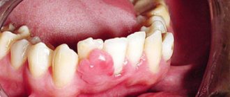
Dental injuries
With any injury to the dental system, you should immediately contact a dentist (if you suspect a fracture of the jaw bone, contact a surgeon). The specialist will conduct an examination, study the X-ray data and give an opinion on the possibility of restoring the tooth or the need for its removal. There are different types of dental injuries, both adults and children suffer from them, and in most cases the problems could have been avoided if the patient had shown more attention and caution.
Causes of dental injuries:
- mechanical damage received during a fall, fight, accident;
- a foreign object that has entered the oral cavity with food;
- habit of biting nails and biting threads when sewing;
- using teeth for purposes other than their intended purpose, for example, to crack nut shells;
- installation of a metal pin of the wrong size or errors made during the manufacture or installation of an orthopedic inlay into the tooth root;
- untimely treatment of caries, periodontitis, hypoplasia, fluorosis and other dental diseases.
Symptoms of dental injuries
The most common tooth injury is a partial or complete fracture of the crown (supra-gingival part); bruises and dislocations of teeth are less common. The loss of the crown is visible to the naked eye; the root may also suffer from the injury, receiving a longitudinal or diagonal fracture, but only a doctor can determine this. If the matter is limited to a bruise and the tooth visually retains its integrity, its trouble, in particular the rupture of the pulp (neurovascular bundle), is indicated by aching pain that intensifies when the jaws are closed. If the injury causes hemorrhage, the supragingival part of the affected tooth sometimes takes on a red tint. Dislocation of the latter relative to the socket indicates dislocation. If a tooth falls out of its socket, the dislocation is called complete (typical for the frontal group, especially the central incisors).
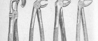
Treatment of dental injuries
The most important question that arises during an injury is whether the tooth can be restored. In case of partial loss of the supragingival part, but maintaining the integrity of the root, artistic restoration with filling material is possible (if at least 50% of its own tissues are preserved). If the supragingival part is completely destroyed or little remains of it, then the tooth is restored using an inlay, onlay or crown. It is worse if the root could not withstand the load and cracked - in this case, the tooth can only be removed and an implant installed in its place, and this can be done (in the absence of inflammation) at the same time, combining the removal operation with implantation and prosthetics. The same treatment is relevant for complete dislocation, but if the tooth has simply moved, it is returned to the socket and wait for healing. During the treatment of injured teeth, it is recommended to exclude too hard foods from the diet.
Advantages of the GrandMed clinic
- Dentistry has created an excellent diagnostic base that allows identifying non-carious dental pathologies in the early stages. Doctors have modern equipment at their disposal (radiovisiograph, orthopantomograph, computed tomograph), and all necessary clarifying tests can be done in the clinic’s biochemical laboratory.
- We employ professionals of the highest level, whose experience and qualifications allow us to treat complex non-carious pathologies, which often require the use of special techniques and non-standard approaches. Clients are provided with high-quality therapeutic and orthopedic care, including prosthetics on implants. So even in the most advanced cases, our patients can count on restoration of the beauty and functionality of their dentition.
- This disease is treated using local anesthesia. Patients of our clinic do not experience pain and feel comfortable during the procedure after injection of one of the modern anesthetic drugs that do not have side effects (Ultracaine, Ubestezin, Scandonest). You should not be afraid of the injection itself - it is performed quickly and accurately, and therefore is absolutely painless.
- Our clinic adheres to the highest sanitary standards, which is confirmed by the use of the latest sterilization equipment, which completely eliminates infection during treatment; Dentists have all the necessary consumables and disposable dental instruments at their disposal.
Note: computer necrosis of teeth
Experts started talking about this non-carious lesion of teeth relatively recently; it definitely did not exist in the 20th century, however, like people who spent most of their working and free time at the computer. The death of dental tissue threatens patients who are ready to sit at a computer for more than 8 hours a day, without days off and a full night’s rest. After 3–5 years of living in this mode, a person (and these are mainly young people under 35 years old) are guaranteed to have problems with the front teeth located in the path of the electrostatic radiation of the monitor.
Symptoms of computer necrosis of teeth
Experts, comparing computer and post-radiation lesions, find much in common between them: dying areas in both cases spread over the entire surface of the supragingival part of the teeth, which acquires a dark, almost black color. The focus of necrosis is filled with softened brown tooth tissues; the dentist easily removes them with special instruments, and the patient does not feel pain. As for the teeth of the chewing group, which are exposed to relatively less computer radiation, they also do not look the best: the enamel loses its shine and acquires a gray tint. Studies have shown that not only hard tissues undergo negative changes, but also the pulp. Thus, during electroodontometry, the neurovascular bundle does not respond to an electric current of 25-30 μA. In addition, changes in the functioning of the salivary glands are observed.
Treatment of computer necrosis of teeth
The “newness” of the disease complicates diagnosis, since the dentist may never have encountered it. An X-ray examination is required (sight image, orthopantomogram, computed tomogram), in the images the teeth look translucent and have unclear contours, which indicates a deficiency of mineral substances.
Treatment of computer necrosis is complicated by the fact that during the removal of damaged tissue with a bur, the tooth falls apart; in such conditions, it is impossible to preserve the vitality of the pulp, and often the tooth itself. If recovery is possible, it will take a lot of time, as it will be carried out in several stages, including local and general therapy, preceding filling or prosthetics. At the first stage of treatment, dead tissue is removed and healthy tissue is remineralized, and the patient is recommended to take mineral-vitamin complexes containing calcium, antioxidants, and vitamin A. After the condition improves, therapeutic linings are placed in the dental cavity and a temporary filling is performed. The result of this stage of treatment, which takes up to two months, is the restoration of dentin, after which the tooth is finally filled.
Prevention of computer necrosis of teeth
Computer necrosis of teeth can be prevented by a rational approach to working at the computer. Firstly, comply with sanitary standards, which require organizing a work area of at least 6 sq.m., located in a room with an area of 20 - 24 sq.m. The monitor should be located no closer than 60 - 70 cm from the face; if there are several computers installed in a room, the distance between them should be at least 2 m. Every two hours you should take a break of 15–20 minutes, during which the room is ventilated. Secondly, monitor your diet; if you don’t get enough antioxidants, vitamins and microelements with food, then take vitamin-mineral complexes. Thirdly, use new models of monitors that have minimal electrostatic effect on the body.