A sinus cyst is a fluid-filled formation with elastic walls; the location of the cyst affects the symptoms of the disease. Most often, cysts form in the maxillary sinuses. The reasons for the development of cysts in the nasal sinuses are varied; there are a number of reasons for the appearance of a neoplasm:
- Blockage of the ducts of the mucus-secreting glands.
- Dental diseases, tooth roots protruding into the paranasal sinuses.
- Allergic diseases of the nose.
- Frequent inflammatory processes.
- Anatomical features of the structure of the nose.
- Mechanical injuries of the nose.
In the oncology department of the Yusupov Hospital, you can undergo examination using MRI, CT, ultrasound, diagnosis of diseases of the paranasal sinuses if a nasal tumor is suspected. The patient will be able to undergo a full examination at the diagnostic center, take tests in the hospital laboratory, and receive advice from an otolaryngologist, oncologist and other specialists.
Cyst in the sinus: symptoms and consequences
Sinus cyst, symptoms of the disease depend on the location of the cyst, there are false and retention. The cyst can increase in size and eventually block the sinus cavity. A false cyst in the sinuses can develop due to an inflammatory, allergic process, while a true cyst develops due to blockage of the ducts of the mucus-secreting gland. A cyst of the paranasal sinuses may not manifest itself as pronounced symptoms for a long time, then nasal congestion, headache, and soreness in the face, on the side of the affected sinus cyst, begin to bother you. The pain can intensify when diving, and inflammatory processes in the nose often develop.
As the cyst of the maxillary sinuses develops, it manifests itself in the following disorders:
- Constant nasal congestion.
- Respiratory dysfunction.
- Pain in the area of the sinus affected by the cyst.
- Unpleasant sensations and false symptoms of increased intraocular pressure.
- Severe headaches.
- Purulent discharge (concomitant development of sinusitis).
- Partial or complete loss of smell.
The consequences of cyst development are negative:
- A growing cyst provokes an inflammatory process and can lead to the development of sinusitis.
- Reduces the patient’s quality of life and causes constant severe headaches.
- A large sinus cyst can lead to destruction of nasal tissue, reach the nasopharynx, impair respiratory function, and deform the face.
- Some sinus cysts can develop into a malignant tumor.
Causes
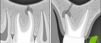
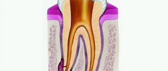
The key reason for the formation of a maxillary sinus cyst is dental disease. Especially when treatment was not carried out or performed poorly. Pathology develops against the background of advanced caries, periodontitis and pulpitis on chewing units. This is due to anatomical features. Premolars and molars are located closest to the paranasal sinuses and are separated from them by a thin septum.
Until recently, teeth with cysts were subjected to extraction. Today, dentists prefer to perform organ-preserving surgeries. All stages of work are performed under control using a dental microscope.
An endodontist removes the cyst, performs sterilization and filling of the canals.
The formation of a tumor can be caused by a deviated nasal septum, jaw abnormalities, or chronic blockage of the ducts of the nasal glands due to rhinitis or sinusitis.
Diagnosis of the disease
A neoplasm measuring up to 15 mm is diagnosed only with a CT scan. A dentist can suspect the presence of a pathology during a routine examination of the patient’s oral cavity based on existing complaints.
Clinical symptoms
As the cyst grows, the patient develops alarming symptoms similar to those of acute sinusitis. These include:
- acute headache;
- chronic nasal congestion;
- clear or yellow nasal discharge;
- a feeling of heaviness and fullness in the area under the eyes;
- the presence of a viscous mucous lump in the throat after waking up.
Clinical symptoms become more pronounced as the lumen in the nasal cavity closes. If there are concomitant dental problems, pain in the tooth and swelling of the gums occur.
Potential Complications
If left untreated, the infection spreads to adjacent teeth in the upper jaw. There is a risk of bone deformation or jaw fracture, the development of an abscess, phlegmon and other purulent complications.
The cyst is accompanied by sinusitis and chronic sinusitis. The growth of the tumor is dangerous due to visual impairment. The formation of a purulent cyst can lead to the development of meningitis or encephalitis.
Complex treatment
When treating pathology, dentists use medicinal and surgical techniques. The tumor is removed during surgery. Further drug therapy is designed to eliminate the symptoms of the inflammatory process and prevent complications.
Treatment in dentistry
Most patients go to the dentist when the size of the cyst exceeds 1 cm. The dental surgeon removes the bag of pus by cystectomy or cystotomy. The choice of technique is based on the size of the cyst and clinical indications.
The dentist is faced with a number of tasks:
- remove the source of inflammation in the oral cavity and stop the inflammatory process;
- provide drainage for the outflow of pus;
- if possible, save the diseased tooth;
- fill the resulting cavity with bone material;
- close the fistula between the oral cavity and the sinus.
Inpatient treatment in the ENT department
If the appearance of a cyst is caused by ENT pathologies, you should consult an otolaryngologist. Tumors are removed by surgery, laser or gentle endoscopic techniques. During endoscopy, the doctor gains access to the affected area through the nasal passages or an opening in the facial wall of the maxillary sinus. Control of manipulations on the monitor screen is carried out.
Main sinus cyst: symptoms, treatment
A cyst of the main (sphenoid) sinus develops more often in young people and rarely in older people. The cavity of the main sinus of the nose is covered with mucous membrane. The sheath glands secrete mucus; disruptions in functioning lead to blockage of the gland ducts and the formation of a cyst of the sphenoid sinus. Inflammatory processes, injuries, and allergic reactions have a negative effect on the mucous membrane. A growing cyst of the sphenoid sinus is accompanied by a feeling of fullness in the nose, nausea and dizziness may occur, a headache that radiates to the back of the head, and visual disturbances rarely develop.
If a sinus cyst is detected, treatment is carried out after a complete examination. The doctor prescribes diagnostic tests for the patient using:
- X-rays to determine the location of the cyst, changes in the condition of the facial bones and nasal septum.
- Magnetic resonance imaging, which determines the condition of the skull bones, tissues, and blood vessels. A more informative study than radiography. Computed tomography has the same capabilities. Research is carried out as prescribed by a doctor.
- Research using an endoscope allows you to examine the nasal cavity and perform a biopsy of nasal tissue.
After the studies, conservative or surgical treatment may be prescribed. Conservative treatment is aimed at pain relief, relieving an allergic reaction, and treating the inflammatory process. If conservative treatment is ineffective or the cyst is large, the patient is referred for removal of the formation.
What to do?
When deciding on treatment for a cyst, an ENT specialist usually takes a wait-and-see approach. The fact is that the growth of education has been going on for decades. Therefore, as long as it is small and does not cause any discomfort, there is no need to touch it. In addition, although infrequently, a maxillary sinus cyst can empty on its own. There is no pain, and the contents of the “ball” flow out through the nose.
But if the pathology causes concern, then the only effective way to treat it is surgery. Many patients, fearing surgical intervention, try to get rid of the problem with the help of traditional medicine: they do rinses, instill herbal decoctions into the nose or lubricate the mucous membranes with them. There is no point in such procedures, since they only temporarily make breathing easier, but do not reduce the size of the formation.
Removal of sinus cyst without surgery
If a sinus cyst is detected, treatment without surgery is carried out using decongestant sprays, antibiotics, painkillers, mucolytics, steroids and antihistamines. They treat concomitant diseases - allergies, sinusitis, inflammatory processes of the gums, teeth, nasal mucosa. In combination with these drugs, various means for rinsing the nasal cavity, regenerating and restorative sprays are used. Treatment is prescribed by the doctor based on the results of the patient’s examination.
What is a paranasal sinus cyst?
A paranasal sinus cyst is a formation with an elastic, elastic wall filled with fluid. Its size and location can be very different, which is reflected in clinical manifestations (patient complaints).
According to the classification, there are several types of cysts:
- Retention (true): occur due to blockage of the ducts of the glands of the mucous membrane;
- Pseudocysts (false): appear under the influence of an allergen, due to an infectious disease or due to an inflammatory process in the roots of the teeth of the upper jaw.
In addition, cysts are classified depending on what contents they are filled with, i.e. mucous, purulent and serous.
Cyst in the sinus: surgery, reviews
Patient reviews give preference to the endoscopic method of cyst removal. When performing the classical method and laser vaporization, an incision is required under the upper lip to gain access to the cyst. Classic removal of a sinus cyst involves dissection of the soft tissue under the upper lip from the frenulum to the first molar (Caldwell-Luc method) or using the Denker method, which is also carried out through the facial part. Classic operations are more traumatic, with long postoperative recovery, but these techniques allow you to gain access to a cyst located in a hard-to-reach place. Endoscopic removal is carried out using an endoscope, the cyst is removed through a small burr hole, the operation is performed under local anesthesia.
At the Yusupov Hospital, patients will be able to undergo a full examination using modern diagnostic equipment. The hospital hosts doctors from various fields, there is a clinical laboratory, and a rehabilitation center. In the hospital you can recover from surgery or illness, and patients have a 24-hour inpatient facility.
Cyst of the maxillary sinus
Maxillary sinus cyst
is a benign neoplasm that appears on the mucous membrane inside the paranasal sinuses.
A maxillary sinus cyst is a rounded sac, with a membrane in the form of a thin film, filled with fluid. It happens that the cyst shell spontaneously ruptures, and this is manifested by copious clear discharge from the nose. Retention cysts
in the sinuses are formed due to prolonged disruption of the normal movement of air in the nasal cavity against the background of sluggish chronic inflammation (for example, allergies or chronic sinusitis). Odontogenic cysts occur in the sinus in response to inflammation in the area of the roots of the teeth of the upper jaw or as a reaction to the installation of dental implants.
WHAT IS THE DANGEROUS OF A PARONAL SINUS CYST?
Cysts of the paranasal sinuses are not tumors
, do not become malignant, and do not pose a threat to life.
A small cyst may be located in the sinus and not manifest itself in any way for many years. If the cyst does not grow and does not bother you, no treatment is required. On the other hand, if the cyst occupies more than half the volume of the sinus
, this leads to impaired ventilation and stagnation of mucus. A large cyst completely clogs the sinus anastomosis, preventing the normal movement of air and the outflow of secretions. There are often cases when, against the background of an acute respiratory infection, a maxillary sinus cyst suppurates - acute purulent sinusitis occurs, which is already fraught with complications. For this reason, large cysts are recommended to be removed.
TREATMENT OF MAXILLARY SINUS CYST
Cysts of the paranasal sinuses can be cured only in one way - surgically remove them.
. Once formed, the cyst will no longer be able to resolve on its own or with the help of medications. If the cyst bursts during a sinus puncture or is “cauterized” with a laser, it will grow again in the same place. To avoid recurrence of the cyst, it is important to carefully remove all its membranes. This is only possible under visual control, during endoscopic surgery.
ENDOSCOPIC MAXILLARITY IN THE ENT CENTER
At the ENT Clinic, treatment of paranasal sinus cysts is carried out only by the endoscopic method.
according to the principles of FESS - functional endoscopic surgery of the paranasal sinuses.
This method involves the most gentle approach through the natural anastomosis of the sinuses, small incisions, minimal damage to the mucous membrane and intranasal structures. Endoscopic surgery to remove a maxillary sinus cyst is called endoscopic maxillary sinusotomy
.
METHODS OF TREATMENT OF MAXILLARY SINUS CYST
Cysts of the paranasal sinuses can be cured only in one way - surgically remove them. Once formed, the cyst will no longer be able to resolve on its own or with the help of medications. Traditional methods of treatment (rinsing the nose and sinuses with solutions of salt or medicinal plants), nasal sprays and antibiotics are ineffective against cysts. Non-surgical treatments can temporarily shrink the cyst by draining the fluid. If the cyst is burst with a needle during sinus puncture or cauterized with a laser, it will grow in the same place again in 2-3 months. To avoid recurrence of the cyst, it is important to very carefully remove all its membranes. This is only possible under visual control, during endoscopic surgery.
MODERN OPERATIONS FOR THE TREATMENT OF MAXILLARY SINUS CYST
At the ENT Clinic Center, treatment of paranasal sinus cysts is carried out only by the endoscopic method according to the principles of FESS - functional endoscopic surgery of the paranasal sinuses. Endoscopic surgery to remove a maxillary sinus cyst is called endoscopic maxillary sinusotomy. This method involves the most gentle approach through the natural anastomosis of the sinuses, minimal damage to the mucous membrane and intranasal structures. The advantages of endoscopic surgery are that everything is done inside the nose - there are no swelling, incisions, stitches or scars on the face or under the lip after it. The endoscope gives the surgeon a good overview, a wide field of view and bright light, which allows the entire cyst to be removed completely without leaving any membranes.
HOW THE OPERATION WORKS:
- The anesthesiologist and operating nurse prepare the patient for surgery.
- The surgeon examines the nasal cavity with an endoscope and identifies the natural anastomosis of the maxillary sinus on the lateral wall of the nasal cavity.
- With the help of special instruments, the sinus anastomosis expands to approximately 9-10 mm.
- The internal contents of the sinus are examined with an angled endoscope.
- Special micro-forceps are inserted through the enlarged anastomosis, the cyst is captured and removed entirely along with the membranes.
- The sinuses are washed and examined.
- After the operation, special breathable sponge tampons are installed in the nose.
The result of endoscopic maxillary sinus surgery will be the restoration of the normal volume of the sinus, the natural outflow of mucus and the movement of air in it.
Dear patients! Please note that the stated cost of the operation does not include the cost of laboratory blood tests and other examinations. The cost of treatment depends on the chosen method of anesthesia (local or general) and the length of stay in the hospital room. Only a surgeon after a face-to-face consultation can tell you the exact amount in your specific case.
Endoscopic surgery is used in the ENT Center clinic to treat not only maxillary sinus cysts, but also chronic sinusitis of any nature, polyps and other sinus tumors.
ENDOSCOPIC SURGERY IS INDICATED FOR DISEASES:
- Cysts of the maxillary sinuses
- Polyps of the nasal cavity and polypous rhinosinusitis (from a single polyp to widespread polyposis).
- Chronic and recurrent purulent sinusitis (sinusitis, ethmoiditis, sphenoiditis, frontal sinusitis, or their combinations - polysinusitis, pansinusitis).
- Odontogenic sinusitis and foreign bodies of the maxillary sinuses (after treatment by a dentist and cementing material entering the sinuses).
INDICATIONS FOR ENDOSCOPIC SINUS SURGERY:
A maxillary sinus cyst accidentally discovered on a CT scan does not always require urgent surgery. If it occupies less than 1/3 of the sinus, does not complicate breathing and does not cause pain, observation by an ENT doctor is sufficient. You should think about endoscopic cyst removal if:
- One or both halves of the nose are always stuffy, so you have to breathe through your mouth.
- There is an excess accumulation of mucus in the nose and nasopharynx.
- The constant feeling of heaviness, pressure, distension in the sinus area or headache is disturbing.
- Acute purulent sinusitis occurs on the side of this sinus.
WHAT YOU NEED TO PREPARATE FOR ENDOSCOPIC OPERATION:
1. First, you need to undergo an examination by an ENT surgeon to make an accurate diagnosis and determine clear indications for surgery. 2. Be sure to do a CT scan of the nose and paranasal sinuses for individual planning of the scope of the intervention. CT scans also help the surgeon navigate the procedure. 3. After the ENT doctor has confirmed the need for surgery, you need to undergo a general preoperative examination on an outpatient basis and obtain a physician’s opinion on the absence of contraindications. You can undergo the examination both at the ENT Center clinic and at your local clinic. You can download the list of required tests in PDF format from this link.
REHABILITATION AFTER ENDOSCOPIC SURGERY IN THE NASAL CAVITY
- After endoscopic surgery, special sponge breathable tampons are placed in the nose, which the surgeon removes after 1-2 days. Tampons sometimes cause a feeling of pressure in the nose and a headache, these symptoms quickly disappear after removal.
- The patient spends a day in the hospital and goes home the next day after the operation.
- At the appointed time, the patient visits the surgeon on an outpatient basis: to remove tampons, rinse the nose and monitor healing.
- In the first week after surgery, the patient is not recommended to drink hot and alcoholic drinks, take a hot shower, or perform physical exercise (so as not to provoke dilation of blood vessels in the nose and nosebleeds). It is best to follow a home regime and avoid colds.
- As it heals over the course of 1-2 weeks, clots of mucus and crusts constantly form in the nose - this is normal. The doctor will prescribe saline solutions to moisten the nose and other medications to reduce swelling and speed up healing. Nasal breathing is completely restored gradually, usually by the 3rd week after surgery.
Diagnostics
Sometimes the patient is not aware of the pathology, learning about it when examining the nose for suspicion of a completely different disease, for example, sinusitis.
If certain symptoms are present, the otolaryngologist can make an assumption about the presence of a process in the nasal cavities based on a survey (history) of the patient. Rhinoscopy is a method of examining the sinus to determine the presence of a deviated septum and the condition of the mucous membrane. Although this method does not give an idea of the pathological processes in the cavities (other sinus examinations are used for this), it allows one to determine the presence of inflammation, for example, sinusitis that has developed against the background of a cyst, redness of the membrane, pus and other discharge, if any.
There are examination methods through which the doctor will immediately make an accurate diagnosis. These include:
X-ray – the pictures show an image of the paranasal sinuses (two projections), which can be used to make an almost accurate diagnosis if the cyst is large. It looks like a dark, rounded area. However, to clarify the diagnosis, additional, more informative methods are used;
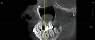
Computed tomography, magnetic resonance imaging and endoscopy are the most accurate ways to clarify the diagnosis. CT provides a layer-by-layer image of the skull; thanks to this method, the diagnosis is clarified, the size and exact location are determined. The method is indispensable if the patient is awaiting surgery on the maxillary sinus to remove a cyst;
The doctor can also refer you for gaimography (a contrast agent is injected into the area where the tumor is located) and biopsy (cells from the inflamed area are examined - for this, part of the diseased tissue is cut off and sent for cytological, microbiological and biochemical analysis);
Puncture (performed using an x-ray) - under local anesthesia, the maxillary cavity is punctured, and if the puncture is large, the fluid is pumped out. This helps to significantly reduce the defective bladder and improve nasal breathing. Fluid from the sinuses can be collected for testing. After the procedure, the improvements are temporary; after some time, the cavity is filled with liquid again.
Differential diagnosis is always necessary. The symptoms of a maxillary sinus cyst are extremely similar to those of a polyp and tumor. An experienced doctor can make the correct diagnosis based on examination results.
If left untreated, vision and more can seriously deteriorate.
Cysts of the maxillary sinuses are dangerous because if they are not treated, they can become a source of very serious health problems.
The infection can spread to the nearby teeth of the upper jaw and along the chain take over the entire row, as a result of which you can lose all your teeth. Pathology can lead to deformation of the skull bones and jaw fracture, the development of purulent processes - abscess, phlegmon, osteomyelitis, meningitis.
The tumor provokes the development of sinusitis and chronic sinusitis. Its presence is always accompanied by a runny nose and inflammation of the sinuses.
If the problem is not addressed, it can affect vision
The growth of the tumor often leads to blurred vision, “double vision.” If the cyst is purulent, it can lead to brain damage and encephalitis. When a tumor grows, there is a risk that it will completely block oxygen and complicate breathing, which is especially dangerous during sleep, since a person may not wake up.
One of the most common causes of tumor is a bad tooth.
A maxillary sinus cyst can form due to a reason such as dental disease. More precisely, when treatment was not carried out, or it was performed poorly. Most often, the development of pathology is provoked by advanced caries, pulpitis and periodontitis on molars and premolars (these teeth are separated from the paranasal sinuses by a thin septum).
Teeth with cysts and granulomas were previously more often removed than treated. Today, in some clinics, high-precision tooth-preserving operations are carried out, where all stages of treatment are accompanied by work on a dental microscope. A competent endodontist carefully removes the cyst under a microscope using special instruments, then the canals are sterilized to their entire length and filled with filling material. Treatment under a microscope is accurate, fast, without risks or complications, and, most importantly, the natural tooth remains intact.
The prerequisite for the appearance and growth of a tumor may be the individual anatomical characteristics of the patient, for example, a deviated nasal septum or jaw abnormalities. Another reason is the systematic blockage of the ducts of the glands located in the nose, against the background of rhinitis, runny nose, and allergic reactions.
Treatment
Making the correct diagnosis of a maxillary sinus cyst is extremely important, as it determines the course of treatment. The peculiarity of the pathology is that it is not amenable to pharmacological influence. No matter what medications the patient takes, the disease will not disappear. The method of successful treatment for a maxillary sinus cyst is its removal.
Surgical method
Surgical removal of a maxillary sinus cyst always occurs in a hospital setting. During surgery, the affected area is opened and drained. Previously, surgery was quite traumatic for the patient and required significant effort and time for complete recovery. Today the situation is different. With the modern endoscopic method of maxillary sinus cyst removal, the incisions are minimal, there is virtually no blood, and the patient recovers in a short time. The patient is discharged from the hospital within the first 24 hours after the intervention.
How the intervention occurs: laser and light-optical instruments are introduced into the surgical area through an endoscope. The doctor carefully makes a small incision, coagulates the tissue (so blood loss is insignificant) and removes the contents of the formation. Removing a maxillary sinus cyst using this method is considered the best.
Pharmacological method
The doctor decides what to do with the resulting bubble individually, taking into account the patient’s complaints, the results of the diagnostics, and concomitant diseases. If several cystic small cavities develop, it is recommended to monitor changes and use remedies to eliminate the causes of the pathology. Sometimes cyst removal is not necessary, especially if the cause of problems with the maxillary sinus is diseased tooth roots. After dental treatment, some formations may resolve.
If the cyst is detected in the early phase of the development of edema in the maxillary sinus, then treatment without surgery is possible, but this only concerns the prevention of complications. The doctor prescribes pharmacological drugs that slow down the growth and rate of development of the pathology:
- anti-inflammatory agents;
- antimicrobial;
- antipyretics;
- drugs that help eliminate toxins.
Almost all medical specialists agree that the best option is endoscopic removal of a maxillary sinus cyst. It is important not to wait for the pathological process to worsen, but to act for sure.
Self-medication is highly discouraged. Any advice that does not come from a doctor can be harmful to your health. When the patient finally turns to a specialist, treatment for a maxillary sinus cyst may take time, especially if complications arise.
The maxillary cyst is most often located in the nose area
The maxillary sinus cyst consists of epithelial cells of the mucous membrane. Inside it can be filled with mucus, serous contents or pus. Most often it is localized in the area of the sinuses, that is, the paranasal sinuses, which is why the pathology is also called a maxillary sinus cyst.
A maxillary cyst is often called a maxillary sinus cyst.
The main method of treating pathology is surgery
To treat a tumor, doctors combine therapeutic and surgical methods. First, surgery is used to remove the tumor, then drug therapy is used to eliminate signs of the inflammatory process and avoid complications, speed up rehabilitation and tissue healing.
If the maxillary sinus cyst is small (less than 1 cm), then drug therapy is used for treatment and dynamic observation is carried out, and there is no need for surgical removal of the tumor. But there are two important points. The first is that rarely do any patients go to the clinic until the cyst has grown and began to cause significant problems. Secondly, therapy can take a long time (4-6 months) and the result will not always be positive.
When treatment is carried out in dentistry
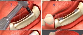
Most often, patients go to clinics when the cyst reaches 1 cm or more in diameter. In this case, its removal can be carried out in different ways. If the cause of the problem is a diseased tooth, then treatment is carried out by a dental surgeon. The doctor removes the tumor by performing a cystectomy if its size is no more than 1–2 cm. Or by cystotomy1, when the size is more than a few centimeters and the tumor has grown into the sinus.
The photo shows an operation to remove a cyst.
The main tasks of the dentist in this case:
- eliminate the source of inflammation in the oral cavity and stop the inflammatory process,
- ensure the outflow of purulent contents, if any, using drainage,
- save the diseased tooth, if possible: in cases where the root is slightly immersed in the tumor, its resection (removal of the apex) is carried out, and the tooth itself is preserved. When the root is immersed in the cyst by more than 1/3, its complete removal is recommended to avoid relapses,
- fill the cavity left after tumor removal with bone material,
- close the communication between the oral cavity and the sinus.
When treatment is carried out at the direction of an ENT doctor in a hospital setting
Maxillary sinus cysts are not always associated with diseased teeth. But regardless of the cause of their appearance, advanced tumors almost always lead to inflammatory processes in the nasal mucosa, congestion, sinusitis, and mucus discharge from the nasal passages. Therefore, many patients with such symptoms turn to an ENT doctor.
In this case, the tumors are removed through surgery. For example, using a classic operation, when a deep incision is made in the upper jaw, through which the formation is removed. This method is the most affordable, but it is quite traumatic, and the patient then requires a long postoperative recovery.
“Two years ago I had a maxillary cyst removed. My background was like this: snot was either pouring out of my nose, or there was constant congestion, and for a long time I could not smell or breathe normally, so I went to an otolaryngologist. The doctor sent me first for an x-ray, and then for tests and surgery. The operation was performed in a hospital and under anesthesia, I was very afraid of how everything would go, and especially how I would recover from the anesthesia, but everything was tolerable. Then I was in the hospital for about 4 days, but some people stay longer. I felt discomfort for another ten days after that.”
[email protected] , fragment of a review from the site woman.ru
However, today the most common surgical method is endoscopy. The operation is performed through an opening in the facial wall of the maxillary sinus, or through the nasal passages. The entire treatment process is monitored on the monitor screen, so the doctor can control his actions. The technology is considered the most gentle, atraumatic; after its use, the patient has no stitches, incisions or scars on his face. The procedure is quick (duration is about 40 minutes), tissue restoration takes about 7 days.
An endoscope is an optical device used in all areas of medicine for high-quality examination and treatment of hollow internal organs. Endoscopes can be inserted through natural body openings (in this case, the nose) or through surgical incisions.
The operation can be performed using an endoscope through the nose
The operation can also be performed using a laser. This approach almost completely eliminates the likelihood of developing postoperative complications. But due to the use of laser equipment, the cost of treatment increases significantly.
There are several types of pathology
Cysts of the maxillary sinuses are different:
- odontogenic: inflammation begins to form at the root of a poorly treated or completely untreated tooth located in the upper jaw. As the tumor increases in size, it destroys bone tissue and gradually grows into the sinuses. Such neoplasms are most often filled with pus,
- retention: formed due to disruption and obstruction of the glands that produce mucus.
Doctors also classify false cysts as a separate category - such formations do not contain epithelial cells and appear due to pathologies and structural disorders of the maxillofacial apparatus.