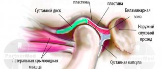
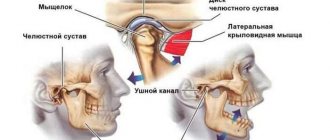
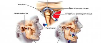
Anatomical structure
The oral cavity is the initial section of the digestive tract, in which food is chewed and saliva is produced to digest food. It is involved in the process of breathing, swallowing, articulation and speech.
The composition of the oral cavity includes:
- vestibule (lips, front side of teeth, inner surface of cheeks);
- gums;
- the bottom on which the tongue lies;
- two thirds of the tongue;
- teeth;
- retromolar triangle - the space on the lower jaw behind the third molar;
- hard and soft palate.
Classification
Oral cancer is divided into three types:
- papillary. The nodule in the mucous membrane increases in size and hangs into the oral cavity. The neoplasm progresses slowly;
- infiltrative. The seal on the pinkish mucosa is distinguished by a whitish color, clear contours and shape, and thinning of the membrane around it. On palpation from the side of the cheek, a dense infiltrate is felt. The tumor tends to grow rapidly. The patient complains of unbearable pain;
- ulcerative The most common form of the disease. Ulcers on the mucous membrane do not heal, they grow, and the border around them turns red. The outline is torn and its edges are bleeding.
Tumor metastases appear quickly. Malignant cells grow into the mental, submandibular, and deep jugular lymph nodes. This process is influenced by the thickness and depth of the tumor. Thus, when the tumor deepens by 4-5 mm, metastases occur in 98% of cases. At the T1 stage of oncology, metastasis is detected in half of the cases, and when the T4 stage is reached, distant spread of cancer cells is observed in 85% of cases.
By what signs can the disease be recognized?
Most often, the patient begins to suspect a deterioration in health based on the onset of symptoms. Although the formation of a cancerous tumor on the lower or upper jaw has some differences in clinical severity, it is still possible to identify the general symptoms of the disease:
- teeth become mobile, shift and begin to fall out;
- There is asymmetry of the face due to edema and a growing tumor;
- pronounced pain in the upper or lower jaw;
- contracture of the lower jaw and upper jaw is observed (limited mobility);
- difficulty swallowing;
- the jawbone is deformed;
- there is an unpleasant odor coming from the mouth;
- there may be discharge of purulent contents from the nose;
- frequent headaches occur;
- there is numbness in the skin in the facial area, etc.
However, more detailed clinical characteristics are described in the table below:
| Form of the disease | Upper jaw lesion | Lower jaw lesion |
| Primary | Clinical severity of a malignant lesion of the upper jaw will have the following symptoms:
| The primary form is characterized by the following symptoms:
|
| Secondary | The secondary form of the disease will be characterized by the manifestation of symptoms that directly depend on the location of the malignant neoplasm. The location of the tumor in the anterioinferior region carries with it the following symptoms:
The postero-superior location of the tumor-like neoplasm is manifested by the following symptoms:
| If cancer of the lower jaw appears, symptoms in its secondary form may be as follows:
|
Symptoms of cancer of the upper or lower jaw
Attention: In the initial stages of the disease, signs of jaw cancer may be completely absent, which significantly complicates timely diagnosis and diagnosis. In this case, it is advisable to conduct an annual preventive examination with a doctor in order to identify the pathology at the very beginning.
Maxillary cancer symptoms
Causes
The prevalence of oral cancer is growing and is currently diagnosed in 2% of patients among the total number of cases. Since 2009, the incidence has increased by 25%, with mostly squamous cell carcinoma being detected and only in isolated cases adenocarcinoma.
Most foci of oncology are observed in the tongue. Slightly less malignant formations on the floor of the mouth. Cancer of the soft and hard palate, gums and cheeks is detected in 20% of cases. Much less frequently diagnosed is damage to the alveoli of the lower jaw - 4%, the arches of the palate, retromolar region and vestibule - 3%.
Based on practice, men are more susceptible to oral cancer than women. This is due to bad habits, for example, the abuse of cigarettes or chewing tonic mixtures increases the production of saliva, which washes away beneficial elements from the mucous membrane. The risk group includes patients with HPV, elderly people, workers in hazardous industries, patients with lichen planus, people whose oral mucosa is systematically injured by fillings, prostheses, and metal objects.
Benign odontogenic tumors
Ameloblastoma. Its characteristic feature is a pronounced change in the shape of the face associated with a violation of symmetry proportions as a result of the development of a tumor located in the lower jaw. The violation of symmetry can be mild or pronounced. The degree of distortion of the face shape is influenced by the size and position of the tumor. For example, the localization of a tumor along the body and ramus of the lower jaw is characterized by a change in the shape of the lower lateral part of the face. The color of the skin does not change, and it can be easily moved in the area of the tumor.
Inflammatory processes accompanying the tumor can give similar symptoms to phlegmon or mandibular osteomyelitis. During palpation, the body of the tumor is felt, which makes it possible to assess the degree of distortion of the shape of the face. The lymph nodes located directly next to the tumor do not change in size, and the deformed area is clearly defined. The formation has a thick filling and a wavy surface. Examination of the oral cavity reveals thickening of the alveolar ridge, soft tissue may be swollen, and teeth tend to shift or move.
Odontoma . Often this type of tumor is diagnosed in adolescence. The tumor has similar symptoms to other tumors localized in the jaw bones. The course of the disease is quite slow and ambiguous. During development, there is a gradual swelling of the jaw bones, which leads to delayed or absent teeth eruption. Large tumor sizes can change the shape of the jaw or contribute to the formation of a fistula. Despite the fact that the course of the disease passes practically without symptoms, the upper layer of the jaw may be damaged, and the tumor itself may contain teeth or their rudiments. When diagnosing, it is necessary to differentiate the tumor from adamantinoma. Odontoma can be simple, complex, soft or mixed.
Odontogenic fibroma . The nature of the development of this neoplasm is very slow; the tumor is mainly diagnosed in young children. A clear symptom of tumor development is impaired teething; pain is not observed during the period of tumor growth. Odontogenic fibroma can be located equally on both jaws and is rarely accompanied by an inflammatory process. It differs from similar neoplasms in its composition, which includes remnants of the epithelium that forms the teeth.
Cementoma . The hallmark of a tumor is the presence of cement-like tissue. The tumor grows quite slowly and is manifested by a change in the shape of the jaw. The tumor is clear and round, has pronounced boundaries, most often affects the upper jaw and is almost always connected to the root of the tooth.
Symptoms
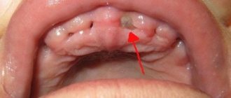
A malignant ulcer from ordinary stomatitis in the mouth can be identified by swelling and swelling of the cheeks, pain and constant discomfort even at rest. You should be wary of prolonged non-healing of the wound and its bleeding.
As the disease progresses, the symptoms intensify:
- swelling increases and spreads to the neck;
- the red or white spot on the oral mucosa intensifies;
- discomfort when chewing and swallowing;
- difficulty speaking due to friction of the mucous membrane on the teeth when moving the jaw;
- the appearance of bad breath;
- feeling of a foreign object in the throat;
- anemia of the mouth.
In the late stages of cancer, teeth fall out and body weight rapidly decreases.
General information
Jaw tumors refer to neoplasms in the cat's tissue. This pathology accounts for 13 to 29% of the total number of dental surgical diseases.
Due to its close location to the teeth, brain and eyes, the pathology is characterized by a special clinical course. Tumor formations in this area lead to functional disorders, aesthetic defects and even death.
Depending on the tissue of which the tumor is composed, it can be osteogenic or non-osteogenic. According to the clinical development of pathologies, there are malignant, benign or locally destructive.
Treatment of jaw tumors is a complex and lengthy task that requires high competence and professionalism from the doctor.
Diagnostics
At the initial consultation, the doctor examines the oral cavity, examines ulcers, erosions, damage to the mucous membrane, and then takes a smear for examination. To confirm the inflammatory process, the patient is sent for a general and biochemical blood test.
The diagnosis is confirmed by the results of the examination:
- MRI and ultrasound of soft tissues of the neck. The images reveal the localization of the pathology, the depth of germination and the structure of the tumor, compaction from blood and lymph, decomposition of the cortical layer of the bone;
- if metastases are suspected, a fine needle aspiration biopsy of the lymph nodes under the chin, under the jaw and in the upper third of the neck is performed;
- positron emission tomography. Shows the depth of the tumor, as well as early metastases;
- osteoscintigraphy. Skeletal bones are examined to look for displaced cancer cells;
- CT scan of facial bones with contrast. The images show the tumor growing into the neck vessels, jaw or base of the skull.
Stages
In most cases, gum cancer is not detected at an early stage, which is typical for all oral tumors. People are not used to looking into their own mouth until there is a good reason for this - pain that occurs when the periosteum is involved in the process.
The height of the gum of an adult is small, however, when staging cancer, the tumor is graded in “steps” of 2 centimeters: less than 2 cm, from 2 cm to 4 cm, and more than 4 cm, because malignant processes of the oral cavity are often non-nodular. , and flat infiltrates and ulcers.
The stages of gum cancer are distributed as follows:
- stage 0 or carcinoma in situ - cancer cells have not penetrated into the deep layers of the mucous membrane, as a rule, the disease is visually manifested by leukoplakia, an oncological diagnosis is established only by microscopy;
- Stage 1 - carcinoma is less than 2 cm and there are no signs of damage anywhere else;
- Stage 2 - the primary lesion is more than 2 cm, but less than 4 cm, grows no more than a centimeter deep, there are no metastases in the lymph nodes;
- Stage 3 involves variability in the size of the primary gum lesion, including more than 4 cm, without metastases or with cancer cells in one lymph node no more than 3 cm in diameter;
- Stage 4 is divided into subgroups:
- 4A - extensive tumor without involvement of lymph nodes, or smaller in size with a large lymph node, but not more than 6 cm;
- 4C - the local condition is not important because there are distant metastases.
4B - cancer growing into the jaw and surrounding tissues without metastases, or a smaller lesion with large or multiple tumor lymph nodes on either side of the neck;
Treatment
The choice of treatment tactics depends on the stage and extent of the tumor. When the tumor grows rapidly, treatment methods are combined.
Operation
The doctor determines the principle of surgical intervention after determining the stage of the tumor and its spread. If cancer cells have penetrated the periosteum and surrounding tissues, a wedge-shaped, planar or sagittal resection of the jaw is performed. If the examination reveals the growth of cancer cells directly into the bone or the defect is noticed during surgery, segmental resection of the lower jaw is performed. The doctor assesses the lesion on site and determines the thickness of the excised layer.
The next stage of the operation is partial or complete excision of the cervical lymph nodes to prevent metastases if the thickness of the tumor is more than 4 mm or the location of the tumor in the floor of the mouth or on the tongue. If the tumor is located in the midline, then the cervical lymph nodes are excised on both sides. The operation ends with the immediate replacement of damaged tissue.
After removal, the tumor is sent for histological examination. Its size, thickness, depth, edges are assessed. Further treatment is affected by cell growth beyond the boundaries of the capsule of the removed lymph node, and the spread of cancer cells to neighboring organs.
Radiation therapy
Radiation after surgery is prescribed when diagnosing T3, T4, N2, T3 stages of the disease no later than six weeks after tumor removal. The need for radiation therapy increases with perineural invasion of the lymphatic vessels. The total focal dose for all sessions is 60 g, and the single focal dose for one session is 2 g. When metastases are detected on the neck, the SOD increases to 66 g, and if there is no risk of metastasis, the SOD decreases to 50 g.
As the main treatment, radiation therapy is used in a total focal dose of 60-70 g. The procedure is performed five days a week and is combined with chemotherapy. Every three weeks, 100 mg of cisplatin is administered.
Chemotherapy
Anticancer drugs are prescribed before surgery or along with radiation therapy to reduce the size of the tumor. Sometimes therapy is prescribed simultaneously with surgery.
Treatment involves the use of a 5-fluoroacyl regimen together with cisplatin or other agents - carboplatin, methotrexate, bleomycin. They cause a number of side effects, for example, vomiting or nausea, hair loss, decreased appetite, and increased bleeding. Symptoms disappear after treatment, but permanent hearing loss is sometimes observed after taking cisplatin.
The prognosis of oral cancer depends on the stage at which the disease is detected. If treatment is started at stage zero, the disease will stop. It is worth noting that smoking provokes relapse or degeneration of the tumor, so repeated surgery or radiation may be required. Surgery at the first stage increases survival rate to 80-85%, and the combination of radiation therapy with surgery at the second stage by 60-80%. Already at subsequent stages of cancer development, the survival rate is no more than 50%, and all three treatment methods are used simultaneously.
Prognosis and survival
The prognosis of the disease depends on the stage, localization, and timely provision of assistance. In the early stages, with adequate treatment, the disease can almost always be overcome. When contacting a doctor was not timely, the prognosis is somewhat worse, and the percentage of cured patients is lower. In assessing the prognosis, the presence or absence of distant metastases is important. Despite enormous progress in oncology, distant metastases sharply reduce 5-year survival; this is a significant problem for a specialist. In addition, the prognosis depends on the histological type of the tumor, because The growth rate of different types of tumors can vary significantly. The age of the patient, the location of the tumor, the presence of concomitant diseases in the patient, the chosen treatment tactics - all these factors significantly influence the prognosis.
Dispensary observation
Since the tumor can recur and metastasize, after completing the course of treatment the patient is registered with the oncology clinic. The first year you should visit a doctor every month, the second year a preventive examination is carried out every 4-6 months, and then once a year or in case of any ailments. The examination involves an examination - ultrasound and contrast MRI of the soft tissues of the neck, PET, osteoscintigraphy. Consultation with an otolaryngologist, dentist and oncologist is required. The doctor may shorten the period of medical examination if there is a high risk of relapse.
List of references on the topic:
- Gantsev Sh.H. Oncology – M, 2012 – P.204-205.
- Golovin D.I. Errors and difficulties in diagnosing tumors, D.: Medicine. Leningr. department, 2015 305 pp.
- Selected lectures on clinical oncology/Ed. IN AND. Chissova, S.L. Daryalova. – M., 2010
- Matyakin E.G., Alferov V.S. Chemotherapy of head and neck tumors // Mat. 2nd Ros. oncol. conf. “Current trends in the development of drug therapy for tumors” December 8–10, 2016 – M., 256 p.
- Tumors of the head and neck: hands/ A.I. Paches. - 5th ed., add. And revised - M.: Practical Medicine, 2013. -478 p.
- Shine A.A. Oncology. M – 2014 365 pp.
- Encyclopedia of Clinical Oncology/Ed. M.I. Davydova. – M., 2014 –P.140-179.
- Bityutsky P.G., Kitsmanyuk Z.D., Trofimov E.I. Diagnosis and treatment of cancer of the oral mucosa // Medical consultations. - 2014. - No. 1. - P. 23-27.
- Byakhov M. Yu. Options for combined and complex treatment of locally advanced cancer of the oral mucosa and oropharynx: Dis. Dr. med. Sci. - M., 2013.