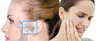
If a person feels pain in the jaw, radiating to the ear and temple, we may be talking about arthritis of the mandibular joint. The pathology is not just painful, but also dangerous. If not diagnosed in a timely manner, it can lead to severe complications, including paralysis of the facial nerves. Therefore, the first thing to do when such symptoms appear is to consult a specialist. In 10-18% of cases, diagnosis and treatment of TMJ arthritis is the responsibility of dentists and occurs in people from 25 to 45 years old.
Characteristics of the disease
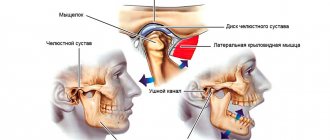
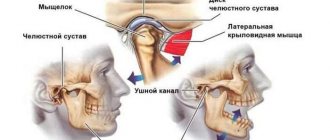
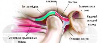
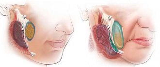
In medicine, the disease is called arthritis of the temporomandibular joint (TMJ). With the help of this joint all movements of the lower jaw occur.
The inflammatory process can occur in acute or chronic form. The main sign of the development of pathology is restriction in the movement of the lower jaw.
Such a pathology requires complex treatment and intervention by specialists of various profiles.
Depending on the area of localization, there are several forms of arthritis:
- disease of the lower or upper joint;
- arthrosis of the upper jaw joint.
Osteoarthritis occurs when a joint is severely deformed. The lesion affects both areas of the jaw node, the cartilage tissue is destroyed. In the first stages, the joint capsule becomes inflamed, then the cartilage tissue is affected.
Comprehensive treatment of TMJ
When the doctor has determined the cause of the joint pathology, or causes, he determines the patient's readiness for a comprehensive treatment plan. In addition to the orthodontist, an osteopath or chiropractor may be involved, or even an orthopedist if more complex correction of the musculoskeletal system is required.
The patient should be aware that it is possible to straighten the jaw using a splint or splint, but this will not solve the problem of malocclusion. Orthodontic treatment will be required to correct the bite. If you have already had orthodontic treatment before, then it is more difficult to decide on repeated treatment.
Therefore, first the problem with the joint is solved using a splint or joint splint, then bite correction is carried out, and, if necessary, prosthetics. In parallel, work is underway with an osteopath to restore the muscular corset of the back and neck.
It happens that a patient refuses treatment with braces after the problem with the joint has been resolved. In this case, we warn him about the need to wear a joint splint constantly in order to avoid the recurrence of old problems with the TMJ. After all, a relapse can happen quite quickly due to stress.
Risk factors
Main reasons:
- Injury. The cause in this case may be a bruise, a blow, or a sudden opening of the mouth.
- Infection of ENT organs. Arthritis of the maxillary joint can be caused by streptococci, chlamydia or tuberculosis.
- Osteomyelitis.
- Untreated caries.
- Periodontal diseases.
- Otitis.
In addition, the following conditions may also affect the development of jaw arthritis:
- Gout.
- Systemic lupus erythematosus.
- Diabetes.
- Thyroid diseases.
- Rheumatoid and reactive arthritis.
Sometimes inflammation can be caused by general hypothermia of the body.
Occlusive therapy for TMJ dysfunction
After diagnosis, the patient is scheduled for an appointment with the orthodontist to determine the central relationship of the jaws (“true” position of the lower jaw, the position in which your joint and chewing muscles will be most comfortable).
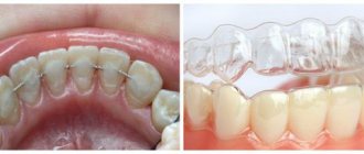
In order to more accurately establish and fix this position, an occlusal splint (splint) will be individually made for the patient from a special plastic, which is erased as it is worn. The splint must be worn constantly (sleeping, talking, eating in it if possible) - this is the meaning of occlusion therapy, which will help the joint and masticatory muscles rebuild into the most comfortable functional state.
Cleaning and caring for the splint is very simple - after eating (as well as while brushing your teeth), brush with a soft brush with toothpaste or soap.
How does the disease manifest itself?
- pain in the jaw area, radiating to other parts of the face and intensifying when opening the mouth;
- restrictions in joint movement;
- dizziness, chills;
- general weakness;
- increased body temperature (may be over 38 degrees);
- sleep problems;
- jaw clicks;
- joint pain on palpation;
- deformation of the oval of the face due to the fact that the chin begins to move to the side;
- hearing problems.
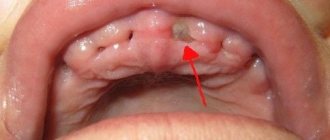
The important thing is that with arthritis of the jaw joint, purulent inflammation can begin! In such a situation, the joint turns red, swells and becomes very sensitive. A compaction is felt in the area of the mandibular joint. The skin changes its color, speech is impaired, and the process of chewing food becomes difficult.
Installation of a brace system for a patient with TMJ dysfunction
Installation of a brace system on the upper jaw is carried out on average after 3 months of occlusion therapy. The splint is adjusted once every 1-2 weeks, or at the discretion of the doctor, until the main complaints from the TMJ are eliminated (in parallel with the alignment of the teeth in the upper jaw), then a brace system is installed on the lower jaw with partial reduction (grinding) of the interfering parts of the occlusal tires, or complete removal. Here the patient needs to be patient - the process may take several months.
At the same time, the new position of the lower jaw is monitored: repeated manual functional analysis, photometry, bite registration is possible, computed tomography of the face during treatment, continuation of orthodontic treatment with a brace system.
Upon completion of orthodontic treatment, final monitoring of the position of the lower jaw follows (manual functional analysis, photometry, bite registration, 3D CT scan of the face upon completion (after) treatment).
Joint splint
Joint splint with braces
Type of pathology
The clinical picture of arthritis depends on the form in which the disease occurs:
Acute form.
It is expressed by swelling of the soft tissues and severe pain in the affected area. As a rule, the acute form is the result of some kind of injury.
Chronic form.
This form is manifested by aching pain and difficulty opening the jaw, especially in the morning.
Infectious form.
Most often it is a consequence of a previous illness (flu, ARVI). Infectious arthritis is characterized by severe pain that makes it difficult to open the mouth. The pain radiates to the ears, temples and back of the head.
Traumatic appearance.
The most common form of the disease that occurs against the background of trauma.
Specific arthritis.
Rare disease. As a rule, it occurs as a consequence of syphilis, tuberculosis or gonorrhea.
Purulent form.
It is a consequence of acute arthritis in an advanced form. A compaction forms in the area of the mandibular joint, and body temperature may increase.
Rheumatoid form.
Constant pain in the maxillary joint area. With this form, the clinical picture may be supplemented by pain in the knee, hip or elbow joint.
Depending on what caused the inflammation, the following routes of infection are distinguished:
- Hematogenous. Bacteria enter the joint along with the blood (syphilis, tuberculosis, measles, etc.).
- Contact path. The disease develops due to damage to areas located near the maxillary joint (otitis, phlegmon, abscess).
- Straight way. Develops due to direct penetration (wound, jaw fracture).
The form of pathology can be identified only after examination and diagnostic testing.
Diagnosis and treatment methods
Inflammatory processes of the TMJ can be diagnosed in the early stages, but this requires timely treatment. Initially, the lesion is localized in the capsule, but as the problem progresses, it spreads to the surrounding tissues. If left untreated, joint mobility may be completely lost.
Diagnosis is made during the initial visit to the dentist. Additionally, consultations with a traumatologist, otolaryngologist, infectious disease specialist and other specialists may be required. The basic methods are CT, radiography, CBCT. Using an x-ray, the dentist is able to determine the presence of arthritis, widening or narrowing of the joint gap. The following types of examinations are also carried out:
- Schüller and Bordes techniques for lateral examination of the affected area;
- plain radiographs;
- layered tomography;
- zonography;
- contrast arthrography;
- magnetic resonance imaging;
- diagnostics ELISA (enzyme-linked immunosorbent assay);
- PCR study.
Treatment begins with analyzing the data obtained and drawing up a treatment regimen. The jaw is immobilized and the affected organ is kept at rest for 2-3 days. If necessary, a sling bandage, splint or interdental plate is applied. The patient should follow a semi-liquid diet, relieving tension on the joint. The treatment regimen depends on the form and degree of the lesion; physiotherapy and myogymnastics are usually prescribed.
In the acute form, injections of antibiotics, corticosteroids, and chondroprotectors are prescribed. In case of purulent form, surgical intervention, opening of the cavity, and drainage through an external incision are indicated. Chronic arthritis requires the use of physiotherapy methods, sanitation of the oral cavity and nasopharynx. In some cases, dental prosthetics are indicated.
Treatment tactics for arthrosis of the mandibular joint
Ideally, starting treatment for TMJ arthrosis is optimal at stages 1 or 2. First of all, it is important to exclude risk factors - excessive mechanical stress, prolonged opening of the mouth, for example at a dentist appointment, etc. Therapy should be continuous and comprehensive - individual orthopedic measures or diet are not enough.
- Diet. To reduce the mechanical load, the patient is recommended to switch to soft foods, and during an exacerbation - to purees, juices, cereals and fermented milk products.
- Daily routine and lifestyle. It is very important to get enough sleep and limit stress, since any stress causes you to reflexively clench your teeth, which can aggravate the situation.
- Dentist consultation. Provoking factors include dental defects, missing teeth, and malocclusion. These problems are easily solved today, and patients with jaw arthrosis are primarily referred to the dentist and orthodontist.
A dentist and an orthopedist always take part in the treatment of jaw arthrosis.
Classification
Based on the results of an X-ray examination, in accordance with the diagnosed changes, it is customary to distinguish between two types of arthrosis:
- sclerosing - changes suggest sclerotic changes in bone surfaces, accompanied by a significant narrowing of the gap between the heads of the bones;
- deforming – there is a significant loss of texture and inherent forms of bone tissue, as well as a significant proliferation of osteophytes.
According to their origin, there are two types of pathology:
- primary – forms independently, without previous diseases/damages, mainly in old age;
- secondary - has a close connection with previous trauma, inflammation or metabolic disorders.
Prevention
Preventive measures require the fulfillment of such conditions as:
- maintaining proper and at the same time maximally balanced nutrition;
- optimization of the patient’s motor activity;
- rejection of bad habits;
- ensuring the proper level of oral hygiene;
- systematic attendance at preventive medical examinations.
Do not delay the treatment of arthrosis of the jaw joint, because in the first stages the disease is treatable, which allows us to talk about the complete restoration of jaw functionality.
What symptoms should you see a doctor for?
The earlier the diagnosis is made, the more favorable the prognosis. So seek help if:
- you experience pain in the lower jaw - intense or mild;
- you cannot fully move your jaw, you feel limited;
- hear a painful clicking or crunching sound when opening and closing your mouth;
- pain and noise in the ears appeared (especially if the otolaryngologist did not find any pathologies);
- There was a feeling of incomplete bite.
Arthrosis of the TMJ is not a very common phenomenon, but quite dangerous. Our usual comfort during conversation, eating, and other household activities is at risk. To avoid this, do not attribute alarming symptoms to fatigue and do not rely on “it will go away on its own”: be sure to find time for yourself and your health!