Bleeding always indicates damage to the vascular wall. In some cases it is harmless, in others it poses a threat to life and health. People often experience bleeding. In most cases, for injuries, cuts, dental treatment. Such situations are considered normal and do not cause panic. Blood from the mouth is something completely different. Such a symptom may indicate many serious diseases. Among them are lung cancer, tuberculosis, stomach ulcers, etc. If even a small amount of blood appears, you should consult a doctor immediately. You should not panic in such a situation, as this can only worsen the patient’s condition. Also, you can’t start any manipulations yourself. Only a specialist can find out the cause of this symptom and help a person.
Types of bleeding from the mouth
Blood from the mouth is not always associated with lung diseases, as many people think. There are many different factors that lead to the development of this symptom. Depending on the source of damage, there are:
- Bleeding from the mouth. It is associated with damage to the mucous membranes and gums.
- Bleeding from the respiratory tract. It is considered the most dangerous and often leads to the death of patients. It is worth distinguishing it from hemoptysis. This symptom is observed in tuberculosis and tumor diseases of the bronchopulmonary system. It is characterized by the release of up to 50 ml of blood when coughing. Despite the fact that this symptom is less dangerous, if it develops, you should also immediately consult a doctor.
- Bleeding from internal organs. This refers to the stomach and esophagus. When the vessels in these organs are damaged, blood enters the pharynx and then into the oral cavity.
A symptom may be the only sign of pathology or be combined with other pathological reactions. These include coughing and vomiting. Depending on the source of damage, venous, arterial and capillary bleeding are distinguished. Sometimes the integrity of several vessels is compromised.
Other causes of oral bleeding
In addition to the gums, there are other reasons that lead to the development of bleeding. Let's look at them in more detail:
- Bleeding tonsils. Tonsils can bleed due to tonsillitis, tonsillitis and a number of other diseases. Inflammation of the tonsils can occur for various reasons, and in this case it is very important to determine what triggered it.
- Diseases of the gastrointestinal tract. Bleeding in the mouth can be caused by a stomach ulcer, erosion of the esophagus or stomach, as well as severe stages of liver cirrhosis. In addition, if the blood has a dark tint, then there may be a risk of cancer, therefore, with these symptoms, it is very important to immediately consult a doctor.
- Coughing up blood may indicate the development of tuberculosis. Despite the fact that this disease has been studied to date, nevertheless, it remains very dangerous, and therefore it cannot be started.
- In addition, the blood itself may not be observed in the oral cavity, but its taste may be acutely felt. This may indicate various disorders, ranging from dry mouth to diseases of the genitourinary system.
The presence of blood is a consequence of negative processes occurring in the body, and therefore at the first symptoms it is necessary to immediately consult a specialist. After all, any disease is easier to treat in the initial stages.
Blood from the mouth: causes
The causes of blood in the mouth can include both minor damage to the mucous membranes and serious diseases of the respiratory and digestive system. To identify the etiology, doctors pay attention to the accompanying clinical manifestations, and also find out the factors that precede the onset of the symptom. What can cause bleeding from the mouth? The reasons for the development of the symptom may be the following:
- Pulmonary tuberculosis. This disease has been known for a long time. Previously, tuberculosis was diagnosed too late, and the main symptom of the pathology was hemoptysis. Currently, such a symptom rarely develops with timely treatment.
- Lung cancer. This pathology is common among older people who smoke. Most cases of cancer are diagnosed in men.
- Damage to the mucous membranes of the mouth.
- Stomach ulcer.
- Damage to the mucous membrane of the esophagus.
- Complication of liver cirrhosis.
- Oncological diseases of the oral cavity.
- Poisoning with various chemicals (acids, alkalis).
- Mallory-Weiss syndrome. Develops in patients suffering from alcoholism.
- Blood pathologies (hemophilia, thrombocytopenic purpura).
Blood from the throat can appear due to inflammatory diseases of the tonsils, oropharynx and nasopharynx. Sometimes this symptom indicates chronic gum disease.
Traumatic lesions of the oral mucosa
- Chronic mechanical injury
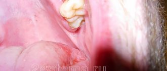
Clinical manifestations of a traumatic ulcer depend on the strength of the damaging factor, the general reactivity of the body, the state of the microbiocenosis of the oral cavity and other factors. As a rule, a traumatic ulcer is single. The mucous membrane around the ulcer is hyperemic and swollen. A traumatic ulcer is always painful, has uneven edges, and its bottom is covered with a fibrinous coating that can be easily removed. With the long-term existence of an ulcer, its edges and base become denser (due to the predominance of the phenomenon of proliferation). The edges of the ulcer are hyperemic, painful on palpation, the bottom is often bumpy, covered with necrotic plaque. The depth of the ulcer varies down to the muscle layer. Regional lymph nodes are enlarged, mobile, and painful on palpation.
Traumatic ulcers can be complicated by fusospirochetosis or candidiasis, and with a long course (2-3 months) they can become malignant.
- A traumatic ulcer must be differentiated from:
- ulceration of a cancerous tumor,
- tuberculous ulcer,
- syphilitic chancre,
- chronic ulcerative necrotic gingivostomatitis Vincent,
- trophic ulcer.
Removal of the traumatic factor also serves differential diagnostic purposes. Rapid healing of the ulcer within a few days indicates its traumatic origin.
The bone tissue of the alveolar process is resorbed in places, the alveolar edge becomes soft, mobile (“dangling”). Angular cheilitis often develops simultaneously. In the occurrence of such conditions, in addition to chronic trauma, a certain role is played by the influence of fungi of the genus Candida, which, as a rule, are found in large quantities on prostheses, and less on the mucous membrane of the prosthetic bed. You should also remember about the possibility of an allergic reaction of the oral mucosa to the base material of the prosthesis (usually acrylic plastic).
- Chemical damage
In case of acute injury, sharp pain usually occurs immediately and is localized at the site of contact with the chemical. The clinical picture depends on the nature and amount of the damaging substance and the time of action.
Burns with acids lead to the appearance of coagulative necrosis on the mucous membrane - a dense film of brown (from sulfuric acid), yellow (from nitric acid) or white-gray (from other acids) color. The films are tightly fused to the underlying tissues and are located against a background of pronounced inflammation of the mucous membrane with swelling and hyperemia.
A burn with bases (alkalies) causes liquefaction necrosis of the mucous membrane, while a dense film is not formed, necrotic tissue has a jelly-like consistency. The damage is deeper than with an acid burn. Necrosis from alkali burns can involve all layers of soft tissue, especially on the gums and hard palate. Burns, especially extensive ones, cause severe suffering to the patient. A few days after the necrotic tissue is sloughed off, slowly healing erosive or ulcerative surfaces are exposed. In mild cases, alkali burns can only cause a catarrhal inflammatory process.
Symptoms of “galvanism” should be differentiated from stomalgia, glossalgia, and allergic stomatitis.
Anamnesis (absence of symptoms of “galvanism” before prosthetics), as well as measurement of the magnitude of microcurrents in the oral cavity, are important in the differential diagnosis.
- Radiation sickness
Radiation sickness (morbus radialis) develops as a result of exposure to ionizing radiation on the entire body or its large parts: chest, abdomen, pelvic area.
Radiation damage can be caused by any type of ionizing radiation: X-rays, beams, neutron fluxes, etc. In irradiated tissues, the morphological structure of the walls of blood vessels changes, the barrier function of connective tissue decreases, and regeneration is suppressed. There are acute and chronic forms of radiation sickness.
Acute radiation sickness (morbus gadialis acutus) develops after a single irradiation of the body with massive doses (1 - 10 Gy, or 100 - 1000 rad). In the first period - the period of primary reactions, which begins soon after irradiation (1-2 hours) and lasts up to 2 days, dryness (or salivation) appears in the oral cavity, taste and sensitivity of the mucous membrane decrease. The mucous membrane of the mouth and lips swells, hyperemia appears, and pinpoint hemorrhages may occur. In the second, latent period, which lasts from several hours to 2 weeks, all these phenomena disappear. The period of pronounced clinical phenomena (third period) is the height of the disease. Against the background of a sharp deterioration in the general condition, changes in the oral cavity reach a maximum. A burning sensation appears, the mucous membrane becomes anemic and dry. A picture of radiation stomatitis appears.
Changes in blood vessels and blood composition during this period cause the phenomena of hemorrhagic diathesis: increased bleeding, multiple petechial hemorrhages. Due to a sharp decrease in tissue resistance, a rapidly developing autoinfection, especially putrefactive one, occurs.
Thus, the clinical picture of radiation stomatitis consists mainly of hemorrhagic syndrome and necrotic ulcerative process. The latter is most pronounced in places of injury with overhanging fillings, sharp edges of teeth, dentures, tartar, and where there were accumulations of microflora before the disease, i.e. First, the edge of the gums and tonsils are affected, and then the lateral surfaces of the tongue and the palate. In areas adjacent to the mucous membrane of metal prostheses and fillings, the damage may be more pronounced due to secondary radiation. The mucous membrane of the mouth, lips and face swell. The gingival papillae loosen, bleed, and then the gum edge becomes necrotic. The bone tissue of the alveolar process is resorbed, the teeth become loose and fall out. Multiple necrosis of the mucous membrane does not have sharp boundaries, the inflammatory reaction of surrounding tissues is mild. The bottom of the ulcers is covered with a dirty gray necrotic coating with a putrefactive odor. In severe cases, necrosis can spread from the mucous membrane to the underlying soft tissue and bone, radiation necrosis of the bone occurs with sequestration, and jaw fractures are possible. This is facilitated by the spread of infection from foci of chronic periodontitis and periodontitis.
The tongue swells, becomes covered with a thick coating, cracks, hemorrhages and necrosis appear, most often in the area of the root of the tongue; taste and sensitivity disappear. Severe necrotizing tonsillitis develops. If the patient does not die, then the fourth period begins - recovery: a slow reverse development of the symptoms of the disease occurs. In the oral cavity, everything also returns to relative normal. During this period, relapses of the disease are possible.
Chronic radiation sickness (morbus radialis chronicum) develops as a result of prolonged exposure to low doses of radiation on the entire body or a significant part of it. The oral cavity is sensitive to the effects of ionizing radiation, so at the onset of the disease, changes in the oral cavity can be especially pronounced. Dryness gradually increases due to damage to the salivary glands. Persistent catarrhal gingivitis occurs, which can subsequently develop into ulcerative gingivitis. Initially, erosions and ulcers may appear along the transitional folds on the vestibular side, followed by damage to the gums and red border of the lips.
Glossalgia may develop, and then glossitis with swelling of the tongue, cracks, and coating on the tongue. The long course of chronic radiation sickness usually leads to a picture similar to periodontitis - the so-called radiation periodontitis.
The reaction of the mucous membrane to radiation develops gradually - from hyperemia and swelling to the appearance of erosion. This reaction has its own characteristics in different areas of the mucous membrane. The first clinical signs on the mucous membrane, which does not have a keratinized layer in the epithelium (cheeks, floor of the mouth, soft palate), are manifested by mild hyperemia and swelling, which increase as the absorbed dose increases. Then the mucous membrane becomes cloudy, loses its shine, thickens, wrinkles appear, and when scraped, the surface layer is not removed. This occurs due to increased keratinization of the epithelium. Some affected areas resemble leukoplakia or lichen planus. If the radiation dose increases, the keratinized epithelium in some areas is rejected, desquamated, erosions appear, covered with a sticky necrotic plaque - focal membranous radiomucositis develops. Then the epithelium is rejected over large areas, the erosions merge and focal radiomucositis transforms into confluent membranous radiomucositis. The mucous membrane of the soft palate is highly radiosensitive, there is no keratinization stage when it is irradiated, and the reaction develops faster than in other parts of the oral cavity. In areas of the mucous membrane that are normally subject to keratinization, the radiation reaction proceeds more favorably and leads only to focal desquamation of the epithelium or single erosions.
The course of pathological processes in the oral mucosa is complicated by damage to the salivary glands (with remote irradiation methods). At the beginning (the first 3-5 days), increased salivation may be observed, which is quickly replaced by dry mouth up to complete xerostomia, which is practically impossible to stimulate.
The consequence of the death of the taste buds of the tongue is a violation of taste. At first, sensations in the tongue may manifest themselves as glossalgia, then a perversion of taste is noted, and later - loss of it.
Radiation changes in the oral cavity are largely reversible. After cessation of irradiation or during a break in treatment, the mucous membrane quickly returns to relative normal. This period lasts 2-3 weeks. With a large absorbed dose (more than 50-60 Gy, or 5000-6000 rad), irreversible changes in the salivary glands and mucous membrane (edema, hyperemia, telangiectasia, atrophy, radiation ulcers) may occur.
Radiation therapy should be preceded by sanitation of the oral cavity, since the reaction of the mucous membrane to radiation exposure is more severe in an unsanitized oral cavity.
Due to trophic disorders, the reactivity of the mucous membrane to mechanical trauma and infection is sharply reduced. The edematous mucous membrane with defective epithelial cover is easily injured by the sharp edges of teeth and dentures, which can lead to the appearance of sharply painful, long-term non-healing ulcers.
- Leukoplakia
The initial manifestations of leukoplakia are usually invisible, since there are no subjective sensations. Leukoplakia begins with the appearance of areas of clouding of the epithelium (grayish in color, with elements of keratinization on the surface). The lesion occurs against the background of an unchanged mucous membrane. Typical localization of the focus of leukoplakia is the mucous membrane of the cheeks along the line of closure of the teeth in the anterior section, the corners of the mouth and the red border of the lower lip without skin lesions. The dorsum and lateral surfaces of the tongue are somewhat less commonly affected. Smokers are characterized by lesions of the palate, described as “Tappeiner smokers' leukoplakia.”
There are the following forms of leukoplakia: flat or simple, verrucous and erosive. These forms can transform one into another. It is possible to combine different forms of leukoplakia in different areas of the oral mucosa in the same patient. The disease begins with the appearance of a flat shape.
Flat, or simple, leukoplakia (leukoplakia plana) is the most common. This form usually does not cause subjective sensations and is discovered by chance. Sometimes patients have complaints of a feeling of tightness, burning, and an unusual appearance of the mucous membrane. In the presence of extensive lesions on the tongue, taste sensitivity may be reduced. The main morphological element of the lesion in flat leukoplakia is a hyperkeratotic spot, which is an area of turbidity of the epithelium with clear contours. During the examination, foci of hyperkeratosis of various shapes and sizes are revealed, not rising above the level of the mucous membrane, but with clear boundaries of the lesion. Elements of flat leukoplakia resemble a burn of the mucous membrane with lapis, thin tissue paper pasted on, or a white coating that does not come off even with intense scraping. The keratinization can vary in intensity, so the color of the affected areas varies from pale grayish to intensely white.
The surface of the area of flat leukoplakia is usually slightly rough and dry. There is no compaction at the base of the lesion, just as there is no visible inflammatory reaction along its periphery.
There is a direct connection between the shape, color, size of keratinized areas and their location. Thus, with hyperkeratosis of the mucous membrane in the area of the corners of the mouth, symmetry of the lesion is observed in 85% of cases. The shape of the lesion has the shape of a triangle, the base of which faces the corner of the mouth, and the apex faces the retromolar space.
When the focus of leukoplakia is localized on the mucous membrane of the cheeks along the line of closure of the teeth, the elements of the lesion represent an elongated line, the continuity of which may be disrupted in certain areas. Leukoplakia on the red border of the lips looks like sticky tissue paper of irregular shape and grayish-white color (Fig. 11.5). Limited areas of lesions form on the mucous membrane of the tongue, hard palate, and floor of the mouth, sometimes resembling wide stripes or solid spots. More extensive lesions are also noted.
The relief of the mucous membrane and its turgor are also reflected in the appearance of the lesion. So, if leukoplakia develops against the background of folding of the tongue, the protruding areas become more keratinized and the surface of the tongue resembles a cobblestone street. A similar picture can be seen with a decrease in turgor and folding on the cheeks. If there is no pronounced folding on the tongue, flat leukoplakia looks like grayish-white, slightly sunken spots, the papillae of the tongue on them are smoothed.
Tappeiner's leukoplakia (leucoplakia nicotinica Tappeiner, nicotinic leukokeratosis of the palate) occurs in heavy smokers (especially pipe smokers). The mucous membrane of the hard palate and the adjacent part of the soft palate is mainly affected. Sometimes the gum margin is involved. The mucous membrane in the affected area is grayish-white in color, often folded. Against this background, mainly in the posterior half of the hard palate, red dots stand out - the gaping mouths of the excretory ducts of cystically dilated salivary glands, looking like small nodules. They are formed due to blockage of the excretory ducts by hyperkeratotic masses. Damage to the palate in smoker's leukoplakia can be combined with the location of elements on the mucous membrane of the cheeks, corners of the mouth, lower lip, etc. This form of leukoplakia is an easily reversible process: stopping smoking (an irritating factor) leads to the disappearance of the disease.
Verrucous leukoplakia (leucoplakia verrucosa) is the next stage in the development of flat leukoplakia. This is facilitated by local irritants: trauma with sharp edges of teeth and dentures, biting areas of leukoplakia, smoking, eating hot and spicy foods, microcurrents, etc.
The main feature that distinguishes this form of leukoplakia from flat one is more pronounced keratinization, in which a significant thickening of the stratum corneum is detected. The area of leukoplakia rises significantly above the level of the mucous membrane and differs sharply in color. Upon palpation, a superficial compaction may be detected. Patients usually complain of a feeling of roughness and tightness of the mucous membrane, burning and pain when eating, especially spicy food.
There are plaque and warty forms of verrucous leukoplakia. In the plaque form, limited milky white, sometimes straw-yellowish plaques with clear contours are identified, rising above the surrounding mucous membrane. The warty variety of verrucous leukoplakia is characterized by dense grayish-white bumpy or warty formations, rising 2-3 mm above the level of the mucous membrane. The warty form of leukoplakia has a greater potential for malignancy compared to the plaque form. On palpation, the lesions are dense, painless, and not fused with the underlying areas of the mucous membrane. The thickness of the raised areas of hyperkeratosis varies from pronounced to barely noticeable during normal examination. With a slight elevation of the elements, the affected area is almost not determined by palpation. A leukoplakic lesion of considerable thickness becomes dense to the touch, and it is not possible to fold it.
Erosive leukoplakia (leukoplakia erosiva) is actually a complication of simple or verrucous leukoplakia due to trauma. With this form of leukoplakia, patients complain of pain, which intensifies under the influence of all types of stimuli (eating, talking, etc.). Usually, against the background of foci of simple or verrucous leukoplakia, erosions, cracks, and, less often, ulcers occur. Erosions are difficult to epithelialize and often recur. Patients are especially concerned about erosion in the keratinized area of the red border of the lips. Under the influence of insolation and other irritating factors, they increase in size without showing a tendency to heal. The pain intensifies.
Pathohistologically, thickening of the epithelium is revealed due to the proliferation of the horny and granular layers. The stratum corneum reaches considerable thickness, especially with the verrucous form of leukoplakia. In it, foci of hyperkeratosis often alternate with foci of parakeratosis. The granular layer of epithelium in the affected area has varying degrees of severity. Verrucous and erosive forms of leukoplakia are characterized by pronounced hyperkeratosis and acanthosis. Acanthosis in leukoplakia can be significant if the keratinization has the character of parakeratosis; conversely, with severe hyperkeratosis, acanthosis is minimal or completely absent. In the connective tissue stroma of the affected areas of the mucous membrane, diffuse chronic inflammation is determined with pronounced infiltration of the surface layers by lymphocytes and plasma cells, the manifestation of sclerosis. The latter explains poor healing and frequent relapses with the appearance of cracks and erosions, which is most typical for the plaque form of leukoplakia.
Leukoplakia is a precancerous condition, since all its forms can become malignant with varying degrees of probability, transforming into spinocellular cancer.
Flat leukoplakia malignizes in 3-5% of patients, and in some the process of malignancy occurs quickly (1-1.5 years), in others the disease can exist for decades without transforming into cancer. Most often, malignancy occurs in verrucous and erosive forms of leukoplakia (in 20% of cases).
Clinical signs of malignancy are increased keratinization processes; rapid increase in the size and density of the lesion; the appearance of compaction at the base of the plaque, erosion; papillary growths on the surface of erosions, bleeding due to injury; the appearance of non-healing cracks.
- Soft leukoplakia Pashkova
Characterized by the presence of areas of peeling, slightly swollen, pasty mucous membrane with unclear boundaries, without inflammation or compaction. The mucous membrane has a grayish-white color. Most often, soft leukoplakia is localized on the mucous membrane of the cheeks along the line of closure of the teeth and lips, and less often on the tongue and gums. The lesion can be limited or diffuse, when almost the entire mucous membrane, and sometimes the red border of the lips, is involved in the process. In patients with neuropathy, predominantly young, with habitual biting or sucking of the cheeks, lips, tongue, the epithelium along the line of closure of the teeth is unevenly desquamated, has a fringed appearance due to the presence of multiple small flaps, which causes the unevenness of the affected surface (it looks like it is moth-eaten). This altered epithelium is partially removed by scraping. In severe cases, painful erosions may occur from biting the epithelium.
Histologically, pronounced parakeratosis and acanthosis are noted. At all levels of the spinous layer there are so-called “light” vacuolated cells, the cytoplasm of which is almost not stained, and the nuclei are deformed (pyknotized). In the connective tissue layer, expansion of small blood vessels, thickening of collagen, and thinning of elastic fibers are detected.
- White spongy nevus of Cannon
White spongy nevus may appear in early childhood or later, then progresses and reaches its maximum development during puberty. The disease may then regress or remain unchanged.
There are no subjective sensations. Patients may complain of an unusual appearance of the mucous membrane.
The typical location of a white spongy nevus is the buccal mucosa. The lesion is always symmetrical. The mucous membrane of the cheeks is white or grayish-white, somewhat thickened, soft, as if spongy, strongly folded. In some cases, the folding and wrinkling of the mucous membrane is so pronounced that the folds hang into the oral cavity. The surface layers of the epithelium are removed by scraping. At the same time, similar lesions may appear on the mucous membrane of the genital organs and rectum.
White spongy nevus should be differentiated from:
- flat leukoplakia;
- candidiasis;
- lichen planus;
- soft leukoplakia.
There is great clinical similarity between white spongy nevus and soft leukoplakia. Some authors consider white spongy nevus and soft leukoplakia to be different clinical forms of the same disease related to nevi, but there are quite pronounced differences between them. White spongy nevus, unlike soft leukoplakia, occurs mainly in childhood and is often hereditary. The development of mild leukoplakia, as a rule, is associated with various kinds of neurotic reactions or with hormonal changes in the body (puberty, postmenopause).
Patients with Cannon's nevus have congenital defects in the structure and maturation of the epithelium of the oral cavity, and in some cases, the genitals and rectum. With mild leukoplakia, the oral mucosa initially has a normal appearance, and its pathological changes are associated with the action of irritating factors against the background of neurological disorders or hormonal dysfunctions. In addition, damage to the oral mucosa with white spongy nevus is usually generalized.
Damage to mucous membranes
In various diseases, the mechanism of symptom development is clear in most cases. But why does a healthy person bleed from the mouth? In most cases, this is due to damage to the surface of the mucous membrane. Minor capillary bleeding occurs when biting the inner surface of the cheeks, tongue, or lips. The symptom often develops after dental procedures. In most cases, blood appears from the child’s mouth in this way. Children often bite their lips and cheeks after anesthesia, as the tissues lose sensitivity.
With severe inflammation of the tonsils, blood is often detected when rinsing the throat. Also, red streaks in the sputum are sometimes observed in patients with pharyngitis. This symptom indicates minor damage to the vessels of the pharynx. Such pathologies are not accompanied by dangerous bleeding. However, it is recommended to get checked. Blood from the throat may indicate oral cancer. Damage to the gums can also be dangerous. In some cases, it indicates the development of diseases such as thrombocytopenic purpura, hemophilia or scurvy.
3. Symptoms and diagnosis
Iron deficiency anemia is characterized by dullness of taste, pallor of the mucous membrane, painful reaction of the mucous membrane to sour or spicy foods, various false sensations (tingling, tingling, etc.), swelling and, less often, bleeding of the tongue, as well as inflammation along the red border of the lips (cheilitis) and multiple caries.
With hypoplastic anemia, hemorrhagic foci, swelling and cyanosis of the interdental papillae, and a tendency to expose the periodontal pockets are observed; in more severe cases, erosions or ulcers and areas of local necrosis appear on the oral mucosa.
B12-folate deficiency Addison-Biermer anemia is characterized by a yellowish-pale tint of the mucous membrane, glossitis (inflammation of the tongue), and in some cases, pinpoint hemorrhages on the mucous membrane.
As for leukemia, their existing classifications (based on etiological, cytogenetic and other criteria) include several dozen clinical forms, the analysis of which is beyond the scope of this material. Even the traditional division in medicine according to the type of course - into acute and chronic leukemia - in this case takes on a slightly different meaning: in fact, these are two different diseases, and they never transform into each other.
If we do not touch upon the main symptoms and limit ourselves only to changes in the oral mucosa (as in the case of anemia, see above), then acute leukemia is manifested by severe bleeding of the mucous membrane on the gums, cheeks (along the line of dental occlusion), on the upper palate, and tongue. Loose hyperplastic growth of the gums in the form of ridges that bleed at the slightest touch or spontaneously is often observed. In addition, pale necrotic areas surrounded by a bluish halo with an off-white or gray foul-smelling coating are often observed. Swallowing is difficult, eating causes pain in areas of necrotic ulceration; trophic disorders are often accompanied by activation of chronic infections (herpes, candidiasis).
Chronic leukemia, which can be latent for a long time, is also characterized by bleeding of the oral mucosa, but it is less pronounced than in acute leukemia, and hemorrhages occur mainly due to trauma or microtraumatization. A diagnostically informative sign is the combination of bleeding, cyanosis, and pallor of the mucous membranes in the absence of signs of inflammation.
In myeloid leukemia, erosive and ulcerative lesions of the oral mucosa often predominate; in chronic lymphocytic leukemia, infiltrating nodes with a loose consistency and bluish tint predominate.
If, during an examination by a dentist, orthodontist, otorhinolaryngologist, therapist or other specialist, there is a suspicion that specific changes in the oral mucosa may be caused by diseases of the hematopoietic system, a consultation with a hematologist is strictly necessary, followed by an in-depth laboratory blood test , the results of which are the most informative and decisive in making a diagnosis. As necessary, other laboratory and instrumental studies are prescribed (imaging diagnostic methods, bone marrow puncture followed by histological analysis, etc.).
About our clinic Chistye Prudy metro station Medintercom page!
Bleeding due to lung pathology
The most common pulmonary pathologies that are complicated by bleeding include tuberculosis, cancer and bronchiectasis. All these illnesses are very dangerous and require long-term treatment in specialized institutions. Pulmonary hemorrhage indicates progressive cavernous tuberculosis. It often occurs against the background of a coughing attack.
In lung cancer, bleeding indicates tumor disintegration and vascular damage. It can be very difficult to stop him. Particularly dangerous is profuse bleeding, in which 500 ml of biological fluid is released. Such a symptom can be fatal. In addition to bleeding from the mouth, patients complain of difficulty breathing, coughing, and weight loss.
Bronchiectasis is a progressive destructive-inflammatory pathology of the lungs. It is characterized by a constant cough with purulent sputum and shortness of breath. Diagnosed in childhood or young age. With severe damage to the bronchi, it can be complicated by a violation of the integrity of blood vessels.
Signs of gastrointestinal bleeding
If there is blood coming from the mouth, this may be a symptom of diseases of the digestive system. The most common of these are liver cirrhosis and stomach ulcers. In the first case, the source of bleeding is the dilated veins of the esophagus, in the second - the vessels of the stomach wall. Diseases of the digestive tract have characteristic features. The blood with these pathologies will have a dark color. It often occurs during vomiting. In addition, there is severe pain in the upper abdomen, nausea, and deterioration in health. In some cases, scarlet blood enters the oral cavity from the digestive tract. This may indicate the development of Mallory-Weiss syndrome, which consists of a linear rupture of the superficial vessels of the gastric mucosa. Other causes of bleeding include polyps, acute erosive gastritis and esophagitis. Regardless of the source of the damage, assistance must be provided immediately.
2. Reasons
Of all diseases of the hematopoietic system, the oral mucosa (OM) is most sensitive to two groups of diseases, one of which concerns red blood cells (erythrocytes) and the hemoglobin associated with them, the other - white blood cells, or leukocytes.
The group of anemia includes a number of diseases and pathological conditions in which the number of red blood cells decreases and the hemoglobin content in the blood decreases.
The group of leukemias includes malignant diseases of the hematopoietic system, in which the proliferation (abnormal multiplication and growth) of blood cells or bone marrow, as well as the activity of the hematopoietic parenchyma, precedes the maturation and differentiation of blood cells (i.e., their acquisition of one or another functional type), therefore, a large number of altered, atypical, immature leukocytes - leukoblasts, myeloblasts, etc. - appear in the blood.
Both anemia and leukemia, with all their etiopathogenetic and clinical diversity, which certainly deserves separate consideration, affect the state of the oral mucosa in more than 90% of patients, and often a preliminary diagnosis is made precisely on the basis of specific changes in the oral mucosa.
Visit our Oncology page
Bleeding in lung cancer: features
Lung cancer is considered one of the most common cancer pathologies among men. In most cases, it develops against the background of chronic inflammatory diseases of the respiratory system. The main risk factor is considered to be tobacco abuse over many years. The source of bleeding in cancer is damaged blood vessels or the tumor itself, which is in the decay phase.
Signs of the disease are a prolonged dry cough that cannot be treated. After a few months, hemoptysis occurs. In addition, fever, severe weakness, shortness of breath and weight loss are noted. Bleeding can have a different volume: from small (50-100 ml) to profuse (more than 0.5 liters). In most cases it is accompanied by a cough. In this case, there is a high risk of aspiration and the development of hemorrhagic shock.
Diagnosis of bleeding from the mouth
If a person has bleeding from the mouth, it is necessary to urgently call an ambulance. To identify the cause of this symptom, the doctor must find out from relatives what diseases or complaints the patient had previously. If bleeding from the digestive tract is suspected, an FGDS is performed. If the source of damage is in the lungs, bronchoscopy is performed in the hospital. An x-ray of the chest and abdomen is also required. To determine the degree of blood loss, the level of hemoglobin and red blood cells is checked. A coagulogram is also required. It is needed to exclude blood pathologies.
Causes of blood taste in mouth
Changes in taste during pregnancy
Pregnant women are characterized by sharp hormonal changes, which cause an increased sense of smell, changes in taste preferences and sensations. As a result, a specific metallic taste, perceived as the taste of blood, occurs when eating fully cooked meat products, fish or even vegetable dishes. The symptom occurs periodically and often goes away on its own after some time.
Anemia
The main reasons for changes in taste are a drop in hemoglobin and coagulation disorders, leading to capillary fragility. The taste of blood in the mouth is especially common in young women if anemia develops during pregnancy. Its persistence throughout the day, combination with painful nausea, the urge to vomit, indicates concomitant pathology (coagulopathy, hypovitaminosis). In the absence of gestation, an anemic change in taste is observed with chronic bleeding, neoplasia, etc.
Gingivitis
Chronic inflammatory disease of the gums occurs in 20-30% of the adult population. Usually, the taste of blood appears after brushing your teeth with hard toothbrushes, which cause trauma to the mucous membranes and bleeding gums. When gingivitis worsens, an unpleasant taste in the mouth is felt at any time of the day for no apparent reason. The symptom is accompanied by redness and swelling of the gums, and the formation of difficult-to-remove plaque on the teeth.
Other dental causes
A bloody taste in the mouth is detected in various pathological processes: medium and deep caries, pulpitis, periodontitis. The symptom can occur spontaneously, but is more often provoked by eating solid food and brushing teeth. A specific unpleasant taste of blood develops against the background of intense pain in the area of the diseased tooth. Odontogenic causes are dangerous due to their complications, so you need to visit a specialist as soon as possible.
ENT pathology
The taste of blood in the mouth often occurs with chronic rhinitis and sinusitis, in which atrophy of the mucous membrane occurs and scanty bleeding periodically begins. The bloody taste bothers you in the morning, when the blood that has accumulated overnight begins to flow down the back wall of the throat. The person experiences severe nausea and aversion to food. The discomfort disappears after rinsing your mouth and brushing your teeth.
Respiratory infections
Most often, the symptom occurs with influenza, for which a toxic effect on capillaries and spontaneous bleeding from the oral mucosa is pathognomonic. The taste occurs periodically in the first 2-3 days of infection, then disappears on its own. A similar clinical picture also develops in severe ARVI, pharyngitis and laryngitis.
Diseases of the gastroduodenal zone
GERD and gastritis are characterized by a taste of blood in the mouth after drinking alcohol, stress and heavy physical activity. In these diseases, the main reasons for the unusual taste are the formation of erosions in the mucous membrane of the digestive canal, which, under the influence of unfavorable factors, begin to bleed. Patients experience severe burning and pain in the chest, accompanied by an unpleasant sensation of blood in the mouth.
Cirrhosis
Fibrous liver damage provokes dilation of the esophageal veins and thinning of their walls, which is fraught with spontaneous bleeding. In the mouth of patients with cirrhosis, a strong taste of blood suddenly begins to be felt, and with massive bleeding, vomiting of dark red blood begins. The symptom occurs more often in men who abuse alcohol. If there is a suspected rupture of esophageal varicose veins, a person needs emergency medical care.
Cardiovascular disorders
The symptom is determined by hypertension, vasculitis, which is accompanied by thinning and fragility of the capillaries. A specific blood taste appears after physical activity or stress, leading to a rise in blood pressure and rupture of small blood vessels. During an attack of angina, accompanied by pain in the heart and shortness of breath, sometimes there is a feeling of an unpleasant taste of blood in the mouth.
Rare causes
- Diseases of the respiratory system
: bronchial asthma, chronic obstructive pulmonary disease, tuberculosis. - Poisoning with heavy metals
: lead, mercury, cadmium, etc. - Gastrointestinal pathology
: pancreatitis, cholecystitis, hepatitis. - Tumors
: malignant neoplasms of the oral mucosa and tongue, lung cancer.
Tactics at the prehospital stage
Before the ambulance arrives, you should try to calm the patient and apply something cold to the suspected source of bleeding (upper abdomen or chest). If the symptom is not severe, you can give the patient to swallow pieces of ice or ice cream. If the source of bleeding is in the oral cavity, the wound should be washed and cold applied. Further measures are carried out by the doctor.
Help with bleeding in the hospital
A dangerous sign is blood from the mouth. What to do if this symptom develops in a patient? Hemostatic and infusion therapy is carried out in a hospital setting. To stop fluid loss, the drugs “Aminocaproic acid”, “Dicinone”, “Vikasol”, “Calcium gluconate” are administered. To replenish the blood volume, systems with saline and glucose are installed. In case of hemorrhagic shock, infusion of blood substitutes and vascular agents are required. In some cases, surgery is performed.
"Diclofenac"
Another NSAID drug. Relatively safe, good pain relief.
Tablets are recommended for toothache. They contain from 25 mg to 100 mg of diclofenac sodium.
The medicine is prohibited:
- children under 6 years of age (if the dosage is more than 25 mg), and adolescents under 18 years of age (if the dosage exceeds 100 mg);
- for erosions and ulcers of the digestive organs;
- if the patient is intolerant to non-steroidal anti-inflammatory drugs.
Contraindicated for pregnant and lactating women.
Prescribe with caution to people with heart, kidney and liver failure.
- Bleeding in the middle of the cycle: causes
Diclofenac will relieve pain faster and more effectively if you take it half an hour before meals. The maximum dose per 24 hours is 150 mg, divided into 2 to 3 doses.