The clinics of the German Implantology Center are equipped with the most modern diagnostic equipment necessary to prepare the patient for dental implantation. According to dental market experts, both clinics of the Research Center, located in the Ramenki and Dorogomilovo districts of Moscow, occupy the TOP 1 rating for 2017-2019.
private dental clinics in Moscow and Russia.
Let's see what diagnostics are carried out before implantation in our clinics, without what equipment, procedures and diagnostic data it is currently impossible to prepare qualitatively
for implantation surgery.
The chief physician of the Research Center, implant surgeon Dakhkilgov Magomed Umatgireevich answers these questions:
As an implantologist with more than 20 years of experience
Magomed Umatgireevich, who has made a serious practical contribution to the development of this area in Russia, makes great efforts to ensure that the equipment of clinics meets
leading world standards.
He brought clinics to the TOP1 ratings in Moscow and Russia, which today makes it possible to successfully perform dental implantation even in the most complex clinical cases.
Please note that some of the material is accompanied by a video from our YouTube channel, we recommend spending a little of your precious time and watching the videos, it will be useful. More than 1,000 patients use the recommendations of medical specialists from the Scientific Research Center on YouTube every day
.
Classic implant installation protocol
Let's consider the case when dental implantation is carried out only on the basis of data obtained from an orthopantomogram (OPTG).
From the OPTG image we obtain the following information:
- based on the contrast of a 2-dimensional image, the presence of bone in the proposed implant installation site;
- the approximate height of the bone (the fact is that the picture is taken at an angle)
do not get complete information from the OPTG image
- about the actual distance from the bone crest to the mandibular canal or maxillary sinus;
- about the real cross-sectional profile of the bone.
Thus, using only the OPTG image, the doctor receives about 50% of the necessary information (conditionally), and the patient must rely solely on the experience and qualifications of the surgeon performing the implantation. A doctor's mistake can lead to the following:
- a complication may occur in the form of damage to the mandibular nerve, and, as a result, paresthesia (numbness of the lip and chin).
- Another error associated with the lack of information is illustrated in the following OPTG - perforation of the maxillary or nasal sinuses is shown, after such an “operation” the patient receives a referral to ENT specialists instead of teeth.
- Another type of complication is perforation of the cortical plate by drills or implants and, as a result, bone resorption. Unfortunately, what you see in the pictures is a real case.
For example, in the right figure we have modeled the correct direction of the implant axis that needed to be selected.
Cross-sectional sections show that the apical part of the implant is located in the mandibular canal and damages the nerve.
2) Now consider the case when, in preparation for implantation, the doctor has at his disposal a Computer Tomogram (CT) of the patient
Because From a CT scan it is possible to obtain all the data on bone volume in all planes and sections, then the completeness of information can be conditionally estimated at 75%. Where is the remaining 25%? Our experts believe that 25% is a prosthetic plan that was previously thought out and modeled in a 3D program. Let us remember the main purpose of installing implants – restoration of defects in the dentition. Each implant carries the function of supporting a future crown, as well as supporting a bridge for its fabric.
Incorrect installation of the support, and the structure becomes loose, loses strength, and as a result, the bridge collapses, and in the case of teeth, reimplantitis and other complications develop
Of course, an experienced orthopedist can mentally imagine exactly how 1, 2, 3 or 4 teeth will be located in the dentition, but what to do when there are more missing teeth? And if the orthopedist only has an approximate idea of what the teeth will ultimately look like, then how can the surgeon understand where he should place the implants?
Still, let’s assume that the orthopedist is experienced and, having a CT scan in hand, he made markings for implantation for the surgeon (indicated on the CT scan the exact location of the implants).
Now it's time for implantation. As a visual example, imagine a photograph of a dental model, indicating the sizes and positions of the holes that should be drilled.
The surgeon must complete the orthopedist's task, working by eye. Not that easy, right? If the holes are drilled with a deviation of only 1-2 millimeters, the treatment plan that the orthopedic surgeon has in mind will not be implemented, and the model will be thrown into the trash. In real life it is more difficult, the surgeon works in cramped conditions, his blood pressure may drop, and the patient may move involuntarily. At best, the orthopedic design will not be installed perfectly; at worst, harm to the patient’s health will be caused.
The photograph shows a case where the implants were installed in the wrong direction. How to get prosthetics now? But the conditions made it possible to install them at the desired angle.
Another clinical case: with missing teeth 45 and 46, one implant was installed because... at the time of its installation, due to bone deformation, the distance between teeth 44 and 47 was only 12.5 mm. There is a lot of space for one tooth, but not enough for two. This is a case where the surgeon performed implantation without prior orthopedic planning. In such a situation, the orthopedist can no longer change anything, so he is forced to provide prosthetics “as best he can.”
Our clinic’s specialists believe that the most important part of dental restoration using implantation is a properly developed strategy. In its absence, the probability of an error is very high, which is what happened. The exhausted patient subsequently contacted us with complaints about the unsatisfactory appearance of the teeth and inconvenience in use. It’s sad, but to correct the situation, you will need to remove the previously installed implant.
So, successful installation of implants using the standard method often depends 100% on the qualifications of the orthopedist and implant surgeon, the patient’s patience and other factors. The cost of an error can be either a “crooked” (in other words, there is no way to describe it) final design, or harm to the patient’s health.
How to avoid such complications? A new development by Russian scientists, implantation using templates, comes to our aid - the Implant-Guide technology.
How serious is the procedure?
To understand why you need to carefully prepare for a sinus lift, you need to have a general understanding of this option of bone grafting. This method is optimal for filling the lack of bone in the lateral parts of the upper jaw due to the anatomical features of its structure. One of the most important paranasal sinuses, the maxillary sinus, is located in this bone. The presence of this cavity formation allows specialists to free up the necessary space for tissue expansion by performing a sinus lift.
The doctor performing the procedure carefully lifts the lower wall of the maxillary sinus, and inserts osteoplastic material or installs an autograft into the freed space. The manipulation can be carried out in an open or closed way, therefore there are two main types of sinus lifting - open and closed. Depending on the initial condition of the patient’s upper jaw, the doctor chooses the optimal option for bone augmentation, however, both of these approaches to performing the intervention are considered equally safe and effective and each of them has its own advantages.
Open sinus lifting is considered more traumatic than closed sinus lifting, but regardless of which treatment option your doctor recommends, you need serious preoperative preparation. Any, even minimal, surgical intervention is accompanied by the risk of complications. Competent preoperative examination and surgical planning can effectively prevent them and guarantee good final results.
3D implantation planning and navigational implantation using individual templates
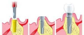
To obtain the remaining 25% of data, when planning implantation, we use special radiopaque templates during CT scanning. Already at this stage, using templates, we predict future orthopedic designs. The template is somewhat reminiscent of a removable denture resting on your own teeth.
Now from the CT image we receive additional data: the shape and type of the future orthopedic structure allow us to choose the best direction for installing the implant, and information about the thickness of the mucosa in the implantation area will allow us to predict the fit of the gum to the crown. Thus, before implantation, we select the surgical protocol and the optimal implantation technique. We know exactly, and do not decide “on the spot”, what type of incision will be made (straight, beveled or WITHOUT A CUT AT ALL), what protocol we will use (one-stage or two-stage), what to prepare in advance (for example, a healing abutment or a plug). Is implantation possible with immediate loading, etc. Based on the table of implants, we will prepare in advance the necessary implants and drills for the correct operation.
This technology allows us to virtually plan and play out implantation, based on the location and type of future orthopedic structure.
At the end of the calculation, an implantological template is made, into which special titanium guide bushings are installed, according to which the doctor will accurately install the implants.
What's the difference in price?
The difference in price is the cost of producing radiopaque and implantation templates. For assessment, for one jaw, regardless of the number of implants installed, you will need to pay an additional 26,000 rubles. to the planned cost of the implantation operation. Often, when it comes to a simple case of installing a single implant, we can do without making templates. An implantological template is used when installing 2 or more implants, then the price increase will be 13,000 per implant. If there are even more implants, the cost of the template ceases to play a significant role in the price of treatment.
Why is preoperative examination necessary?
Preoperative examination is the most important step in performing any type of sinus lift. A thorough diagnosis of the condition of the oral cavity and the entire body as a whole allows us to promptly identify and, if possible, eliminate contraindications to the upcoming surgical intervention.
Preoperative examination is aimed at identifying or excluding the following relative and absolute contraindications to the procedure:
- Inflammatory gum diseases;
- Caries;
- Neoplasms in the maxillary sinuses;
- Acute or chronic sinusitis in the acute stage;
- Previous operations in the area of the upper jaw and maxillary sinus;
- Anomalies of the anatomical structure of the upper jaw and maxillary sinus;
- Acute rhinitis (including allergic etiology);
- ARVI;
- Pathologies of the blood coagulation system;
- Severe mental disorders;
- Diseases of the heart, lungs, kidneys in the stage of decompensation;
The above pathological conditions can become an obstacle to performing a sinus lift or delay it during treatment (inflammatory gum diseases, acute respiratory viral infections, allergic rhinitis and others).
So, what are the advantages of the ImplantGuide technology?
- Full mutual understanding between the orthopedist, surgeon, and dental technician in choosing the best design, the ability to involve other specialists before and at the stage of implantological treatment;
- Optimal placement of the implant as a support for the future orthopedic structure;
- Selection of the optimal individual operating technique in each clinical case;
- Precise positioning of the implant in the planned location;
- 2-5 times reduction in the time of dental implantation surgery;
- Minimal trauma, pain and swelling after surgery, reducing the likelihood of dental complications;
- 100% predictable and guaranteed final aesthetic result;
- Possibility of installing implants without incisions (bloodless implantation method);
- Allows you to perform precise and safe operations.
Dental treatment before dental implantation
Preliminary treatment is necessary in the presence of diseased teeth affected by caries, as well as if there are foci of infection, teeth or tooth roots that require removal. The endodontist evaluates the condition of the dental tissues under a microscope to eliminate the possibility of rejection of the prosthesis after installation, since over a long period of implant healing, infected tissue can provoke disease, which will negatively affect the result.

