The eruption of wisdom teeth is often accompanied by various complications. This is due to the lack of space in the dentition for the number eight, which erupts last. In dental clinical practice, third molar retention is often encountered - incomplete eruption of the crown above the gum. Removing a wisdom tooth in the gum is a small operation that requires not only extraction of the tooth, but also the creation of access and an incision in the gum. Our dental clinic performs extractions even in the most complex clinical cases. Dentists have extensive experience and high qualifications, and perform procedures painlessly and safely.
A wisdom tooth remains in the gum - is this critical?
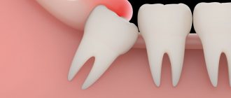
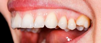
Retention or partial eruption is characterized by incomplete emergence of the crown above the gum. Only 1-2 bumps or part of the tooth crown may appear. Retention can be independent or together with dystopia - incorrect positioning of the molar. Symptoms of the disease will depend on the individual clinical situation.
Depending on the location, retention can be vertical, horizontal or angular. With a vertical crown, the tooth crown is located normally in the bone, in accordance with other teeth. Horizontal is characterized by the location of the tooth perpendicular to the others, horizontally to the arch of the jaw. With angular retention, the crown is tilted to the side. According to the depth of its location, the tooth can be covered only by gum, but by a bone plate.
Retention is accompanied by the ingress of food debris and plaque under the gum. This causes inflammation, redness, tissue swelling, and pain. Pericoronaritis may occur - acute inflammation of the gums and mucous hood above the crown of the impacted tooth. The disease can be serous and purulent, causes acute pain, hyperemia, tissue swelling, and makes it difficult to open the mouth.
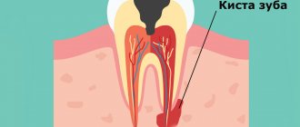
In most cases, a tooth in the gum is a source of various complications: pericoronitis, the formation of caries and its complications, cysts, stomatitis, periostitis, abscess. Therefore, in case of pathology, dentists recommend removal.
To date, exact methods for preventing retention are unknown. General measures include proper hygienic care of the oral cavity and monitoring the correct development of the child’s jaws and bite. As well as timely and correct orthodontic treatment of malocclusions.
How does gum grow into a tooth hole?
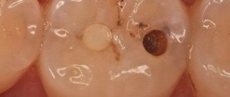
The gum grows into the hole of the tooth if left untreated for a long time. At the initial stage, the enamel is damaged by superficial caries, then medium and deep. The carious process destroys the arch of the coronal cavity, which leads to the proliferation of fibrous connective tissue.
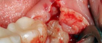
At an early stage of the disease, the tissue filling the tooth hole has a bright red tint, which indicates the presence of an inflammatory process. If the sprouted gum has been in the tooth hole for a long time, it acquires a natural pinkish tint, characteristic of the oral mucosa.
Indications for removal
Retention can manifest itself as a stage of eruption. Over time, the crown completely appears above the gum and the tooth can function normally. Therefore, the decision on the need for removal should be made by a doctor only after examining and diagnosing the condition of the dental system. Indications for surgery are the following clinical cases:
- Constant pain;
- Complications of eruption;
- Trauma to an adjacent molar;
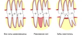
- Pressure on the dentition and its deformation;
- Horizontal or angular position of the third molar in the jaw;
- Dental crowding, which can be aggravated by wisdom teeth;
- Orthodontic and orthopedic indications, that is, the need for extraction to correct the bite or prosthetics.
Treatment methods for ingrown gums in teeth
Patients faced with a problem want to get an answer to the question of what to do if gum has grown in a tooth hole. The chosen treatment methods will depend on the reasons that provoked the ingrowth of soft tissues into the damaged dental cavity.
- The initial stage of eliminating pathology includes eliminating carious lesions of the tooth and removing all dead tissue.
In case of extensive damage to the dental crown and destruction of the tooth walls, extraction is carried out along with excision of the tissue surrounding the roots.
- If there is hyperplastic pulpitis in a tooth, it is necessary to eliminate the overgrown mucous membrane and remove the inflamed pulp. Treatment includes the following steps:
- Excision of tissue with a scalpel or coagulation method.
- Preparing a tooth hole affected by caries and cleaning the canals.
- Medical treatment with antiseptics and antibiotics.
- Adding an anti-inflammatory paste that enhances soft tissue regeneration.
- Sealing.
- When treating epulis, local irritating factors are eliminated - dental plaque and carious areas are removed. In the case when the gum has grown too much into the tooth hole and the neoplasm has reached a large size, the epulis is excised within healthy tissue using a scalpel.
- If the pathology appears as a result of overgrowth of the papillae, surgical removal of the overgrown gums using a carbide bur with a turbine attachment or a laser is indicated. The operation consists of stages:
- Application of anesthesia.
- Excision of overgrown periodontal tissue.
- Using electrocoagulation to stop bleeding.
- Applying a silicone membrane to the operated surface.
- Treatment with saline and betadine.
- If the appearance of gums that have emerged on the surface of the tooth is caused by inflammatory diseases of the oral cavity, drug therapy is prescribed, which includes the use of the following drugs:
- antiseptic solutions (Chlorhexidine, Furacilin, Miramistin);
- anti-inflammatory drugs (Nimesil, Ibuprofen, Ketoprofen);
- dental gels (Holisal, Kalgel, Kamistad);
- antibiotics (Erythromycin, Amoxicillin, Ciprofloxacin);
- painkillers (Parodontocide, Asepta).
The process of removing a wisdom tooth in the gum
The removal operation consists of the following steps:
- Preparation. To reduce the risk of postoperative deposits, sanitation of the oral cavity and professional hygiene are carried out with the removal of dental deposits. Elimination of pathological microorganisms and foci of infection promotes rapid wound healing without complications.
- Anesthesia. Before the operation, local anesthesia is performed using a carpule syringe and modern painkillers. The anesthetic is selected for each individual, taking into account individual sensitivity, age, and medical history.
- Creating access to the tooth. In case of retention, the dentist must provide good access so that the forceps can be securely fixed. To do this, size the gums and, if necessary, remove the bone and gingival hood.
- Extraction. The difficulty of molar removal depends on the type of retention and the individual situation. The tooth is fixed with forceps and dislocated; if necessary, additional instruments are used.
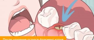
- Procedures after removal. After extraction, the tooth socket is inspected and bone fragments and protrusions are removed. The doctor applies a gauze pad and, if necessary, medications to stop bleeding. Sutures are placed on the gum to speed up healing.
- After surgery, the patient is given instructions, advice, and recommendations in the postoperative period.
How is an overgrown tooth removed?
Root extraction is a fairly common dental operation. It is performed under local anesthesia and, as a rule, does not cause complications. An overgrown root cannot be removed using traditional methods, since it is completely covered with gum tissue and there is no access to it. Treatment is carried out in several stages:
- Examination by a dentist, x-ray examination. X-ray allows the doctor to see the size, position, and depth of the root. The dentist assesses the risk of damage to adjacent teeth during surgery.
- The operation is performed under anesthesia. First, the doctor makes an incision in the gum, providing himself with access to the root. After this, the root is removed and sutures are applied.
- During the rehabilitation period, the doctor selects anti-inflammatory drugs and agents that accelerate wound healing.
The process of removing an overgrown root is individual in each case. The complexity of the operation is influenced by many factors:
- Tooth location. Removal of roots on the upper and lower jaws has its own characteristics and is carried out using various dental instruments.
- The degree of damage to the root and nearby tissues, the presence of fragments. Sometimes, for a successful and as painless removal for the patient as possible, the root is destroyed by a drill into several parts. Separation is required in cases where the tooth has several strong, deep roots. As a rule, after destruction, individual parts are easily removed using ordinary tools.
- The presence of pathology in the surrounding bone tissue, inflammation of the gums.
If you have a problem similar to that described in this article, be sure to contact our specialists. Don't diagnose yourself!
Why you should call us now:
- We will answer all your questions in 3 minutes
- Free consultation
- The average work experience of doctors is 12 years
- Convenient location of clinics
Single contact phone number: +7
Make an appointment
If there is no inflammation, the gum is carefully separated from the alveolus and the tooth is removed using suitable dental forceps. The inflammatory process “melts” the surrounding tissue, so for successful removal it is enough to provide access to the root by making an incision in the gum. Application of forceps does not require labor, and there is no need to additionally separate the gingival tissue from the alveoli. In difficult cases, elevators are used.
Prevention after surgery
The rehabilitation period after removal of an impacted wisdom tooth lasts longer than after a normal one. Healing of the gum socket can take up to 2-3 weeks. During this period, it is important to follow the doctor's recommendations to prevent complications. It is necessary to carefully but delicately carry out hygienic care of the oral cavity. For the first days after the intervention, take painkillers and follow a gentle diet. Too hard, burning foods can damage the gums and cause tissue inflammation.
Dentists also recommend rinsing the mouth after each meal using antiseptic solutions, herbal decoctions, or just water. During the week, you must avoid physical activity, visiting the gym, baths, saunas. If any complications occur, you should not self-medicate; you must immediately seek help from a doctor.
If it hurts, what should you do?
In case of acute pain in the area of the impacted tooth or after extraction, you can take an anesthetic. For this purpose, non-steroidal anti-inflammatory drugs are best suited - Nimessil, Ibuprofen, Paracetamol, Ketanov. At home, you can also carry out hygienic cleaning and rinsing the mouth with antiseptics. For a professional examination and treatment, you must visit a dentist.
Causes of gums appearing in a decayed tooth
Gum ingrowth into a hole occurs due to many reasons. They are associated with the presence of dental diseases, the development of pulpitis, epulis or fibromatosis.
Tooth caries
One of the main reasons for the growth of mucous membrane in the cavity of the affected tooth is the presence of advanced caries, which contributes to deep destruction of the crown. In this case, the walls of the tooth are practically absent, which provokes the accumulation of food debris in the hole and the development of inflammation.
In the gum area appears:
- swelling and soreness;
- protrusion of soft tissues beyond the crown;
- discomfort due to mechanical impact on the gums;
- irritation of the mucous membrane when exposed to spicy, sour or salty foods.
Deep caries is the most severe stage of destruction of hard tissues. Lack of treatment at this stage can lead to the development of complications - pulpitis and periodontitis.
Inflammatory gum diseases
The reason that gums have grown in the tooth hole can be inflammatory dental pathologies. Hypertrophic gingivitis is characterized by the proliferation of gum tissue.
Clinical manifestations of pathology:
- swelling;
- burning;
- presence of hyperemia;
- bleeding
The growth of tissue leads to the formation of false gum pockets that cover the crown of the destroyed tooth. In this case, the mucous membrane grows not only in the hole, but also between the teeth.
Ingrown gingival papilla
The pathological growth of the gingival papilla occurs due to damage to it by the sharp edges of the carious cavity during the destruction of the tooth walls from the side. With regular trauma to the mucosa, its pathological growth and increase in size occurs.
The process of inflammation of the interdental papillae is characterized by:
- bleeding;
- increased sensitivity.
Hyperplastic pulpitis
The gum grows into the hole due to the development of hypertrophic pulpitis. When the dental crown is destroyed, the pulp is exposed and is subject to regular mechanical trauma. This leads to the formation and proliferation of granulation tissues.
If there is a hole, patients experience periodic pain syndromes while eating, as well as bleeding in the area of growing soft tissue. Excess granulations can be soft or dense in consistency, and protrude beyond the pulp chamber into the carious cavity.
Factors provoking the development of pathology:
- hole in the tooth near the gum;
- mechanical irritation of soft tissues;
- the presence of a bacterial infection.
Exacerbation of hypertrophic pulpitis leads to gangrene of the pulp.
Epulis gums
Epulis, formed at the gums, is a benign neoplasm that affects the vestibular side of the mucosa or grows through a hole in the tooth.
Pathology is provoked by chronic soft tissue injuries, which result in the development of an inflammatory process with the formation of granulations that develop into fibrous tissue.
There are 2 types of epulis:
- fibrous – appears on the vestibular side of the gum and is characterized by:
- irregular shape;
- the presence of a hyperemic edge;
- pinkish tint;
- spreading through the interdental space.
- angiomatous - a limited formation on the mucosa, which is distinguished by a brighter color, softer consistency and the presence of bleeding.
Prevention of tooth loss
It is easier to prevent a disease than to treat it - this truism is remembered more often when the disease has progressed far and complex treatment is required. Tooth loss can be prevented by following these recommendations:
- Maintain personal oral hygiene: brushing your teeth should be done at least twice a day, morning and evening;
- use dental floss to clean the spaces between your teeth from food debris;
- Visit your dentist regularly, even if your teeth are not causing concern;
- strictly follow your dentist’s recommendations for dental care and disease prevention;
- take care of chronic diseases (if any);
- protect your teeth from injury.
Recommendations after tooth extraction
To reduce the likelihood of complications, the patient needs to follow simple rules at home.
- The gauze swab applied by the surgeon must be removed from the wound as carefully as possible so that the clot covering the bleeding wound does not accidentally come off. If you carelessly remove the tampon along with the clot, the wound may become infected, which will cause inflammation. It is recommended to remove the tampon 20 minutes after surgery, but not earlier. If the gauze has dried to the wound, you need to soak it with a chlorhexidine solution.
- Pain after tooth extraction is normal. To reduce pain, you can cool your cheek with an ice cube or a cold water bottle. A good folk remedy is to place a coin on the outside of your cheek. The cold cannot be kept for long, 10 minutes is enough. After an hour break, the cold manipulation can be repeated.
- It is not recommended to rinse your mouth immediately after surgery. You can resort to rinsing no earlier than 6 hours after tooth extraction.
- You should not eat for 2 hours after surgery. Until the wound has completely healed, food taken should be warm. You need to refrain from spicy and salty foods - this causes irritation of soft tissues.
- On the day of surgery, you should not drink alcohol, which dilates blood vessels and increases bleeding.
- You can smoke only three hours after surgery.
- Any medical procedures in the mouth can be resumed no earlier than 8 days after tooth extraction.
- After surgery, it is not advisable to touch the wound with your fingers or tongue, visit the sauna, or take a hot bath. Heavy physical activity and stressful situations are undesirable during the rehabilitation period.
- It is prohibited until the wound has healed to brush your teeth on the side of the dentition where the amputation was performed.
No dentist guarantees 100% that the patient will not have complications after tooth extraction. The result depends not only on the professionalism of the surgeon, but also on the characteristics of the patient’s body. If there is prolonged (more than a day) swelling, redness, severe pain and bleeding, you should seek medical help.
Prevention
To prevent the mucous membrane from growing into the tooth hole, you need to take some preventive measures to avoid the formation of carious lesions:
- visit the dentist promptly when the first signs of caries appear;
- monitor oral hygiene;
- use a toothbrush of medium or low hardness;
- pay special attention to the selection of toothpaste;
- lead a healthy lifestyle: do not abuse smoking and alcohol;
- eat a balanced diet.
The growth of mucous membrane in a tooth hole is often provoked by carious processes, which lead to the development of various pathologies in the oral cavity. Surgery, as well as the use of drug therapy, will help get rid of the unpleasant symptoms of mucous membrane ingrowth into a tooth hole.
Bibliography
- Barer G.M. – Periodontal diseases. Textbook, M.: GEOTAR-Media, 2008.
- Maksimovsky Yu.M., Mitronin A.V. - Therapeutic dentistry - M.: Geotar-Media, 2012.
- Grigoryan A.S., Grudyanov A.I., Rabukhina N.A., Frolova O.A. — Periodontal diseases, M., 2004.
- O. O. Yanushevich, V. M. Grinin [and others] - Periodontal diseases. Modern view on clinical, diagnostic and therapeutic aspects, M.: GEOTAR-Media, 2010.
- Ijndhe J. - Textbook of Clinical Periodontology. 1995. Copenhagen.
- Ivanov V.S., Urbanovich L.I., Berezhnoy V.P. — Inflammation of the dental pulp, M., Medicine, 1990.
- Stephen Cohen - Pathways of the Pulp - Mosby - 1980
- Ivanov V.S., Urbanovich L.I., Berezhnoy V.P. — Inflammation of the dental pulp, M., Medicine, 1990.
Find a clinic