In modern dentistry, the importance of the dentist is increasing and the dental technician is becoming less and less important. It is the doctor who designs the final result, taking into account the requirements for the aesthetics and functionality of the patient’s maxillofacial system.
Traditionally, aesthetic analysis involves photographing the patient. Photographs are used to evaluate the outcome of treatment, but it is not practical to use them for planning, since a two-dimensional image does not give accurate results. Instead, they resort to 3D visualization, when the face, teeth, and gums are displayed in a three-dimensional projection.
Computer modeling in dental prosthetics - what is it?
Computer modeling, as is already clear from the very name of this technology, allows you to use a regular computer/tablet for dental prosthetics, but, of course, with special “software” (that is, software). Using it, a three-dimensional digital model of the patient’s future smile is created, taking into account all the anatomical features - the shape and color of the teeth, their size and combination with the contours of the face, skin color, gum level, size of the lips and nose, and the width of the smile.
The use of a computer for smile modeling not only speeds up the process of prosthetics, but also optimizes the relationship between the dentist and the patient, as well as the dentist and the dental technician. Patients especially like that they actively participate in the modeling process - together with the doctor they choose the color and shape of their future teeth.
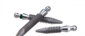
Complex on 4 OSSTEM implants with delayed loading - from RUB 170,000.
Complex implantation Osstem (South Korea) with delayed loading after 4-6 months.
Guarantee for the doctor’s work - unlimited Call now or order a call
Opening hours: 24 hours a day - seven days a week
For computer smile modeling in dentistry, a concept called Digital Smile Design, or DSD for short, is used - which translates to “digital smile design.” It was developed in 2007 by Brazilian dental technician Christian Coachman, and since then it has gained great popularity among the global dental community. By the way, DSD models the future smile, which becomes a kind of aesthetic standard for the prosthesis, but does not model the prosthesis itself - for this they use other solutions, which we will discuss below.
Virtual tooth preparation
3D visualization and planning provide information about the future shape of the tooth. Using it, you can design the shape of the future stump created by preparation, that is, perform “reverse planning”. To do this, the thickness of the future crown (framework and facing material) is taken from the shape of the tooth. Knowing the shape of the diamond bur for preparation, they set the trajectory of movement along the tooth to obtain the desired result.
Since the bur is secured in the handpiece in a unique way, the movement of the handpiece in relation to the dentition is easy to calculate. This can also be done with respect to three reference objects attached to the tip.
The next stage is designing a template, or mouthguard, to be fixed to the teeth. In the body of the mouth guard, it is necessary to virtually “subtract” the grooves according to known specified trajectories. The template is made by prototyping and is used to guide tooth preparation.
The technology could be widely used, but this has not yet happened, or at least it is not yet used for commercial purposes. The development belongs to the American company Origin (2012).
Tasks of computer smile modeling
- select the shape and color of crowns/veneers in accordance with the patient’s requests,
- see your future smile with new teeth in advance and “try on” it: this happens on a computer screen or on a tablet,
- assess the performance of the prosthesis in advance: how the jaws will close, whether the bite relationship will be disrupted,
- rejuvenate the face through Dental face lifting and the application of the principles of the “golden” section and “beauty mask”: these parameters are taken into account during digital modeling (colors and shapes of crowns) - as a result, the face looks more attractive and younger,
- form and study a model of the patient’s jaw before implantation.
What is 3D modeling and planning in dentistry
Digital technologies are integrated into many processes during dental restoration; often the implantation itself using them is called 3D implantation (from the English three-dimensional). It is not a separate method or protocol; rather, it is a qualitatively different approach to the process of dental restoration.
3D technologies include those that help volumetrically visualize information about the condition of the jaw or create three-dimensional models, both virtual and real, at the stage of developing the implantation process:
- computed tomography (CT): using X-ray scanning, creates accurate digital copies of the maxillofacial apparatus,
- programs for visualization and 3D modeling: for virtual study of the stages of implantation and prosthetics,
- 3D printing: printers that create highly complex real-life models of jaws and surgical templates (as well as prosthetics and mouth guards) from specialized materials. Templates are required at the stage of surgery to install implants or to work out the process - in fact, to hone manual skills.
%akc54%
The basis of the system is a computer program that processes incoming information from a computed tomograph and is connected to a 3D printer, oral scanners, and robotic machines. The software allows you to create a very accurate treatment plan, assess the quality and size of bone tissue, select the location of the implant, and much more (more on this below).
There are many different programs that dentists use. Among them are universal and specialized: NobelClinician, SimPlant, Blue Sky Plan, coDiagnostiX.
Indications and contraindications for digital modeling
The scope of computer smile design in dentistry is quite wide. Digital smile design is suitable for patients who need prosthetics with restorative inlays, veneers (as well as their variations - ultraneers, nanoeers, etc.), crowns, bridges, and, less commonly, removable dentures.
The DSD concept is used in the development of implant prostheses. Moreover, these can also be either single crowns on implants or extended bridge-like structures with acrylic gum (its color and shape are also digitally modeled).
Don't know what type of prosthetics to choose?
We will help in the selection, advise where to read more information and compare types of prosthetics.
Consultation with an orthopedic doctor in Moscow clinics is free! Call now or request a call
Working hours: from 9:00 to 21:00 - seven days a week
The only contraindications to computer modeling are contraindications to the installation of dentures. This includes caries, the presence of serious inflammation in the oral cavity and massive deposits of tartar. For example, untreated swelling of the gums or installing a filling after the “smile parameters” have been removed will lead to the orthopedic design being uncomfortable. Therefore, before creating a prosthesis, even a virtual one, existing dental diseases should be cured.
Is it possible to do without 3D technology during implantation?
No, the success of implantation directly depends on proper planning of the operation. Neither an orthopantomogram nor an x-ray can serve as a basis for implantation. Only CT, in combination with data processing with a special computer program, can provide enough information to select an implantation protocol and create a step-by-step implantation scheme.
Thanks to its presence, risks are reduced, surgical intervention time is shortened, the highest precision of implant installation and a pre-planned result are achieved. Based on the results of the 3D modeling, the doctor can be confident that the implant will achieve good primary stabilization.
For which restoration method is DSD suitable?
Dental prosthetics is carried out using two methods – direct and indirect. But digital smile design is used only in one case, read about it further.
Direct method - the prosthesis is created in the patient’s mouth
The direct method most often involves applying a liquid filling directly to the patient's teeth while the patient is in the dental chair. This is how they restore small chips, cracks, and darkening on the front teeth, and restore chipped enamel of the lateral teeth. Before the filling hardens, the dentist can give the patient a mirror to evaluate the result and tell him whether he is satisfied or not. There is no need for computer modeling here because all the work is done by hand (not in a laboratory).
Indirect method - the prosthesis is created in the laboratory
Dentures created in a dental laboratory are of much higher quality than those produced by the direct method. For the indirect manufacturing method, in addition to the skills of specialists, equipment is required - various forms for real models, firing and baking ovens, milling machines, presses and modern high-quality materials (ceramics, glass ceramics, zirconium dioxide).
Here, the use of 3D modeling of a future smile helps the dental technician better navigate the work being performed, because DSD data is combined with special programs that model the prosthesis itself and control its production. Of course, not all clinics use the original Digital Smile Design program (because they don’t want to purchase it from the developers), and digital modeling, in principle, works the old fashioned way manually. But as practice shows, it is this concept that helps create the most beautiful, comfortable and durable designs.
Advantages of dental restoration using CEREC 3D technology:
- All restorations are highly accurate and perfectly match the treated tooth. At the same time, the distance between the treated tooth wall and the crown/onlay is about 20–60 microns, with such accuracy the possibility of the crown/onlay becoming uncemented, saliva getting under it and infection is practically excluded;
- the ceramics used for restorations do not cause allergies, are varied in shades, transparency and even density of the material, so the restored teeth are indistinguishable from natural healthy ones;
- restoration of teeth in one visit to the dentist. Thanks to the 3D camera, there is no need to take impressions and install temporary fillings or crowns.
Dental restoration using 3D technology is reliable, aesthetic and functional.
Make an appointment with doctors
: Goncharova T.A., Selivanova I.V., Cherevko N.I.
You can find out more detailed information and schedule a consultation through the online form.
Make an appointment
(812) 232-88-25, 233-19-08
Stages of computer modeling and prosthetics
The process of dental restoration includes several stages that follow one after another. Let's look at them further and tell you when computer smile modeling is used and what initial data is needed for it.
Taking impressions or 3D scanning of the oral cavity
Now computerization can accompany the prosthetics process from start to finish. For example, dentists often abandon the usual casts or impressions of the dentition and use digital “casts” (using a 3D oral scanner).
But the usual physical impressions can also be used. True, in this case you will have to transfer them to the laboratory, make a plaster model1 and scan it - in order to create a digital version for its correction in Digital Smile Design. This route takes longer (2-7 days).
Photo and video session, assessment of bite parameters
Photographs (as well as short videos) are taken in order to evaluate the patient’s smile parameters, their relationship with other facial parameters, and also to understand how the facial muscles work. Filming is carried out when the patient is smiling, calm or deliberately “angry”, when he speaks or is silent. All photos and videos, and there can be several dozen of them, are loaded into the program and combined with digital impressions.
Also at this stage, the dentist uses various jaw movement analyzers (articulators, HIP plane analyzer, face bows). After all, in a future prosthesis, not only beauty is important, but also how it will relate to the existing bite.
Virtual smile modeling
All received data (imprints, photos, etc.) are entered into a computer smile modeling program. Then the parameters are processed, combined, and a three-dimensional projection is displayed on the screen that fully corresponds to the patient’s natural dentition. Next, adjustments are made to improve your smile. Moreover, you can immediately make several options for a new smile - with different shapes or heights, widths of teeth, with different shades of enamel, for example. And then the best option is selected, both from the dentist’s point of view and based on the patient’s wishes.
By the way, such a virtual projection can be combined with digital computed tomography of the patient and begin to develop a prosthesis, incl. and for placement on implants. Popular programs for creating prostheses are CAD/CAM systems CEREC and inLab from Dentsply Sirona, NobelProcera from Nobel Biocare.
Wax modeling and fitting of the prosthesis blank
In some cases, patients who need veneers, extended bridges or removable orthopedic structures should go through one more stage - fitting of the prosthesis blank. This is necessary to assess the comfort of the new teeth and their relationship with the neighboring ones. Here, after developing a model, the dental technician makes a “trial” prosthesis (from silicone, plastic, etc.), and then attaches it to a wax model, after which he hands it over to the orthopedic dentist, who tries it on the patient. If necessary, adjustments are made and the layout is sent back to the laboratory.
Creation of the prosthesis and its final installation
The approved version of the prosthesis goes through all stages of manufacturing - milling on a robotic machine, baking, glazing, polishing, etc. The production technology depends on the type of prosthesis, the properties of the material and its volume, which is needed to create each specific structure.
Previously, all production machines were manually adjusted by a person, using measurements also taken by hand. Therefore, there was no need to talk about accuracy, especially the first time. Today, dental devices (namely CAD/CAM systems) are configured largely automatically, in accordance with the parameters set by the computer. Therefore, inaccuracies and human errors are excluded here.
Today, dental crowns, bridges and the thinnest veneers are cut automatically by machines - quickly and the first time. As a result, the doctor simply fixes the prosthetic structure, without first trying it on and subsequent adjustment.
Neuromuscular diagnostics in orthopedics
It is impossible not to mention such an important aspect of preparation for prosthetics as studying the work of the maxillofacial muscles in dynamics. The data obtained as a result of such diagnostics allows not only to make the orthopedic apparatus as comfortable as possible in use, but also with its help to correct existing defects in the functioning of the dental system. As part of such an examination, specialists conduct computer diagnostics of muscle tone, scan and record changes in muscle tissue at the time of jaw movement, and evaluate noise in the joint area.
Rules for caring for a new smile
Despite the fact that computer modeling allows you to create comfortable, beautiful and durable dentures, this does not mean that new teeth do not need to be cared for. On the contrary, proper care will only extend the life of the restoration and preserve its integrity and aesthetics. However, there is nothing difficult about care. Restorations need to be cleaned 2-3 times a day with a brush and paste (only without abrasive particles); you should not bite nuts and candies, chew chewing gum or toffee. And if veneers are installed, then you should not bite off food with your front teeth, especially hard food (carrots, apples, etc.).
Advantages
- The most accurate determination of the optimal installation site and position of the implant while minimizing the likelihood of error (for example, damage to the maxillary sinus).
- Successful implantation of an implant into bone tissue even with its deficiency.
- The ability to install the implant at the desired angle of inclination without additional surgical intervention, which previously could have been performed to prevent damage to surrounding tissues.
- Clear visualization of all stages of prosthetics, increasing the awareness of the implant surgeon and thereby reducing the risk of making mistakes.
- Minimization of postoperative complications during the recovery phase.
- Multiple reductions in the duration of the procedure - from several months to several days.
How much does computer smile modeling cost?
It is worth noting that this type of creation of prostheses increases the cost of overall treatment, and modeling is not available in every clinic. However, many dentists are able to competently build a pricing policy in such a way as to minimize the cost of additional manipulations and equipment used. That is, in one clinic a patient may be charged an additional 10-15 thousand rubles for computer modeling of a smile. And in some places it is already included in the turnkey price and increases the final price of the prosthesis by only 1000-2000 rubles, or does not change it at all.
Patient Questions
QUESTION: I read about trying on a smile on a computer and wanted to clarify whether it is possible to do this if you only need 2 veneers? I have a gap between my front teeth and would like to somehow see the result in advance. And then suddenly I don’t like what the doctor does. Elena
ANSWER: Hello, Elena. Yes, computer smile modeling can be used for any number of necessary prostheses - be it 1-2 veneers or a whole complex for the front teeth. This concept really allows you to “try on” a new smile virtually - the doctor will show suitable options on the computer screen, and you will choose the one you like best. You can also select the color of the veneers so that it matches perfectly with the adjacent teeth and meets your expectations.
- Haug S. Correct modeling, 2006.
Author: Sambuev B. S. (Thank you for your help in writing the article and the information provided)
3D dental implantation – digital technologies for successful and predictable smile restoration
Article navigation
- What is 3D modeling
- When are 3D models needed?
- Stages of implantation in 3D
- — carrying out photometry
- - CT scan
- — taking impressions
- — 3D visualization of the process
- – production of surgical templates
- — surgical stage
- - re-taking impressions
- — production and installation of a prosthesis
- - What is digital smile design?
- Benefits of 3D planning
- Real-time visualization
Question for a specialist
The success of implantation largely depends on high-quality preparation and diagnostics. Today, dental clinics have a whole range of equipment and tools for comprehensive research. And with the development of computer technology, a qualitatively new approach has been introduced in the planning and elaboration of upcoming procedures - 3D planning of dental implantation or 3D implantation. How innovative “three-de” programs and devices help restore teeth, what new methods are being introduced thanks to this, and how 3D modeling affects the quality of implantation, read in this article.
Aesthetic digital smile modeling: analogue and digital virtual planning
Valerio Bini, doctor
Over the past twenty years, the use of computers in dentistry has radically changed the clinical approach, especially at the analysis stage, creating a real information microcosm and turning the dentist into an information systems operator. Digitization of photographs, which makes it possible to instantly process the image, now accompanies the diagnosis of every clinical case concerning aesthetics.
Using a variety of static positions, virtual planning can be carried out and expressed technologically thanks to the achievements of modern dentistry.
Smile designer: a new method in communication
The dentist is increasingly faced with the system of interdisciplinary treatment plan “Face and Smile”, in which the interdisciplinary examination gives the doctor involved in the aesthetic component a very important, if not the main place. The harmonious combination of teeth, periodontal tissue, face and smile creates excellent aesthetics. It is necessary to have artistic ability and various know-how for complex consideration, balanced design and combination of dental elements in the context of “Face”. Today, beauty and aesthetics are increasingly associated with size, proportion, symmetry - constants known to ancient civilizations and forming their basis. Today they are inseparable from modern technologies, which have radically changed with the advent of the digital era.
Modern scientific knowledge puts at the disposal of professionals various therapeutic options: collaboration between specialists from different fields (orthodontist, implantologist, periodontist, dental technician, oral surgeon, plastic surgeon and aesthetic medicine doctor) and examination by these specialists allows the best treatment plan to be drawn up ( Fig. 1).
Rice. 1. Aesthetic team and interdisciplinary examination
In addition, the doctor can work with images taken elsewhere and obtained via video conference via Skype. This opportunity turns the esthetic dentist into the conductor of an orchestra and the main player in the process. Today, with the advent of digital dentistry, it is difficult for a doctor to work while maintaining high quality of work and ergonomics if he does not have an accurate protocol that can predict the result (Virtual Planning). The use of 2D and 3D software with digital image editing and morphing (Digital Image Editing) makes it possible to process data and individual parameters according to the specific requirements of Smile Makeover - smile formation.
Modern digital technologies, combined with the experience and aesthetic sense of the dentist, become the key to success in smile modeling, ensuring predictability of both aesthetic and therapeutic results for the patient.
The combination of concepts such as aesthetic dentistry, interdisciplinary examination, digital dentistry and predictability gives rise to a new phenomenon in the profession - the “smile designer”, the main advantage of which is the possibility of communication between the patient, the main character in aesthetic dentistry, and a team of estheticians, virtual reality specialists. planning. For quite some time now I have been improving the ADSD system - “aesthetic digital smile modeling”, using various software. ADSD can and should serve as a purely auxiliary method of diagnosis and prognosis, the goal of which is the health and well-being of the patient. In addition, since ADSD is a way to simulate an aesthetic treatment plan, it is advisable to obtain the patient's prior consent for the use of this technique, which only when accompanied by real clinical examples, such as a mock-up, will become clear to the patient. It is important to remember that dentistry requires that the dentist observe three basic principles of the profession: caution, diligence, and experience.
ADSD: methodology and protocol for use
ADSD, Aesthetic Digital Smile Design (aesthetic digital smile design), is, first of all, a way to improve communication with the patient, since it is with the help of processed images that you can see before and after photos on a digital monitor, predict the results of treatment and discuss them with the patient. Another important point is the innovative method of aesthetic and clinical planning in aesthetic dentistry, prosthetic dentistry, which is of great importance for analysis and design in the dental laboratory. This technique can also be used for diagnosis and planning in plastic and maxillofacial surgery. First of all, the protocol involves obtaining patient images through digital photographs and full frame video. Video is especially important, since with its help you can see the dynamic phases of a smile associated with physiological characteristics (facial expressions, phonetics, relationship of dentition and lips). Incorporating this important information into the patient's digital aesthetic record complements the clinical history as it is an integral part of an objective intraoral and extraoral examination and aesthetic analysis. Therefore, we can define this technique as the third part of the analytical processing (Fig. 2) of the aesthetic composition of the smile, the morphological components of the face and smile, including the reference points on which the measurement scale will be based (Fig. 3 a - c). The next stage of digital data processing is Virtual Planning using Digital Image Editing (Fig. 4 a - c), then digital and analog diagnostic wax-up, mock-up, temporary prosthesis, final restoration (Fig. 4d - f).
Rice. 2. ADSD, macro-aesthetic, mini-aesthetic, micro-aesthetic analysis
Rice. 3a. ADSD, Scheduling and Virtual Planning
Rice. 3b. ADSD, Scheduling and Virtual Planning
Rice. 3c. ADSD, Scheduling and Virtual Planning
Rice. 4a. ADSD and its application in a clinical case
Rice. 4b. ADSD and its application in a clinical case
Rice. 4c. ADSD and its application in a clinical case
Rice. 4g. Prosthesis model
Rice. 4d. Virtual wax-up
Rice. 4e. Aesthetic and biological integration of the prosthesis
The digital technique, closely related to image editing, is very reliable, especially when working with clinical cases where functional editing and morphological changes are required. This method is much easier for the dental laboratory to understand than oral explanations. Interaction with other digital systems is also very important, as this makes it possible to use ADSD for digital simulation of an orthodontic situation, digitization of impressions, CAD/CAM method, etc.
Thus, the technique becomes multimedia.
Receive and import digital images
Photographs of the patient should be taken with a good digital SLR camera (possibly a semi-professional one), most importantly in good lighting. Today there are many opportunities to take courses in dental photography, and there are many manuals on this extremely interesting topic. It must be remembered that at the analysis stage, photography is a diagnostic clinical and aesthetic element, which will then become part of the patient’s clinical baggage, different specialists will work with it as part of an interdisciplinary examination. Therefore, the dentist must take photographs of the patient's head in the same position to be used in a smile modeling program. The most reliable position for photographing the patient’s head is the so-called aesthetic plane (Fig. 5), that is, the ratio of the perpendicular (frontal) plane to the plane in the center of the angle formed by the Frankfurt horizontal and the Camper line. The same position should be projected vertically at 45° and 90°, because profile photographs are also extremely important, since they make it possible to analyze the face and teeth from an aesthetic point of view: to record the class of occlusion, the position of the lips, aesthetic angles - all points important for orthodontist, maxillofacial surgeon and plastic surgeon.
Rice. 5. Photography and the aesthetic plane
Analog Face Transfer Support - FATS
The ADSD technique involves transferring the actual dimensions of the photographed object with their configuration in accordance with the pixel system that is used in digital photography. In order to do this, you can use measuring instruments such as a square and a ruler, if possible metal (they are easy to wash and sterilize), or their equivalents. When working with facial analog transfer support (FATS) (Fig. 6a), I use a measuring device that includes a regular ruler with millimeter and centimeter markings. The patient can carry it with him as easily as, for example, glasses.
Figure 6a. Analog face transfer support: analog measurements and digital calibration
This instrument can be made by any dentist or dental technician, subject to some special requirements. The ruler should be light, but clearly visible on the patient's face; the eyes must be visible so that it is possible to determine the position of the pupils; The area around the eyes should also be exposed, including the most clinically and aesthetically interesting reference points. Another important point is the nose pad, which should be comfortable enough to allow us to move the analog dial back and forth freely. When assessing the facial profile, it can be noted that the position of the upper incisors may be slightly shifted in relation to the aesthetic plane. Therefore, in order to accurately take analog measurements, you need to install the nose stop at a point as perpendicular to the vestibular surface of the upper incisors as possible, so that it practically continues this vertical plane. In addition, since prototypes (mock-ups, PMMA prototypes, etc.) still need to be shot, it is advisable to use clamps similar to the craniostat stops installed in the headrest of the dental unit chair. As for even more accurate measurements of individual elements of the dentition and gingival parameters, you can use digital calibers by setting their edge on the cervical line and incisal edge (element length) or on the mesial and distal edges relative to the equator of the tooth (tooth width) (Fig. 6b ). Some digital image editors have an analytical system that allows you to convert the size of a digital photo from pixels to millimeters (analog photo), insert all the object data into the photo and the dimensions obtained using FATS (Face Analogue Transfer Support) (Fig. 6c). One of the most important points is to indicate and store the unit of measurement on the photo for the software conversion system so that the correct data is indicated in the clinical situation. If the dimensions have been previously transferred to an analogue/digital scale, excellent indications of reference points can be obtained. For example, the location of the maxillary central incisors may indicate the distance between the incisal or cervical edge and the point between the base of the nose and the upper lip or the interpupillary line. The dimensions transferred in this way will be especially useful when the dentist interacts with the dental technician, because it is thanks to him that the most important results of this technique are achieved. It must be borne in mind that patients are rarely interested in the dimensions in millimeters in a digital photograph and the design of the contours of the teeth - at the first stage, more visual examples are much more important to them. The dimensions of 3D wax-ups and mock-ups, which are tried directly in the patient's mouth, give the patient a much better idea.
Figure 6b. Analog face transfer support: analog measurements and digital calibration
Rice. 6th century ADSD, transfer of analog dimensions to a computer and measuring grid
Editing Digital Images
There are many ways to digitally process images, depending on the requirements for smile modeling: you can use different programs that are easy to find on the Internet - it all depends on aesthetic experience and on the practice of working with digital images. Some of these image processing and photo editing programs are free, others are paid, some are for beginners and some are for professionals. In the simplest programs it is quite easy to simulate a smile with a schematic construction of the contour of the teeth. Aesthetic digital smile modeling involves DDD (digital dental modeling), which, as I already said, is quite simple - you just need to outline the contours of the elements, choosing the most suitable shape for the new smile (Fig. 7).
Rice. 7. Digital Dental Design: Contours
For this technique, it is necessary to make another model in a dental laboratory, which will then be checked by the dentist and possibly used as a prototype.
Only at this stage, having photographed the patient, can we begin to more accurately model the smile. The ADSD technique provides for such aspects of processing as transfer, adaptation, processing of the shape and type of dentition, taking into account the shape in the photographs. To accomplish these important tasks, you need to create a file cabinet that we will call a DDPD (Dental Photograph Database). This may include: A catalog of tooth shapes is perhaps the most user-friendly database. It can include 5 types of teeth according to anatomical shape and color, and can be easily changed depending on the quality of lighting in the photographs provided by the operator. The tooth shapes contained in this catalog must match the actual shape, for example: triangular, oval, rectangular, square, trapezoidal (Fig. 8).
Rice. 8. Personal database: incisal edge
Catalogs of dentition/smiles that can be called aesthetically ideal: there are some catalogs where the elements are already formed in accordance with the morphology of the incisal edge (flat, square, rounded). Catalogs of teeth for removable dentures - they can be found on the Internet on the websites of manufacturers such as Ivoclar, Kulzer, Vitapan, Candulor, etc. Database of own clinical cases - prosthetics, aesthetic treatment, all previous virtual wax-ups, mock-ups and treated teeth of patients who agree to use their photographs for noble purposes.
Catalog of smiles - models, men and women, who may be useful; you can choose the teeth you like from models, who, as a rule, are photographed by professional photographers in fashion style, and “try on” them. Simply download from paid websites such as 123rf.com, Fotolia.com, Shutterstock.com, Fotosearch.com, etc. Another factor in the smile modeling technique is digital deformation/distortion (DDID), which allows modification of the morphology elements of the dentition. This feature is very useful when creating a DDPD database. It is important to use not only vectors of length and width (Fig. 10 a - c), but also vectors of all directions, both external contours and tooth surfaces.
Rice. 10a. Distortion of dental elements in one direction
Rice. 10b. Distortion of dental elements in one direction
Rice. 10th century Distortion of dental elements in one direction
This treatment is often extremely important for highlights on tooth surfaces with micro- and macro-texture, transfer lines, and interproximal spaces. It is also effective in analyzing contact points and interincisal angles, with the morphological characteristics of the incisal edge and the external contour as a whole, which tell us about the age, gender and personality characteristics of the patient (morphopsychology). DDID is the most important part of the entire ADSD technique because you cannot “print” a standard smile on the patient's mouth. This smile can be composed of ideal elements, but you need to use artistic flair and dental know-how to be able to change them. It is necessary to model, sculpt, deform, increase, decrease or remove elements that do not harmonize with the rest (Fig. 9 a - f).
Rice. 9a. ADSD, digital image distortion
Rice. 9b. ADSD, digital image distortion
Rice. 9th century ADSD, digital image distortion
Rice. 9d. ADSD, digital image distortion
Rice. 9y. ADSD, digital image distortion
Rice. 9e. ADSD, digital image distortion
In many clinical cases, it is advisable to use DDCT (digital calibrated dental transposition), that is, the transposition of elements of the dentition necessary for aesthetic modeling and orthodontic movements that need to be analyzed for further aesthetic treatment, as well as orthopedic or prosthetic with the installation of implants. The transfer of elements to the desired position must be precise, it is necessary to preserve the anatomical dimensions, then it is possible to more accurately plan the future structure not only from an aesthetic, but also from a functional point of view. Quite often in orthodontic cases, mesialization/distalization is required for implant placement, so coordination with the implantologist, orthopedist and orthodontist via x-rays (Dicom, Tac3D) will be required. Only after planning the final position of the dentition elements can the smile designer proceed with aesthetic improvements, modeling them using DDID (digital deformation/distortion). In practice, the doctor and dental technician can use their professional experience using a mouse and conventional modeling tools. The same method is suitable for orthodontic simulation, processing data obtained using programs such as ClinCheck from Invisalign (Align Tech). They can be used in virtual modeling. Processing in 3D changes in the position of elements gives us the opportunity to build an ideal dentition on a millimeter grid with the overlay of before and after options, allowing us to perform virtual planning of aesthetic and orthopedic treatment together with orthodontic treatment.
Conclusion
Today, aesthetic dentistry has new ways of developing a treatment plan: Digital Dentistry technology and software for digital processing of clinical images, which are gradually becoming an integral part of the dental profession. ADSD - Aesthetic Digital Smile Design - is a simple and cost-effective way to demonstrate to the patient the expected treatment result, aesthetic and functional improvements using appropriate prototypes already during the second visit, and then transfer it to the entire Face Aesthetic Medical Team ) information necessary for the work of the interdisciplinary team. Thus, a new figure has appeared in dentistry - the smile designer, its aesthetic architect.
The list of references is in the editorial office
What 3D technologies are used in dental implantation?
When they talk about dental implantation in 3D, it is meant that the entire process, from any diagnostic measures to the creation of a prosthesis suitable for all the individual characteristics of the patient, is modeled using three-dimensional visualization. On guard are: computed tomography of the jaw, specialized software NobelClinician, Simplant, Blue Sky, etc., surgical templates, 3D printers, HIP analyzers, milling and robotic machines, Cerec, Procera, CAD/CAM devices and others. Don’t be intimidated by complex names – we’ll tell you about everything in more detail, read on!
Important! Often in advertising or on the websites of many dental clinics you can come across such a concept as “3D implantation”. Here you need to understand that this is not a separate treatment method, but only a qualitative improvement of the protocols that already exist today. In particular, advanced technologies are used primarily in one-stage implantation methods, when there is atrophy of bone tissue, but its extension is not carried out. That is why planning the entire treatment process comes to the fore here - the doctor has no right to make mistakes.
In order to understand what these technologies are, how they work and are consistently used in dental implantation, it is worth considering the stages of the procedure.