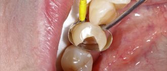
What is a dental emergency?
Emergency dental care is understood as medical care provided for acute diseases, painful conditions, exacerbation of chronic diseases WITHOUT obvious signs of a threat to life, delaying the provision of which for an indefinite period of time will lead to a deterioration in the patient’s condition. Medical services can be provided in an outpatient setting.
Conditions requiring emergency dental care include:
- pulpitis (acute or exacerbation of chronic)
- periodontitis (acute and exacerbation of chronic)
- maxillofacial injuries (such as a dislocated or fractured tooth)
- exacerbation of odontogenic and non-odontogenic inflammatory diseases of the maxillofacial area
- stomatitis
- other acute painful conditions
- treatment requiring surgery
Emergency dental care can be provided both in a public dental clinic and in a private dental office.
First aid at an outpatient dental appointment
A. M. Kokhan , anesthesiologist at Medical On Group (Odintsovo, International Medical Center)
Patient emergencies that occur in the dentist's office are typical in most cases. They can be classified to facilitate diagnosis and subsequent urgent therapy, since before you begin to assist the patient, you must first determine the nature of this acute condition.
Most often, during an outpatient dental appointment, patients develop collapse - acute vascular insufficiency, called fainting by ordinary people.
This condition can occur in both vertical and horizontal positions of the patient's body. In most cases, it is iatrogenic in nature: a painful examination, pain from manipulations, including injections of local anesthetic, puncture of a vein if treatment is carried out under sedation, the sight of blood, etc. In particularly sensitive people, collapse develops from one type of medical equipment and even during a preliminary conversation with the doctor. This kind of patient excitement leads to a predominance of the influence of parasympathetic innervation and causes a decrease in heart rate, as well as a decrease in vascular tone, which causes a redistribution of blood in the body due to a sharp expansion of the vascular bed and leads to bleeding of the brain.
Loss of consciousness is accompanied by uncoordinated movements of the eyeballs, dilated pupils, local or generalized convulsions
Signs of approaching collapse are the patient's complaints of nausea, weakness, sometimes suffocation, and sudden thirst. The patient's skin turns pale, is often covered with perspiration, the pulse in the peripheral arteries is threadlike or absent, and blood pressure when measured by the indirect method is sharply reduced or not determined. Loss of consciousness is accompanied by uncoordinated movements of the eyeballs, dilation of the pupils, local or even generalized convulsions; the monitor may show a sharp decrease in heart rate, a drop in blood oxygen saturation.
According to the stage of collapse, therapy should be gradual.
The first thing to do is to give the patient the Trendelenburg position (F. Trendelenburg) - a position in which the pelvis and lower limbs are located above the head, which is achieved by tilting the head end of the chair by 30-45°. This will ensure blood flow to the brain and eliminate circulatory hypoxia. It is incorrect to bend the patient forward from a sitting position. Some dentists justify this action by saying that a confused patient lying on his back may aspirate the contents of the oral cavity. Flexion leads to increased intra-abdominal pressure, which negatively affects blood circulation. In addition, the effectiveness of such a position in the fight against collapse is very doubtful, because the head with the hypoxic brain located in it is located above the pelvis and legs, and aspiration can be prevented by placing the patient in the Trendelenburg position on his side [1]. Next, to stimulate breathing and recover from a fainting state, carefully bring a small piece of gauze or cotton wool moistened with an official aqueous solution of ammonia to the nostrils. The physiological effect of ammonia is due to its locally irritating effect: vapors of the substance excite the sensitive endings of the nerves of the upper respiratory tract (the endings of the trigeminal nerve), which leads to reflex stimulation of the respiratory and vasomotor centers of the brain, causing increased respiration and increased blood pressure. It should be used with caution; in high concentrations, ammonia vapor can cause reflex respiratory arrest [2].
In most cases, these measures are enough to stop the acute condition.
However, similar symptoms are accompanied by other acute conditions that require urgent hospitalization of the patient in the intensive care unit for intensive care: acute ischemia of the heart muscle, acute cerebrovascular accident of the hemorrhagic type. The hypoglycemic state of the patient can also simulate collapse.
Differential diagnosis with anginal attack
With myocardial ischemia, loss of consciousness is sometimes preceded by paroxysmal pain behind the sternum, often in its lower third. The description of pain is usually emotional: pressing, squeezing, burning. In the vast majority of cases, there is an appropriate medical history; patients often independently warn the doctor about the presence of a chronic disease and have medications with them.
With such symptoms, the patient can be given one to three nitroglycerin tablets to dissolve under the tongue for fifteen minutes, placed in the Fowler position - a half-sitting position in which the shoulders are raised 45 degrees and the knees are slightly bent. (The position is named after the surgeon GRFowler, who in the nineteenth century proved the positive effect of this position on the well-being of patients with cardiogenic circulatory disorders.) The Fowler position reduces congestion in the lungs, relieves tension in the abdominal muscles and facilitates the work of the heart [1]. Painkillers available to the dentist will not significantly alleviate ischemic pain, however, an injection of any non-narcotic analgesic - a non-steroidal anti-inflammatory drug (NSAID), for example, Ketorol, preferably intravenous, 30-60 milligrams - will help determine if the effect of the injection will actually occur. and not the severity of the attack manifested by the patient.
The patient’s blood pressure and pulse should be monitored at least once every 5 minutes. It is recommended to immediately call an ambulance. In case of circulatory arrest, take measures to maintain the vital functions of the body using the Basic Cardiac Life Support system, and, if appropriate equipment is available, Advanced Cardiac Life Support until the arrival of the team [3].
Painkillers available to the dentist will not significantly alleviate ischemic pain, however, an injection of NSAIDs, for example, ketorol, will help determine the real severity of the attack
Differential diagnosis of acute cerebrovascular accident of hemorrhagic type (intracerebral hemorrhage)
Unlike the ischemic type of cerebrovascular accident, hemorrhagic one often develops suddenly, very quickly and even lightning fast (within minutes). Focal neurological symptoms appear in accordance with the affected and involved areas of the brain. Also, depending on the nature, location of the stroke and the degree of its severity, cerebral and meningeal symptoms are observed.
For the development of intracerebral hemorrhage, as a rule, a combination of arterial hypertension with a lesion of the arterial wall is necessary, which can lead to rupture (atherosclerosis, aneurysm) and the development of hemorrhage such as a hematoma or hemorrhagic impregnation.
Patients often have a history of risk factors such as primary and secondary arterial hypertension (70-80% of cases), old age, smoking, excess body weight, cerebral atherosclerosis, arterial hypotension, heart disease (myocardial infarction, endocarditis, valvular lesions, rhythm disturbances), cerebral vascular dysplasia, vascular aneurysms, vasculitis and vasculopathies (angiopathies), blood diseases and a number of other diseases.
When should you suspect a stroke?
- When a patient develops sudden weakness or loss of sensation in the face, arm, or leg, especially if it is on one side of the body.
- Sudden visual impairment or blindness in one or both eyes.
- If you develop difficulty speaking or understanding words and simple sentences.
- If you experience sudden onset of dizziness, loss of balance or loss of coordination, especially when combined with other symptoms such as impaired speech, double vision, numbness or weakness.
- When the patient suddenly develops depression of consciousness up to coma with weakening or absence of movements in the arm and leg of one side of the body.
- With the development of sudden, unexplained intense headache [4].
Actions in case of suspected development of a hemorrhagic stroke are reduced to calling an ambulance, and in the event of circulatory arrest, the doctor and nursing staff are required to take measures to maintain the vital functions of the body using the Basic Cardiac Life Support system, and if appropriate equipment is available, Advanced Cardiac Life Support until the arrival of the team [3].
Differential diagnosis with hypoglycemic state
This kind of acute condition can develop both in patients with diabetes and in healthy people who have not eaten for 12 hours or more. First, let's focus on the second group of patients.
Careless eating behavior, cerebral atherosclerosis and associated memory impairment, excessive zeal in following the recommendation to “come on an empty stomach,” etc. lead to starvation of patients and, as a consequence, to a decrease in blood glucose levels.
The first group of patients includes those suffering from hyperinsulinism due to endocrine obesity and similar diseases and patients with diabetes mellitus.
The hypoglycemic state refers to the clinical symptoms that appear in patients when the glycemic level decreases below 3 mmol/l.
In patients with diabetes mellitus who are on insulin therapy, symptoms of hypoglycemia may also appear during sharp fluctuations in glycemic levels, when blood glucose quickly decreases from high levels, but does not reach a level below 3 mmol/l. In addition, hypoglycemia can also develop during treatment with tableted hypoglycemic drugs (glibenclamide group - Maninil and its analogues) in elderly diabetic patients.
Deep hypoglycemic coma usually occurs when glycemia decreases to 1-2 mmol/l. The pathogenesis of hypoglycemia is based on a sharp decrease in the supply of glucose to the neurons of the brain (carbohydrate starvation of the brain), which leads to disturbances in the psyche and consciousness - up to the development of deep coma [5].
Hypoglycemic coma develops quickly, suddenly, within 15-30 minutes, however, in the clinic one can distinguish the initial stage, the stage of mental disorders with or without agitation, and the stage of hypoglycemic coma itself.
Actions in case of hemorrhagic stroke come down to calling an ambulance, and in case of circulatory arrest - to maintaining the vital functions of the body
The initial stage of hypoglycemic coma is characterized by sudden general weakness, profuse sweating, trembling of the whole body, and a feeling of hunger; There may be headache, palpitations, numbness of the lips, tongue, paresthesia, diplopia. Children often experience nausea, vomiting, depressed mood, sometimes agitation, and aggressiveness. In the absence of help, a state of psychosis develops within a few minutes: the patient’s behavior may resemble alcohol intoxication, aggressiveness, negativism, and unmotivated actions are often observed; Patients may have auditory and visual hallucinations. The patient in this stage of coma is insane. Then clonic-tonic convulsions occur, stupor, a soporous state and complete loss of consciousness - coma. It is characterized by the following symptoms: smooth breathing, moist skin, muscle hypertonicity, moderate tachycardia, sometimes bradycardia, blood pressure is normal or slightly increased. Hypoglycemic coma can be complicated by the development of cerebral edema, which is observed either with delayed diagnosis and, accordingly, late treatment, or as a result of inadequate therapy. If the patient does not have a diabetic card or other medical documents with him, it can greatly complicate the diagnosis. If you suspect the development of hypoglycemia, to confirm the diagnosis, it is permissible to examine the patient’s pockets and bag: diabetics often carry foods made from easily digestible carbohydrates (cookies, sweets, sugar). For therapeutic and diagnostic purposes, 40-60 ml of 40% glucose can be administered intravenously. If no more than 1 hour has passed since the development of hypoglycemic coma and cerebral edema has not developed, then usually after the administration of 40-60 ml of glucose the patient’s condition improves until consciousness is restored. In an occasional case, if the coma is hyperglycemic in nature, administering such an amount of glucose to the patient will not harm.
The diagnosis of hypoglycemic coma can be definitively confirmed by blood glucose testing when low glycemic levels are detected, so every dental practice would benefit from purchasing an easy-to-use, inexpensive household glucometer.
Emergency care for hypoglycemic coma
In the initial stage, hypoglycemia can be stopped by ingesting easily digestible carbohydrates: juice, sweet tea, jam, sugar, sweets - patients retain their swallowing reflex. At the stage of mental disorders or with the development of a deep coma, emergency care is provided by injecting 40-60 ml of 40% glucose solution into a vein [5].
At the same time as these measures, an ambulance team should be called. In the event of circulatory arrest, the doctor and nursing staff are required to take measures to maintain the vital functions of the body using the Basic Cardiac Life Support system, and, if appropriate equipment is available, Advanced Cardiac Life Support until the arrival of the team [3].
Exacerbation of hypertension
A rise in blood pressure above 140/90 millimeters of mercury by 40-50 millimeters in a patient suffering from hypertension, accompanied by a deterioration in health, is called a hypertensive crisis.
A common occurrence at dental appointments is arterial hypertension, which should be distinguished from hypertensive crises. The author of the article has repeatedly witnessed a significant increase in blood pressure caused by fear of treatment in patients of any age, including adolescence.
For the differential diagnosis of these conditions, it should be borne in mind that a hypertensive crisis - an exacerbation of hypertension - is characterized, first of all, by the presence of this disease in the anamnesis. In addition, a hypertensive crisis is accompanied by a deterioration in the patient's condition. Possible headaches in the occipital region, nausea, vomiting, numbness of the lips, forehead, facial flushing, flickering of spots before the eyes, and confusion.
The danger of hypertensive crises lies in the possible acute damage to target organs. Regional circulatory disorders are defined as acute hypertensive encephalopathy, stroke, acute coronary insufficiency and acute heart failure. Damage to target organs can occur both at the height of the crisis and with a rapid decrease in blood pressure, especially in the elderly [5].
This condition should not be treated in a dental office; it is necessary to transfer the patient to a called emergency team, while waiting for which the patient should be given antihypertensive drugs, such as Corinfar, every 15 minutes under the tongue (up to 3 tablets), which gradually reduce blood pressure or prevent it from increasing.
To prevent hypertensive crises during an appointment, chronic antihypertensive therapy should not be canceled in patients suffering from hypertension. Only according to strict indications should you use anesthetics and retraction threads with the addition of adrenaline; after an injection, before administering the anesthetic, always pull the syringe plunger towards yourself, controlling the location of the needle cut. When blood enters the carpula, be sure to change the position of the needle to avoid intravenous administration of anesthetic with adrenaline.
Epileptic seizure
As in the case of hypertension, the patient has a history of the disease, so the differential diagnosis of a seizure with seizures that occur during collapse is not difficult. Hypo- and anoxic, hypoglycemic, toxic, metabolic, hypnic (occurring when falling asleep or during sleep, with twitching in the whole body or part of it), psychogenic and, finally, unclassifiable seizures should be distinguished from epileptic ones.
A seizure is characterized by transient disturbances of any brain function - sensory, motor, autonomic, visceral, mental.
Not only among the population, but also among doctors, there is an opinion that an epileptic seizure is a convulsive attack. In reality, not all seizures are epileptic, and among epileptic seizures, especially in children, many, if not most, are nonconvulsive.
An epileptic seizure occurs in susceptible individuals as a result of excessive neural discharges of nerve cells in the epileptic focus. A seizure typically develops when a discharge spreads from a focal point to part of the brain (partial seizure) or to the entire brain (generalized seizure).
An undeveloped convulsive seizure manifests itself either as only tonic or only clonic convulsions. The post-ictal period is usually not accompanied by coma; the patient may become agitated or immediately regain consciousness. Infantile seizures may be accompanied by vomiting.
A hypoglycemic state manifests itself in patients when the glycemic level decreases below 3 mmol/l. In the initial stage, it is stopped by the ingestion of easily digestible carbohydrates
Generalized seizures, accompanied by motor phenomena involving both sides of the body simultaneously, also include massive generalized myoclonus - lightning-fast synchronous symmetrical myoclonic jerks, manifested by shudders and usually repeating one after another in the form of series.
The second type of generalized epileptic seizures is non-convulsive, respectively called petit mal. The correct name is absence, since minor seizures are also usually called partial non-convulsive seizures, which are similar in appearance to absences (pseudo-absences). With absence seizure, the patient resembles a statue with a blank look, action and movement are interrupted, consciousness is completely absent. There is moderate dilation of the pupils, paleness or redness of the skin of the face. Absence seizures usually go unnoticed as they last for seconds. The patient himself is not aware of the attack, and then may not know about it. This picture is typical for simple absence seizure.
With complex absence, short-term elementary automatisms are added - rolling the eyes, fidgeting with the hands, etc. (absence of automatisms), myoclonic twitching of the eyelids, shoulder girdle (myoclonic absence) or falling (atonic absence). Attacks are facilitated by a decrease in the level of wakefulness, so they occur in the morning after sleep (more often), after eating or in the evening before bed [5].
Helping the patient comes down to the following: loosen a tie or other object that fits the neck, place something soft and flat under the head, free the mouth from foreign objects (tools and materials), and lay the patient on his side if possible to avoid aspiration.
An ambulance should be called if:
- the attack lasts longer than five minutes,
- a new attack begins soon after the first, i.e. there is a threat of developing status epilepticus,
- consciousness does not recover after an attack,
- the person was injured
- an attack occurs in a pregnant woman.
Foreign body aspiration
Symptoms of foreign body aspiration are violent from the very beginning: an attack of reflex cough appears, which, depending on the nature and size of the foreign body, is accompanied by breathing difficulties of varying degrees up to asphyxia, phonation disturbances, vomiting, and agitation. Pallor of the skin and visible mucous membranes develops, turning into cyanosis over time. Continued asphyxia causes loss of consciousness and death. The speed of development of the clinic is avalanche-like, calling an ambulance is mandatory, but before its arrival it is necessary to take measures to maintain the patency of the airways.
It should also be taken into account that irritation of the vocal cords by a foreign body can lead to laryngospasm.
First, you need to examine the accessible part of the oral cavity and pharynx, remove all foreign objects, instruments, materials from there, and collect liquid (saliva, blood, etc.) by suction. After this, you need to try to perform indirect laryngoscopy: heat the viewing mirror to body temperature, placing its reflective surface towards you on the back wall of the pharynx, pull the patient’s tongue toward you and examine the condition of the vocal cords. If they are involuntarily closed, you should pull the patient’s lower jaw towards you and upward to slightly open the ligaments. If you find a foreign object or liquid, try to remove it using suction.
Not all seizures are epileptic, and among epileptic seizures, especially in children, many, if not most, are nonconvulsive.
If there is no laryngospasm and the foreign object is not detected, you should use the Heimlik maneuver (Harry Heimlik is an American doctor and scientist): standing behind the patient and clasping him with your hands horizontally at the level of the lower ribs, clasp your hands and sharply squeeze the patient, simultaneously hitting him against your chest. The technique allows you to create the necessary air flow to remove almost any foreign body from the respiratory tract.
If it is impossible to remove the foreign body using the above methods, you should try to push it deeper from the trachea into one of the bronchi. Even if one bronchus is obstructed and, accordingly, one lung is turned off, the second will provide the necessary ventilation for the period until the foreign body is removed.
If this fails and the symptoms of suffocation continue to worsen, an urgent conicotomy should be performed: pierce the thyroid-cricoid ligament with a needle or cut across it and insert any tube there that will provide sufficient air flow, for example a needle with a large lumen of the Dufault type, the tip of a saliva ejector, a vacuum cleaner, etc. etc. If possible, fix the tube securely - hem it, glue it with a plaster, etc.
In the event of cardiac arrest, the doctor and nursing staff are required to take measures to maintain the vital functions of the body using the Basic Cardiac Life Support system, and, if appropriate equipment is available, Advanced Cardiac Life Support until the arrival of the ambulance [3].
Asthmatic attack
Choking due to bronchial asthma develops extremely rarely due to the success of symptomatic treatment of this disease; patients carry individual inhalers with them, however, the doctor should always be prepared for the development of such a complication and know what should be done in case of its development to stop it.
In addition to a history of bronchial asthma, signs of an asthmatic attack of suffocation are the appearance of a dry cough, and then with a small amount of glassy sputum, wheezing, and a significant lengthening of exhalation in relation to inhalation. As the attack progresses, the patient takes a forced “asthmatic” position: he sits, leaning on his hands, thus involving additional muscles in the act of breathing. Due to insufficient oxygen saturation of the blood, cyanosis appears, and chest pain may develop. If the attack is not stopped by using an individual inhaler, the patient should be given intravenous 10-20 ml of a 2.4% solution of aminophylline, 90-150 mg of prednisolone in solution or a similar dose of other corticosteroids, and given, if available, inhalation of humidified oxygen. The last stage of prehospital treatment of an asthmatic attack of suffocation should be considered intravenous administration of a solution of adrenaline 1 mg per 10 ml of isotonic sodium chloride solution.
If the treatment of asthmatic suffocation requires additional measures other than the use of a personal inhaler, treatment by a dentist should be postponed and the patient should be hospitalized in a specialized department of the hospital.
Let me interrupt this review of the most common somatic problems of dental patients. Coverage of the diagnosis and treatment of such a formidable complication as drug intolerance requires a separate article.
The list of references is in the editorial office
What is emergency dental care?
Emergency dental care refers to medical care provided for sudden acute diseases, conditions, exacerbation of chronic diseases that pose a threat to the patient’s life. Such assistance can ONLY be provided within a hospital setting. If, based on the results of the examination, the dentist made an objective decision that it is impossible to provide assistance on an outpatient basis, he has the right to refuse the patient and send him by ambulance to the hospital.
Conditions requiring emergency dental care include:
- cellulitis (an acute bacterial infection of the skin and subcutaneous tissues, most often caused by streptococci or staphylococci)
- abscess
- furuncle
- carbuncle
- osteomyelitis.
- tooth extraction requiring external access
- other conditions for which assistance can only be provided in a hospital setting
Emergency_condition_in_the_practice_of a dentist
Yes, man is mortal, but that was not so bad.
The bad thing is that they are sometimes suddenly mortal, that’s the trick!
M. Bulgakov, “The Master and Margarita”
Introduction
In modern dental practice, the occurrence of emergency conditions is a fairly common predictable phenomenon. This is due to various specific factors of outpatient dental appointments.
Firstly, this is a massive type of outpatient medical care, which is in second place in terms of use after general therapeutic care, and often there is not always enough time for a comprehensive examination of the patient.
Secondly, the percentage of patients with concomitant somatic pathology is high.
Thirdly, dental intervention in many patients is carried out under significant psycho-emotional stress associated with a long-lasting pain syndrome, which causes a decrease in the threshold for the perception of irritations and increases the stress reactions of the body to a pathological level.
Fourth, a significant proportion of today's patients have negative emotional memories of past visits to the dental office.
Among other things, we should not forget about the possibility of toxic effects of anesthetic drugs, which can cause severe complications that are life-threatening to the patient. Every dentist must be able to recognize the most common emergency conditions and be able to provide first aid. However, in some cases the dentist is unable to help the patient. This is due to the lack of theoretical and moral preparation of a specialist for an emergency situation. Often, a dentist who has encountered this problem once and experiences feelings of panic
and inability to control the situation, refuses to perform anesthesia, entrusting it to another person, or changes specialization.
All this can be avoided by thoroughly studying the most common emergency conditions, their clinical presentation, diagnosis and first aid. Only when the doctor knows step by step his every step when a particular complication occurs, only then can he calmly and confidently receive patients. To determine a particular emergency condition and learn step-by-step methods of providing assistance, this guide was created to help students
and young doctors.
Content
INTRODUCTION CHAPTER 1. ACUTE DISEASES OF THE CARDIOVASCULAR SYSTEM Angina pectoris Myocardial infarction Cardiogenic shock Cardiac asthma Pulmonary edema Disturbances of cardiac rhythm and conduction Extrasystole Paroxysmal tachycardia Atrial fibrillation Complete atrioventricular block Hypertensive crisis CHAPTER 2. BRONCHIAL ASTHMA CHAPTER 3. DRUG ALLERGY Lesions of the skin and mucous membranes Swelling Quincke Anaphylactic shock CHAPTER 4. COMATOUS CONDITIONS IN DIABETES MELLITUS Hyperglycemic coma Hypoglycemic coma CHAPTER 5. ACUTE NEUROLOGICAL CONDITIONS Fainting Transient cerebrovascular accidents Stroke Epileptic seizure CHAPTER 6. LOCAL ANESTHESIA IN DENTISTRY AND E E SIDE EFFECT CHAPTER 7. RESUSCITATION MEASURES IN THE EVENT OF SUDDEN DEATH
additional literature
Bichun A.B., Emergency care for emergency conditions in dentistry
/ A.B. Bichun, A.V. Vasiliev, V.V. Mikhailov - M.: GEOTAR-Media, 2017. - 320 p. — ISBN 978-5-9704-4126-8 — Text: electronic // EBS “Student Consultant”: [website]. — URL: https://www.studentlibrary.ru/book/ISBN9785970441268.html (access date: 06/10/2020). — Access mode: by subscription.
Anesthesiology
: national leadership / ed. A.A. Bunyatyan, V.M. Mizikova - M.: GEOTAR-Media, 2022. - 656 p. — ISBN 978-5-9704-3953-1 — Text: electronic // EBS “Student Consultant”: [website]. — URL: https://www.studentlibrary.ru/book/ISBN9785970439531.html (access date: 06/10/2020). — Access mode: by subscription.
Intensive therapy
/ ed. B. R. Gelfand, I. B. Zabolotskikh - M. : GEOTAR-Media, 2022. - 928 p. — ISBN 978-5-9704-4161-9 — Text: electronic // EBS “Student Consultant”: [website]. — URL: https://www.studentlibrary.ru/book/ISBN9785970441619.html (access date: 06/10/2020). — Access mode: by subscription.
Bazikyan E.A., Local anesthesia in dentistry
: textbook manual for university students / Bazikyan E. A. et al.; edited by E. A. Bazikyan. - M.: GEOTAR-Media, 2016. - 144 p. — ISBN 978-5-9704-3603-5 — Text: electronic // EBS “Student Consultant”: [website]. — URL: https://www.studentlibrary.ru/book/ISBN9785970436035.html (access date: 06/10/2020). — Access mode: by subscription.
Pathophysiology
. In 2 volumes. Volume 1: textbook / ed. V.V. Novitsky, E.D. Goldberg, O.I. Urazova - 4th ed., revised. and additional - M.: GEOTAR-Media, 2015. - 848 p. — ISBN 978-5-9704-3519-9 — Text: electronic // EBS “Student Consultant”: [website]. — URL: https://www.studentlibrary.ru/book/ISBN9785970435199.html (access date: 06/10/2020). — Access mode: by subscription.
Pathophysiology
. In 2 volumes. Volume 2: textbook / Ed. V.V. Novitsky, E.D. Goldberg, O.I. Urazova - 4th ed., revised. and additional - M.: GEOTAR-Media, 2015. - 640 p. — ISBN 978-5-9704-3520-5 — Text: electronic // EBS “Student Consultant”: [website]. — URL: https://www.studentlibrary.ru/book/ISBN9785970435205.html (access date: 06/10/2020). — Access mode: by subscription.
