High-quality complete dental prosthetics with crowns in Moscow implies not only the restoration of their chewing function. Teeth are the most important component of a beautiful, attractive smile that evokes the sympathy and trust of others. Therefore, both affordable metal-ceramic crowns and more expensive aesthetic crowns should fit perfectly into the dentition and serve for a long time without causing undesirable consequences. Modern medicine has all the materials and technologies necessary for this.
Filling or crown: which option to choose?
The crown of a tooth refers to both its part located above the gum mucosa and an artificial structure in the form of a case, put on the natural crown destroyed by caries. The need for such tooth restoration arises after treatment of deep caries and pulpitis with the passage of root canals, and sometimes after mechanical trauma to the jaw and tooth.
Many patients are afraid of having a crown installed and beg the dentist to limit themselves to a filling. But if you fill a pulpless tooth, of which less than 50% of its own tissue remains, then it will most likely have to be removed very soon. After all, hard tissues after removing the pulp become very fragile and easily crack under load. Prosthetics after removal will become inevitable, but will cost much more.
What is a metal-ceramic crown?
A metal-ceramic crown is a permanent dental prosthesis that is fixed to the remaining tooth. Consists of a metal frame and layered ceramic coating. More than 500 alloys are used in dentistry; alloys based on:
- gold from 70% in alloy;
- gold and other precious metals (25-50%);
- palladium (more than 50%).
The strength of the finished product depends on the frame; if it is thin, the overall resistance to occlusal load will decrease. A thick base worsens the appearance, because... over time we will shine through the ceramics. Dental technicians adhere to the rule that the minimum thickness of the frame is 0.3-0.4 mm, the maximum depends on the tooth being replaced. The thickness of the metal-ceramic prosthesis is 1.1-2.0 mm.
These crowns are designed to cover individual teeth, as well as to secure bridges in visible areas of the dentition. They are installed as a replacement for the front and chewing teeth.
Attention: if part of the crown veneer breaks off, it will not be possible to restore the integrity of the patient’s oral cavity. She is being removed.
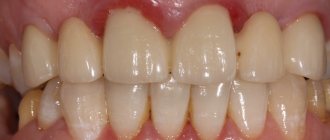
Metal-ceramic dental crowns are not stained by food and do not have a harmful effect on surrounding tissues. The approximate service life is 5 - 15 years, depending on the materials used and manufacturing technology. The more expensive the materials and the more elaborate the manufacturing technique, the longer the crown will last (a gold-platinum alloy as a frame increases the service life to an average of 20 years).
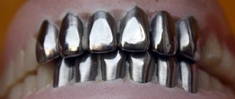
Metal crowns with ceramic coating
Why do they grind down a tooth for a crown?
The dentist must remove part of the tissue to form a stump of a certain shape:
- a ledge is formed at the edge of the gum, on which the crown will be placed in the future;
- The edges of the tooth should remain almost parallel, and not cone towards the chewing edge.
The service life of the artificial crown largely depends on the quality of preparation and compliance with these two conditions. If there is no smooth and even ledge, its edge will not adhere to the tooth, but will hang over the gum, forming a gap. Soft plaque will accumulate around, and colonies of pathogenic microflora developing in it will cause caries and gum inflammation.
If the tooth stump takes a cone-shaped shape after filing, it will be difficult for the dental technician to produce a functional structure that can withstand the chewing load. This is fraught with depressurization (or decementing) of the crown. Residues of food and soft plaque with microflora will penetrate under it, which will eventually lead to its loss.
Preparation for the crown of a living, non-pulpless tooth should be carried out with plenty of cooling. After all, overheating of the tissues will cause inflammation of the pulp, which may appear after the installation of the crown. Due to the need to treat pulpitis, it will have to be removed and redone after treatment of the canals.
Fitting Features
Tissue factors
• Bone or soft tissue obstructions, combined with misdirected force, prevent normal crown retention. This especially applies to the mucous membrane of the palate in the frontal area. • Targeting or flap of soft tissue may be required to detect obstructions. • In some cases (upper anterior restorations), not only gingival reamer and oblique release incisions may be necessary, but also removal of the lunate fragment of the palatal mucosa.
Interdental contacts
•
All interproximal contacts of integrated restorations must be passive.
If the contacts are too tight, the orientation, fit and operation of the locking mechanism are disrupted.
• An orientation (bite) template facilitates the correct positioning of the restoration. However, before fixation, it is necessary to evaluate the aesthetic and functional characteristics, on which the final position of the crown depends. • In maxillary anterior restorations, special attention should be paid to passive interproximal contact
and proper orientation of the post relative to the implant site.
If the abutment is not oriented correctly, tapping will cause deformation of the implant bed.
Fixation force
• Use a special sizing tray to make a bite guide. The template is especially important when restoring the upper front teeth. • To fix the crown, tap it several times with a hammer (250 g). It is extremely important to direct the force along the long axis of the abutment and implant site. • Do not tap crowns until you are sure the force is aligned with the long axis of the implant site. The displaced force will cause permanent deformation of the abutment and implant.
Abutment Post and Implant Site
• Do not handle the abutment post with your hands or tools. Any change in its contours or contamination can lead to retention failure. • Incorrect implantation technique can compromise the integrity of the implant and interfere with the restoration. • Deviation of the integrated restoration from the long axis of the abutment and implant (eg due to bony obstructions) may cause deformation of the implant site and prevent placement of the restoration. • To achieve the best retention of the abutment to the implant, it is recommended to slightly rotate the abutment within the implant site (clockwise and counterclockwise) and then tap the crown several times in the long axis direction.
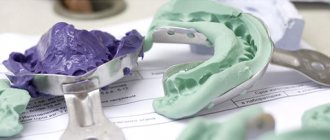
How to take impressions correctly?
If the mucous membrane was damaged when grinding the native crown, the procedure for taking impressions should be postponed until it is restored.
- In some patients, the injured gum can give recession - move away from the neck of the tooth and expose its root.
- Due to the presence of blood, the materials used to obtain impressions may not clearly depict the formed ledge.
As a result, the edge of the manufactured and installed crown will stick out above the gum, which will significantly reduce the aesthetics of prosthetic treatment of the front teeth.
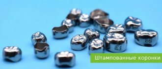
Fitting and cementing of stamped crowns
Single stamped crowns arrive from the laboratory to the clinic in finished form on plaster posts for the patient’s second visit. If several crowns were made for one patient, then the serial number of the tooth and the side (right or left) should be indicated on these posts. Before fitting, the patient’s teeth and artificial crowns are carefully treated with alcohol and ether, since plaque and dental wax from artificial crowns make it difficult to accurately fit the crown.
After applying the open part of the crown to the occlusal part of the tooth, its further advancement encounters some resistance, which slightly increases as the edge of the crown moves towards the clinical neck of the tooth. With a sharp increase in resistance, further advancement of the crown is accompanied by clicks. This is explained by the fact that the edge of the crown stops on an unpolished burr on one of the surfaces of the tooth, and after a certain force “slips” and often injures the round ligament of the tooth.
In these cases, you should stop fitting the crown and re-grind the tooth with soft abrasives.
The edge of the crown is brought to the level of the gums slowly and evenly on all sides. The parallelism between the edge of the crown and the gum is most often disrupted in the area of the dental papillae. This occurs because the dental papillae, which are looser than the rest of the gum, are pressed out to a greater extent by the impression material, which entails a relative lengthening of the edge of the crown in this place. If you do not pay attention to this, you can detach the dental papilla and rupture the circular ligament of the tooth in this place.
Removable defects include shallow folds on the approximal and oral surfaces, lengthening or shortening of the edge of the crown, improper processing of the crown, and poor polishing. In some cases, shallow folds can be straightened on a dental anvil or ground, which after a little polishing has virtually no effect on the quality of the crown. An attempt to eliminate deep folds in the same way leads to distortion of the anatomical shape of the tooth and the formation of hardening with subsequent cracks.
Lengthening the edge of the crown is an easily removable defect. In this case, the edges of the crown, made of expensive metal, are first trimmed with special scissors and then ground. Conventional crowns are simply ground off. To avoid excessive shortening of the crown, a small plaster impression is prepared on the crown being corrected and the edges of the crown are ground according to the imprint of the gums surrounding the neck of the tooth.
Poor polishing of the crown is an easily removable defect, but it should be taken into account that after each polishing the crown becomes thinner.
Disadvantages such as loose coverage of the tooth neck, lack of occlusal and proximal contacts are irremovable. Narrowing the neck of the crown with crampon forceps is unacceptable and impracticable. At the same time, folds form in some places, and in others, most often between teeth, the edges of the crown lag behind the neck of the tooth. The lack of contact between the occlusal surface of the artificial crown and the corresponding surfaces of the antagonist teeth cannot be eliminated due to the fact that the masticatory cusps of the prepared tooth were not modeled. Such crowns are subject to alteration with the obligatory modeling of the occlusal surface in an occludator or occlusal block.
An artificial crown can increase the bite if it is not tightly placed on the tooth. The crown can also move along the vertical axis or in the vestibulo-oral direction. In this case, it is necessary to lift the crown and then, with light force, move it along the path of minimal resistance towards the clinical neck. The crown finds its place, and thus the increase in bite is eliminated.
An increase in the bite can also occur due to the fact that it hangs on sharp edges formed as a result of incomplete preparation of the approximal or vestibulo-oral surfaces. These edges cannot always be determined visually, but they can be easily felt with a probe during palpation and are easily sanded off with soft abrasives.
The most common cause of overbite is insufficient clearance between the chewing surfaces of the prepared and opposing teeth. In this case, you need to complete the tooth preparation, take a new impression and re-stamp the crown.
Careless modeling of masticatory cusps is also a common cause of increased bite on stamped crowns. To identify this reason, you need to accurately bring the crown to the level of the clinical neck, and then ask the patient to clench his jaws tightly, having first placed a cotton roll on the occlusal surfaces of the opposing teeth. If even after this the crown increases the bite, and all other possible causes have already been eliminated, then it is necessary to make a more accurate modeling of the occlusal surface. This deficiency cannot be eliminated in a clinical setting, and therefore new impressions (working and auxiliary) should be obtained and a new crown made.
A correctly fitted crown is secured to the corresponding teeth using special cements. A loose crown injures the mucous membrane, becomes deformed under the influence of pressure during chewing, and becomes looser (especially gold crowns), which deteriorates its quality.
Metal stamped crowns are fixed in a certain sequence. After fitting, the crown should be degreased again and thoroughly dried. The cement is mixed to a consistency at which it slowly flows from the end of the spatula. The use of thicker cement is dangerous, because its excess may not be squeezed out from under the crown and this will lead to an increase in the bite.
Before the final application of the crown with cement, the tooth should be dried again with a stream of warm air. At the same time, the physiological pocket is also freed from the remains of the contents of the oral cavity. Then, applying considerable force, the open part of the crown is brought to the level of the clinical neck and the patient is asked to clench his teeth tightly in a state of central occlusion. Excess cement is removed with a cotton swab and the relationship between the crown edge and the physiological pocket is checked. If the edge of the crown does not reach the level of the physiological pocket, the crown is removed and fixed again with thinner cement. The crown is fixed to cement in the following sequence.
- 1. Degreasing and drying the artificial crown.
- 2. Degreasing and drying the natural tooth and covering it with cotton rolls.
- 3. Mixing cement and filling the crown with it.
- 4. Repeated drying of the natural tooth and the surrounding mucous membrane with warm air and application of a crown under bite control.
- 5. Checking the relationship between the edge of the crown and the clinical neck of the tooth and the physiological pocket.
- 6. Providing conditions for crystallization of cement with a minimum influx of saliva to artificial crowns and removing excess cement after it has hardened.
The duration of use of artificial stamped crowns depends on the material, the correctness of their manufacture, as well as articulation conditions. Under equal circumstances, crowns made of stainless steel are stronger than those made of gold, silver-palladium alloys, etc. Crowns made of platinum and gold alloys occupy an intermediate place. Articulatory factors that influence the duration of use of crowns include the nature of chewing, type of bite, periodontal reserve capabilities, the number of articulating teeth, the presence of other prostheses in the mouth, etc.
Depending on their constitutional characteristics, people chew food in different ways: some slowly but reliably, with a clear predominance of grinding movements (mysseterial type of chewing), others very quickly process the food bolus with predominantly vertical movements (temporal type of chewing). In patients of the first type, the crowns are “eaten away”, mainly on the chewing surfaces, while in the second type, the crowns on the vestibular side, closer to the equator, become thinner. For people with a straight bite, the timing of using different crowns varies little. With an orthognathic or deep bite, crowns on the front teeth last longer than on molars and premolars. The reserve forces of the periodontium, which depend primarily on its usefulness, determine the intensity of the participation of the crowned tooth in the act of chewing, as well as the timing of its mechanical wear.
Antagonist teeth and prostheses that replace them also play an important role.
In what cases does a tooth need a crown?
Indications for the use of a crown can be for a variety of reasons:
- Elimination of tooth defects at the crown part, where it is impossible to place a filling or onlay
- Preserving a tooth from destruction
- Protection of a healthy tooth on which a prosthesis will be placed or to which a bridge will be attached
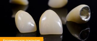
Unlike a porcelain crown, which looks much more aesthetically pleasing, a metal crown is thin. Therefore, its installation does not require extensive cutting of the tooth. It is this circumstance that makes it possible to use stamped crowns to protect healthy teeth.
If you have a problem similar to that described in this article, be sure to contact our specialists. Don't diagnose yourself!
Why you should call us now:
- We will answer all your questions in 3 minutes
- Free consultation
- The average work experience of doctors is 12 years
- Convenient location of clinics
Single contact phone number: +7
Make an appointment
Features of fixing crowns on cement
Before starting the procedure, hardware diagnostics are prescribed. An X-ray image allows you to verify the correct integration and make the necessary corrections to the configuration before installing the crown. A prerequisite is the removal of excess dental cement, which can cause irritation of the mucous tissues adjacent to the area being restored.
Among the advantages characteristic of cementing are high structural stability, as well as the possibility of using passive fitting technology. Soft tissue support is provided by the abutment - the crown is installed so that its edge is located either at the same level or apical to the gum contour.