The most popular classification of prints according to E.I. Gavrilov . It was based on the following basic principles. 1. The principle of the sequence of laboratory and clinical techniques for the manufacture of prostheses. On this basis, a distinction is made between preliminary (indicative) and final impressions. Preliminary impressions are taken with a standard spoon. Diagnostic models of jaws are cast from them, allowing one to study the relationships of the dentition, alveolar ridges of edentulous jaws, the relief of the hard palate and other features that are important for making a diagnosis, drawing up a plan for preparing the oral cavity for prosthetics, and the prosthetics plan itself. The same technique allows you to determine approximately the border of the prosthetic bed and make an individual tray . Based on the final impressions, a working model is cast.
2. A method of designing the edges of the impression, allowing the prosthesis to have a closing circular valve that provides one degree or another of its fixation. In accordance with this, anatomical and functional impressions . According to the method of edge design E.I. Gavrilov subdivides functional impressions , designed using: a) passive movements; b) chewing and other movements; c) functional tests. between anatomical and functional impressions . Essentially, there are no purely anatomical impressions. When taking an impression with a standard spoon, when forming its edges, they always use functional (though not sufficiently substantiated) tests. On the other hand, the functional impression represents a negative reflection of the anatomical formations (palatal ridge, alveolar tubercle, transverse palatal folds, etc.), which do not change their position during movements of the lower jaw, tongue and the functions of other organs. Therefore, it is completely natural that a functional impression has signs of an anatomical one, and vice versa. 3. The degree of pressure or the degree of squeezing of the mucous membrane.
According to the degree of its pressing, functional impressions are divided into:
1) compression or obtained under pressure, which can be arbitrary, chewing, dosed;
2) differentiated (combined);
3) decompression or obtained at minimum pressure.
Under any clinical conditions, only a functional impression using an individual tray.
Individual spoons can be made from : 1) metal (steel, aluminum) by stamping; 2) plastics: a) base (fluorax, ethacryl, yarocryl) by polymerization method; b) quick-hardening (redonta, protacryl) using the free-forming method;
c) standard plastic plates AKR-P; d) light-curing plastic; 3) solar-cured materials with polymerization in special chambers or using a solar lamp; 4) thermoplastic impression masses (Stens); 5) wax. Individual spoons are made in the laboratory or directly in front of the patient.
Making an individual plastic spoon in the laboratory.
In this case, an anatomical cast is taken with a standard spoon and a plaster model is cast from it. On the model, the dental technician draws the boundaries of the future individual tray . On the upper jaw, the border of the tray passes from the vestibular side along the transitional fold, not reaching the deepest point of its arch by 1-2 mm. On the distal side, it overlaps the maxillary tuberosities and runs along line “A” behind the palatine fossae by 1-2 mm. On the lower jaw, the border of the spoon passes from the vestibular side along the transitional fold, not reaching 1-2 mm to the deepest point of its arch, while bypassing the cords and frenulum of the lip. In the retromolar region, it is located behind the mucous tubercle, overlapping it by 1-2 mm. On the lingual side, the border of the spoon overlaps the area corresponding to the retroalveolar region (muscleless triangle), not reaching the deepest place of the sublingual space by 1-2 mm and going around the frenulum of the tongue. From the above it is clear that on both the upper and lower jaws the border of the individual tray is 2-3 mm less than the borders of the prosthesis. This is done so that there is space left for the impression material. The extruded impression material forms the edges of the impression. And, conversely, the distal boundaries of the tray must be larger than the boundaries of the prosthesis so that the anatomical formations, which are landmarks of the distal edge of the prosthesis, are well imprinted when taking an impression.
After drawing the boundaries, the dental technician coats the model with Izokol insulating varnish and begins making an individual tray from quick-hardening or base plastic. To make an individual spoon from quick-hardening plastic, the required amount of material is kneaded to a dough-like stage and a plate is made from it in the shape of the upper or lower jaw, which is pressed onto the model along the outlined boundaries. Then small pieces of plas are used to make a handle perpendicular to the surface of the spoon, rather than tilted forward. This position of the handle will not interfere with the design of the edges of the print. If the alveolar part of the lower jaw is significantly atrophied and the spoon turns out to be narrow, then the handle is made wider, almost up to the premolars: with such a handle, the doctor’s fingers will not deform the edges of the impression when they hold it on the jaw. After the plastic has hardened (10-15 minutes), the spoon removed from the model and processed with cutters and carborundum heads ( the individual spoon is not polished), making sure that the edges of the spoon correspond to the boundaries marked on the model. The thickness of the edge of the spoon should be at least 1.5 mm, because With a thinner edge, it is difficult to obtain the volume of the edge of the print. An individual spoon can be made from base plastic using the polymerization method. To do this, the heated wax plate is pressed tightly onto the model, giving it the shape of an impression tray, and excess wax is cut off with a spatula along the marked boundaries. The wax mold of the spoon is plastered into the cuvette in the reverse way and the wax is replaced with plastic. When making a spoon from AKR-P plastic, standard plates are softened in hot water and crimped according to the model. The excess is cut off with scissors after the corresponding area has been softened. The handle is made from scraps of material and glued to the spoon with a hot spatula (the heat melts and welds the plastic).
Individual spoons made of plastic are classified as rigid spoons. They can be used, as well as thermoplastic trays, to take compression impressions. Advantages and disadvantages of custom plastic impression trays . Plastic spoons are rigid and do not deform in the oral cavity, but, like any laboratory-made spoons (in two visits), they require subsequent correction in the oral cavity. In addition, spoons made in this way provide a modified display of soft tissues, because they are compressed and stretched during the taking of the anatomical impression.
Individual wax trays for the upper and lower jaws
Individual wax spoons can be made either in the laboratory or directly in the mouth. Wax trays using the CITO method are made in one visit directly on the jaw of the prosthetic patient. Such spoons are more accurate than individual ones made from an anatomical cast, because they display the soft tissues of the prosthetic bed at rest. The disadvantage of such trays is that the soft wax is deformed during fitting in the oral cavity and when taking an impression (it cannot withstand pressure), so the wax spoon can only be used to remove decompression impressions. Individual spoons , regardless of the method and material they were made from, must be placed in the oral cavity. A correctly fitted spoon sticks to the jaw and does not lag behind it when moving the lips and cheeks. , the technique of fitting individual spoons using Herbst functional tests has become widespread Functional Herbst tests are recommended when fitting individual trays and obtaining functional impressions .
Five tests are used on the lower jaw:
1) swallowing and wide opening of the mouth;
2) movement of the tongue to the sides along the red border of the upper and lower lips;
3) touching the cheeks with the tip of the tongue with the mouth half closed;
4) movement of the tip of the tongue forward beyond the lips towards the tip of the nose;
5) pulling the lips forward. Three tests are used on the upper jaw:
1) wide mouth opening;
2) cheek suction;
3) shifting the lips forward (pulling).
Obtaining a functional impression.
After fitting an individual tray, they begin to obtain a functional impression.
Obtaining an impression consists of the following steps:
1) fitting an individual spoon;
2) applying the impression mass to the tray;
3) inserting a spoon with the mass into the oral cavity;
4) forming the edges of the impression and conducting functional tests;
5) making an impression and evaluating it. It should be accepted as a rule that a functional impression that ensures good fixation of the prosthesis can only be obtained if the anatomical impression reflects all the structures of the prosthetic field and some functional features of the tissues surrounding the prosthetic bed. When a functional impression , they are only refined. There are unloading or decompression and compression impressions. Typically, the value of a compression or relief impression is associated with the fixation of the prosthesis and its effect on the mucous membrane of the prosthetic bed. However, the value of a particular impression-taking technique is determined by the influence of the prosthesis on the course of the process of atrophy of the alveolar process. Unloading (decompression) impressions are obtained without pressure or with minimal pressure of the impression mass on the tissue of the prosthetic bed. The disadvantage of the unloading impression is that the buffer zones of the hard palate are not subject to compression, and all the pressure from the prosthesis is transferred to the alveolar process, increasing its atrophy.
When receiving a decompression impression, the impression material must reflect every detail of the oral mucosa without distortion so that the microrelief of the prosthesis base exactly matches the surface structure of the prosthetic bed. Therefore, such impressions can only be obtained using impression compounds that have high fluidity and do not require much effort to remove the impression. Such masses include low-viscosity silicone pastes: exaflex, xanthoprene, alfazil, as well as zinc oxide eugenol pastes. The impression obtained using liquid plaster (according to Brachman) usually provides just such a perception of the surface relief of the tissues of the prosthetic bed. Some authors believe that if several holes are drilled in the impression tray to drain excess impression material, then the pressure of the impression mass on the mucous membrane can be reduced. It is known that the fixation of prostheses made using decompression impressions is weak, but they can be used if there are certain indications.
Such indications include:
1) significant or complete atrophy of the alveolar processes and mucous membrane;
2) increased sensitivity of the mucous membrane;
3) uniformly pliable mucous membrane of the prosthetic bed. Compression impressions are designed to take advantage of the pliability of the mucous membrane, so they are removed under high pressure to compress the buffer zones. When they talk about a compression impression, they primarily mean compression of the vessels of the prosthetic bed. The decrease in tissue volume and its vertical compliance are directly dependent on the degree of filling of the vascular bed. The use of compression impressions is recommended in the presence of loose mucous membranes with good pliability.
A prosthesis made using a compression impression does not load the alveolar ridge; outside of chewing, it rests only on the tissues of the buffer zones, like pillows. When chewing, under the influence of masticatory pressure, the vessels of the buffer zones are emptied of blood, the prosthesis settles somewhat and transfers pressure not only to the buffer zones, but also to the alveolar part. Thus, the alveolar process is unloaded, which prevents its atrophy. A prosthesis made using a compression impression has good fixation, because the pliable mucous membrane of the valve zone is in closer contact with the edge of the prosthesis. The compression impression is taken under continuous pressure , which ensures compression of the vessels of the mucous membrane of the hard palate and their emptying. To obtain such a print, certain conditions must be met:
1) you need a hard spoon;
2) the impression must be taken using a low-flow mass or thermoplastic mass;
3) compression must be continuous, stopping only after the mass hardens. Continuity can be ensured by manual force (voluntary pressure). But it is more convenient and correct to take a compression impression under the chewing pressure of the muscles that elevate the mandible, i.e. under bite pressure, which is created by the patient himself, or with the help of special devices that make it possible to create a strictly defined pressure (dosed) taking into account the individual characteristics of the tissues of the prosthetic bed and masticatory muscles. To obtain a functional impression, thermoplastic materials are used, such as dentofol, otrokor, orthlast, etc.
The convenience of using thermoplastic masses is explained by their following properties: 1) they have an extended plasticity phase, which allows for functional tests necessary to obtain a high-quality impression;
2) during impression taking they always have the same consistency; 3) they do not dissolve in saliva; 4) distribute pressure evenly; 5) allow you to repeatedly insert an impression into the oral cavity and carry out correction, because new portions of the mass merge with old portions without deforming the print. However, thermoplastic masses have certain disadvantages. These include: inaccurate print due to low fluidity; deformation in the presence of retention points. When cooled with water, they harden unevenly and may become deformed when removed from the mouth. It should be recognized that when using the above methods for obtaining an impression, in some cases it is not possible to ensure a complete functional reflection of the prosthetic field. The tissues of the prosthetic field and the active muscles surrounding it are not the same in relief, relative volume, physiological status during chewing or speaking, as well as during the day. The physical and emotional state of a person also has a great influence on the condition of the prosthetic bed and the surrounding muscles. Whatever method of taking an impression is used, further adaptation of the prosthesis base to the tissues of the prosthetic field, the relationship of the dentition and the force of chewing pressure is necessary, as well as adaptation of the patient and fitting of the prosthesis over a certain period of time. The wide variety of clinical conditions encountered for prosthetics necessitates the use of a differentiated impression. We should proceed from the general position that there is no single method shown in all cases. In this regard, the method of obtaining an impression in each specific case must be chosen taking into account the patient’s age, constitutional and individual characteristics of the jaw tissues, i.e. in all cases a differentiated approach is required. In cases where the tissues of the prosthetic bed in different areas are not identical in their relief and structure, the biophysical properties of each element of the prosthetic bed should be taken into account. When taking an impression, tissues with pronounced spring properties should be under greater load, while tissues of unloaded zones (in the area of the torus, incisive papilla, etc.) should not be overly loaded. Selective pressure on the underlying tissues, depending on their anatomical and functional characteristics and biophysical properties, may be important due to the need to prevent premature atrophy of the soft and bone tissues of edentulous jaws by redistributing the chewing pressure of the prosthesis base. Consequently, depending on the anatomical and physiological characteristics of the prosthetic bed, it is possible to obtain images of the mucous membrane in various functional states. At the same time, it is recommended to obtain unloading impressions in case of thin, atrophic and excessively pliable (“dangling” ridge) mucous membrane. Compression casts are indicated for loose, highly pliable mucous membranes. The best effect can be achieved only by using differentiated casts obtained with different degrees of compression of the mucous membrane, taking into account its compliance in different areas of the prosthetic bed.
<<backThe choice of impression material is extremely important in the manufacture of all types of denture designs. It should be based on the correspondence of the properties of the material to the impression taking technique chosen by the doctor in a given clinical situation.
To begin with, we are obliged to give several fundamental definitions that will allow us to further discuss such an important topic.
An impression is a negative (reverse) reflection of the surface of hard and soft tissues located on the prosthetic bed and its boundaries.
The prosthetic bed is a complex of organs and tissues that are in direct contact with the denture (E.I. Gavrilov). The concept of prosthetic field also includes tissues of the maxillofacial region located in the zone of indirect action of the prosthesis.
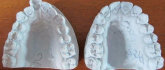
Based on the impression, a model is cast that repeats the anatomical formations in the oral cavity and is a positive reflection of the prosthetic bed (Fig. 1). Models have different purposes.
Fig.1. The impression is a negative representation of the prosthetic bed, the model is a positive one.
Working models are used directly for the manufacture of orthopedic structures. They must be cast from durable plaster and reproduce the prosthetic bed as accurately as possible.
Diagnostic models serve to clarify the diagnosis in complex clinical situations and plan treatment.
Control models are necessary to evaluate the effectiveness of the treatment.
Auxiliary models are needed to display antagonist teeth and fully reproduce the clinical situation in the oral cavity.
Depending on the purpose of the model, clinical conditions in the oral cavity, and the required level of reproduction of the details of the prosthetic bed, one or another type of impression is chosen (Fig. 2).
Fig. 2. Classification of prints.
Anatomical impressions are a static representation of the prosthetic bed and its surrounding tissues. They are obtained in the manufacture of all types of orthopedic structures. During the process of taking an anatomical impression, the soft tissues that limit the edges of the impression are at rest. To obtain anatomical impressions, both standard and individual trays are used.
Functional impressions are taken during the manufacture of removable dentures, when it is necessary to ensure their fixation using the method of functional suction with the creation of a valve zone. In this case, hard individual spoons are used, carefully placed in the oral cavity. In the process of taking a functional impression, active and passive formation of its edges by soft tissues that are in the process of function is necessary.
According to the degree of pressure exerted by the impression material on the tissue of the prosthetic bed, impressions are divided into:
a) o compression b) o unloading c) o differentiated
The choice of the degree of mucocompression depends on the characteristics of the oral mucosa:
- In most cases, it is recommended to take differentiated impressions, since in different parts of the prosthetic bed the pliability of the mucous membrane is usually different.
- Decompression areas are created in areas with atrophied or excessively pliable mucous membrane, as well as in the presence of a “dangling ridge” - an alveolar process devoid of a bone base.
- In the presence of a mucous membrane with a uniform, moderately pronounced submucosal layer, compression impressions are indicated.
Different degrees of compression are achieved by creating perforations in the impression tray, as well as using impression materials with different mucocompression properties.
Print quality requirements:
- A high-quality impression must accurately reflect all elements of the prosthetic bed and adjacent tissues. This is necessary to clearly define the boundaries of the prosthetic bed and form an adequate edge of the prosthesis.
- There should be no bubbles, pores, stretch marks or other defects on the surface of the print.
- The representation of the dentition or alveolar process in the impression should be located in the middle between the sides of the tray.
- The edges of the print must be clearly defined.
<<back