Author of the article:
Soldatova Lyudmila Nikolaevna
Candidate of Medical Sciences, Professor of the Department of Clinical Dentistry of the St. Petersburg Medical and Social Institute, Chief Physician of the Alfa-Dent Dental Clinic, St. Petersburg
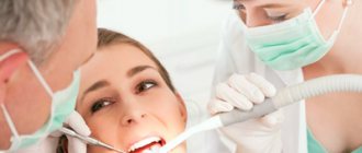
Candidiasis or, otherwise, thrush is an infectious disease, the main cause of which is infection with Candida fungi. These fungi belong to the same species as yeast and are part of the natural microflora of the vast majority of healthy people. The maximum concentration of these opportunistic microorganisms is observed in the intestines, nasopharynx, and vagina; Some fungi can also be found on the surface of the skin.
In a normal, healthy state of the body, the fungus does not cause any unpleasant symptoms. However, when the immune system is weakened and other provoking factors appear, Candida actively multiplies, resulting in discomfort, itching, burning and other manifestations of thrush. This disease can affect various tissues and organs; Candidiasis of the oral mucosa is also common.
Most often, infants suffer from this disease: according to statistics, up to 20 percent of children under the age of one year have suffered from candidiasis at least once. About 10 percent of people over 60 also suffer from symptoms of this infection. It occurs more often in women than in men; very often, signs of the disease appear in smokers. There are many other factors that contribute to the development of candidiasis in the mouth.
Causes of oral candidiasis
The main reason for the occurrence is a decrease in immune status, as a result of which the uncontrolled proliferation of microflora begins. Reduced immunity is observed in older people and infants, in patients suffering from HIV, AIDS and other diseases associated with immunodeficiency, in those who regularly expose the body to excessive stress, neglect the rules of a healthy diet and violate work and rest patterns. Risk factors include reasons such as:
- Use of medications.
Taking antibiotics, immunosuppressants (drugs that suppress the immune system) and some other medications leads to disruption of the immune system and the natural balance of microflora in the body. Oral contraceptives, which affect hormonal levels, have a similar effect. - Pregnancy.
During pregnancy, a sharp and significant change in hormonal levels occurs, which can lead to a surge in the activity of pathogenic and opportunistic microflora. - Radiation and chemotherapy.
Often occurs in patients undergoing drug and radiological treatment for cancer. - Injuries to mucous membranes.
Violation of the integrity of the mucous membranes leads to loss or deterioration of the barrier function, as a result of which the fungus enters deep into the tissues, causing inflammation and other symptoms. Small, but constantly recurring injuries are especially dangerous - for example, when wearing incorrectly fitted dentures or braces. - Overwork and stress.
Prolonged stress of physical and mental forces leads to a deterioration in the protective function of the body. Similar consequences are caused by hypothermia or overheating, regular lack of sleep, insufficient, excessive or simply unbalanced nutrition, abuse of alcohol, nicotine, and narcotic substances. - Hypo- and vitamin deficiency.
May be caused by a lack of nutrients, in particular vitamins B and C. - Somatic diseases.
Frequent companions of candidiasis include tuberculosis, dysbacteriosis and other pathologies of the gastrointestinal tract, diseases of the adrenal glands and other endocrine glands. Candidiasis is a contagious disease. A large number of pathogenic microorganisms are transmitted through kissing and sexual contact, through the use of shared dishes, towels and other household items. Infection can also occur during childbirth (vertical transmission from mother to fetus). In addition, there is a risk of infection through contact with infected pets.
Pathogenesis
Yeast fungi in the human body are considered to be opportunistic microflora . They may be present in the microflora, and if the immune system is strong, then the fungal infection does not cause diseases, for example, urogenital, respiratory, skin, or, as in this case, the mucous membranes of the oral cavity. Normally, in almost 70% of the population, yeast fungi are present in the resident microflora; they are inactive and do not cause candida or other fungal diseases. Reproduction usually occurs by budding and spores. Proliferation occurs not due to the departure of daughter cells from the mother cells, but through elongation and formation of a pseudomycellar tree. As a result of vital activity, toxic substances are released that weaken the human immune system.
Model of a fungus cell of the genus Candida
alcohol and drug abuse, oncology, and the use of contraceptives contribute to imbalance of the oral microflora and excessive proliferation of opportunistic fungi . Fungus in the mouth is most often a superficial disease that affects the mucous surface of the cheeks, the corners and borders of the lips, the back of the tongue, the palate, etc., but with significant spread and penetration into the bloodstream it can cause systemic chronic and generalized disorders.
In neutropenic or severely ill patients, candidal fungal infection can enter the bloodstream, where it causes widespread visceral dissemination. Generalized mycosis can lead to candida sepsis and even death.
Types of disease
The clinical picture of oral candidiasis is classified:
- For clinical and morphological.
- With the flow.
- By localization.
Clinical and morphological is divided into:
- Hyperplastic.
- Erosive-ulcerative.
- Pseudomembranous.
- Atrophic.
The clinical picture, classified according to the course, is divided into:
- Chronic.
- Spicy.
By localization:
- Cheilitis.
- Gingivitis.
- Glossitis.
- Stomatitis.
- Tonsillitis, etc.
Based on the clinical picture, oral candidiasis comes in several types:
- Chronic hyperplastic.
- Candida infection.
- Chronic atrophic.
- Acute pseudomembranous.
- Chronic pseudomembranous.
- Acute atrophic.
CANDIDIASIS OF THE ESOPHAGUS
Candidiasis is an infectious disease of the mucous membranes, skin and internal organs caused by yeast-like fungi of the genus Candida. Esophageal candidiasis (OC), which is a manifestation of visceral candidiasis, occupies a prominent place among infectious lesions of the esophagus. In recent years, there has been a tendency to increase the frequency of CP, especially in patients with impaired immunity. The growth of candidiasis infection is largely due to an increase in the number of patients with HIV infection, advances in transplantation and immunosuppressive therapy, and the uncontrolled use of antibiotics. KP occurs in 0.7-1.5% of gastroenterological patients [5, 6].
The problem with severe fungal infections caused by opportunistic pathogens is that they are difficult to treat and can be fatal. The mortality rate for invasive candidal infections has been found to be 34% [16].
Etiopathogenesis. Candida species are the most common esophageal pathogen, most notably Candida albicans, with occasional occurrences of C. tropicalis, C. parapsilosis, C. glabrata, C. lusitania, and C. krusei. These microorganisms are normal components of the oral flora and their growth is inhibited by bacterial commensals. Infection with fungi such as Candida, which are widespread in the environment, occurs through endogenous or exogenous routes. Endogenous infection is associated with the activation of saprophytic fungi; exogenous infection can occur through direct contact with carriers of infection or from the environment. If the host's body is not weakened, many fungi do not exhibit their pathogenic properties. Research in recent years has shown that the source of fungal dissemination is the intestines, and candidiasis of the oral cavity, genitals, and esophagus is a manifestation of systemic candidiasis. The likelihood of developing systemic damage depends both on the properties of the microorganism itself (their number, virulence, genetic and species heterogeneity of the population), and on the state of the macroorganism, especially its immune system, nutritional status and abdominal blood flow [3, 17].
Favorable conditions for the development of the infectious process are created by various violations of the physiological, anatomical and immunological mechanisms of the body's defense. Factors that provoke the occurrence of esophageal candidiasis include the use of antibiotics, inhaled or injected corticosteroids, antacid therapy or a hypochlorhydric state, diabetes mellitus, alcoholism, the consequences of intoxication, malnutrition, old age, impaired motility of the esophagus or esophageal obstruction, organ and bone marrow transplantation , enteral and especially parenteral nutrition, etc. A weakened immune system can lead to candidiasis infection. In diabetes mellitus, elevated blood glucose levels promote fungal growth because hyperglycemia impairs granulocyte function. Hypofunction of the parathyroid glands and adrenal glands leads to disruption of calcium-phosphorus metabolism, which causes hidden spasmophilia of the esophagus, thereby reducing its local protective capabilities [9]. Impaired nutritional status due to a lack of protein in the body and low calorie food affects the state of the immune system and creates the preconditions for the development of candidiasis [3]. Risk factors for candidiasis include a decrease in the acidity of gastric juice (pH 7.4 is optimal for the growth of Candida fungi, and when the pH shifts to 4.5, fungal growth is completely inhibited) [3, 4, 7].
The pathological manifestations of KP are varied. At first, the affected areas of the esophagus have the appearance of individual whitish or yellowish lesions raised above the mucous membrane. Later, these lesions can merge, forming dense plaques with the introduction of the fungus into the submucosa or pseudomembranous deposits with the penetration of the fungus into the muscular layer and blood vessels [9]. Films that form on the esophageal mucosa in especially severe cases can almost completely close the lumen of the esophagus. Plaque consists of desquamated epithelial cells that mix with fungi, inflammatory cells and bacteria. Microscopic examination reveals uniformly colored yeast-like cells and filaments of mycelium of Candida fungi [9]. True ulceration is observed infrequently and in most cases is observed in immunosuppressed patients with granulocytopenia [29]. Sometimes necrosis of the esophageal wall occurs and phlegmonous inflammation of the esophagus and mediastinum develops, which can become one of the causes of death of the patient [1].
There is a morphological classification, according to which all cases of KP are divided into three groups depending on the severity of the process, that is, depending on the depth of damage to its wall: 1st group - individual whitish plaques with the introduction of pseudomycelium of the fungus between the epithelial cells; 2nd group - membranous plaques merging with each other and forming vast fields, while filaments of pseudomycelium grow not only the mucosa, but also the submucosa; Group 3 - pseudomembranous overlays, combined with deep changes, in which the threads of the fungus penetrate deeply into the thickness of the muscle tissue [10].
Clinical manifestations and complications. Symptoms of the disease are practically absent in 25-30% of patients suffering from KP, especially in immunocompetent individuals. However, most patients present with complaints related to damage to the gastrointestinal tract. The most typical clinical manifestations of KP are dysphagia and, somewhat less commonly, odynophagia. The severity of esophageal symptoms ranges from moderate difficulty swallowing to severe pain, resulting in the inability to eat and the development of secondary dehydration. In severe odynophagia, there may be other causes or co-infection, especially in patients with AIDS. Much less frequently, patients may complain of chest pain not associated with swallowing, heartburn, nausea, sometimes vomiting with the release of films (pseudomembranes), decreased appetite and weight, and the appearance of loose stools with mucus (see figure) [4, 9, 29].
| Symptoms of candidal esophagitis (RS Orlando, 1996) |
Physical examination may be helpful in KP. Approximately two thirds of patients with
AIDS and esophageal candidiasis have candidal stomatitis. KP is observed in patients with chronic mucocutaneous candidiasis, which is a severe form of candidal infection and is more often observed with dysfunction of the adrenal glands and parathyroid glands [29].
Complications of esophageal candidiasis are rare. Esophageal bleeding can be observed in severe cases of the disease, accompanied by the formation of erosions, ulcers, and be associated with coagulopathy; perforation may develop. Secondary obstruction of the lumen by mycetoma has been described. Necrosis rarely occurs with the development of phlegmonous inflammation of the esophagus and mediastinum [1]. In severe cases, specific esophagitis can be complicated by the development of candidiasis sepsis [6].
Diagnostics. Suspicion of esophageal candidiasis should arise in any patient if there are risk factors for the development of esophageal infection and complaints of dysphagia and odynophagia. The presence of candidal stomatitis confirms this diagnosis, but in its absence, damage to the esophagus is also not excluded.
Barium x-ray of the esophagus is usually used for initial evaluation before endoscopy. However, in the early stages of candidal esophagitis, X-ray examination of the esophagus does not have much diagnostic value, since it reflects only nonspecific changes common to all esophagitis [2]. Classic radiographic signs of acute esophagitis caused by Candida spp. are linear or irregular filling defects with clear edges. In severe cases of candidal esophagitis, fusion of lesions occurs, which is why large filling defects sometimes form clusters in the form of bunches of grapes [2]. In this case, the esophagus acquires a “shaggy” (“hairy”) appearance, simulating ulceration [25]. The presence of large, well-circumscribed ulcers is not a sign of candidal esophagitis. Impaired motility and narrowing of the lumen of the esophagus due to pseudomembranes may occur. It should be remembered that a normal barium radiograph of the esophagus does not exclude esophageal candidiasis. Due to severe odynophagia, the patient will not be able to drink barium, which makes X-rays of the esophagus difficult [29].
The double contrast radiological method is considered more informative for the diagnosis of candidal esophagitis, the effectiveness of which reaches 70% [26].
A cytology brush and balloon catheter are used to quickly diagnose esophageal infections without endoscopy. These instruments can be easily inserted through the nasal passages or the mouth through a protective probe that prevents contamination. The material obtained from the protected brush or balloon catheter after it is removed from the esophagus is evaluated cytologically and culturally. The technique using protected brushes has a sensitivity of 88% and a specificity of almost 100% [26].
The cytological method involves staining impression smears or swab sediment from a cytological brush in search of active forms of Candida - budding yeast cells, pseudomycelium and mycelium. The cultural method involves placing the test material on Sabouraud's glucose-enriched medium or other media, in order to then judge the etiology of the infectious process in the esophagus by the nature of the colonies formed.
Endoscopic examination of the esophagus is the most sensitive and specific method for diagnosing esophageal candidiasis. The endoscopic picture of KP is most often characterized by the presence of easily removable fibrinous loose overlays of white or yellow color, under which easily wounded and/or edematous mucosa is found. Catarrhal and erosive-ulcerative esophagitis are less common [19]. Candida spp. rarely causes true ulceration. The presence of an ulcer in candidal esophagitis is often a sign of an additional pathological process in the esophagus [29]. There are various endoscopic classifications of esophageal candidiasis (Tables 1 and 2).
During endoscopy, affected areas of the mucosa may be subjected to brush biopsy for cytological examination or biopsy for histological diagnosis. When ulcers are identified endoscopically, repeated biopsies help rule out the presence of coexisting pathological processes. Cytological examination of brush biopsy material has a higher sensitivity level than histological examination of biopsy specimens for mild superficial candidiasis because microorganisms may be washed off the tissue surface during processing of the biopsy material [19]. In rare cases, positive cytology in the presence of negative histology indicates colonization rather than infection. For more severe candidiasis of the esophagus, the greatest diagnostic value is histological examination of mucosal biopsies using special staining for neutral mucopolysaccharides according to Schiff PAS (CHIK reaction) or according to Gomori with silver hexamethylenetetramine. Only histological examination demonstrates invasion of the mycelium or pseudomycelium of the fungus deep into the tissue of the esophagus.
Skin testing and serological tests are not very informative for diagnosing esophageal candidiasis.
Treatment. There are many oral and intravenous medications that are used to treat candidiasis esophagitis. Despite the relatively wide choice of drugs, the treatment of KP is an urgent problem, since some drugs are not effective enough, others have serious side effects; In addition, there is currently an increase in resistance to antifungal drugs.
When treating KP, oral therapy should initially be prescribed; intravenous administration is used only in case of refractory disease or if there are contraindications to oral use of medications. Patients with moderate severity of the disease and minimal immunocompromise require a short course of therapy using systemically absorbed drugs such as oral azole. Immunocompromised transplant patients and AIDS patients with KP are best treated with longer courses of azole. In patients with granulocytopenia, when there is a significant risk of dissemination of Candida infection, the use of intravenous systemic drugs (azoles, amphotericin B) is justified [29].
The arsenal of modern antifungal agents is quite wide. Antifungal drugs of several groups are used to treat esophageal candidiasis. The most effective drugs are from the azole group. Non-absorbable azoles (clotrimazole, miconazole) are used orally; however, systemic drugs from this group (ketoconazole, fluconazole and itraconazole) are more effective. These drugs, like others in the azole group, alter fungal cell membrane permeability through cytochrome P450 (CYP)-dependent interference with ergosterol biosynthesis, resulting in fungal cell damage and death. New triazoles (itraconazole and fluconazole) have higher affinity similarity than imidazoles (miconazole and ketoconazole) for fungal CYP enzymes [14]. Although other drugs, such as miconazole, clotrimazole, and nystatin, can be used to treat candidal stomatitis, as well as to prevent esophageal lesions, these drugs are less effective as the main group of drugs for the treatment of KP [24].
Clotrimazole and miconazole are imidazole drugs. Clotrimazole tablets and miconazole for oral use are currently available. However, they are not absorbed from the gastrointestinal tract. These drugs can be used for mild candidiasis of the esophagus in people without immunodeficiency.
Ketoconazole (nizoral, oronazole) is an imidazole derivative and, when taken daily in a dose of 200 to 400 mg, gives a good effect in the treatment of esophageal candidiasis. In AIDS patients who usually require higher doses of ketoconazole, the daily dose can be increased, if nausea does not occur, to the maximum (800 mg). Ketoconazole penetrates well into various organs and tissues, but poorly through the blood-brain barrier. The drug is well absorbed from the gastrointestinal tract, but requires an acidic environment for optimal absorption. With gastric hypochlorhydria and the use of antacids, its bioavailability decreases. To improve absorption, ketoconazole should be taken 2 hours before taking antiulcer medications. Approximately 10-25% of AIDS patients experience decreased gastric acid secretion. Ketoconazole can cause a temporary blockade of the synthesis of testosterone and cortisol [6, 8, 29].
Itraconazole (Sporanox) belongs to the group of triazoles, like ketoconazole, and is prescribed at a dose of 200 mg per day. Further increases in the dose lengthen the half-life of the drug and increase its effectiveness. The absorption of intraconazole decreases when the pH of gastric juice decreases [23]. Ketoconazole and itraconazole are metabolized in the liver and excreted in the bile. The half-lives of these two drugs are 7 to 10 hours and 24 to 42 hours, respectively [14]. No dose adjustment is required in patients with renal failure.
Fluconazole (Diflucan, Diflazon, Forkan, Flucostat - domestic fluconazole) is a water-soluble triazole and is prescribed at a dose of 100 mg per day. Fluconazole is a drug whose absorption is independent of gastric pH and is significantly more effective in the treatment of esophageal candidiasis in AIDS than ketoconazole (200 mg daily) [21]. Fluconazole is available for oral and intravenous use. It is minimally metabolized and excreted unchanged in the urine. Fluconazole has a high tissue tropism and does not affect the synthesis of androgens and penetrates well through the blood-brain barrier. Unlike ketoconazole and intraconazole, it is highly soluble in water and minimally protein bound. The drug has a long half-life (approximately 30 hours, unless renal function is impaired and the presence of food or hypochlorhydria does not alter absorption), allowing it to be taken once daily. It has been shown that the administration of fluconazole improves immune parameters in the T- and B-systems [18]. Both fluconazole and itraconazole can be taken orally as solutions. These forms may be more effective than tablets because they enhance the local effect and improve absorption.
Adverse effects of ketoconazole, fluconazole and itraconazole are primarily dose dependent and include nausea, hepatotoxicity, decreased steroid production and cyclosporine metabolism [14]. In rare cases, ketoconazole can cause fatal hepatitis [12]. A slight increase in aminotransferases is a common side effect of all three drugs, but this should not be used as an excuse to discontinue them. The effect on steroidogenesis is most pronounced with ketoconazole. Reversible inhibition of gonadal and adrenal steroid synthesis by ketoconazole may occur when the dose exceeds 400 mg per day [27]. At recommended doses, fluconazole and itraconazole do not affect steroidogenesis. As a result of their effects on hepatic microsomal enzymes, all three azoles inhibit the metabolism of cyclosporine, which leads to an increase in the level of cyclosporine in the blood; this effect is most pronounced with ketoconazole [14].
Another main group of antifungal agents is polyene antibiotics, represented by amphotericin and nystatin. These drugs irreversibly bind to sterols in fungal cell membranes, thereby altering the permeability properties of the membrane, disrupting its barrier function and causing cell death. Nystatin (anticandin, mycostatin, fungicidin) is practically not absorbed from the gastrointestinal tract. It is used to treat candidal stomatitis, but is less effective in cases of esophageal candidiasis. In addition, the effectiveness, safety and ease of use of azole derivatives make it possible to consider nystatin as a second-line therapy. Amphotericin B (amphostat, fungizone) is the only polyene antibiotic for parenteral administration. It is not absorbed in the gastrointestinal tract, is used intravenously, penetrates well into various organs and tissues, and is excreted from the body by the kidneys. The half-life is 24-48 hours, but with systematic use it can increase to 15 days due to accumulation in tissues [8]. Although amphotericin B is the most effective drug used to treat systemic mycoses, its use in the treatment of KP is limited due to serious side effects. Side effects of amphotericin include neurotoxicity, hematoxicity, nephrotoxicity, local irritation (phlebitis), allergic reactions, dyspeptic disorders, fever, etc. [8]. The most adverse side effect resulting from long-term use of amphotericin is nephrotoxicity, which is usually reversible. This medication is now available as an oral solution and lozenges. In patients with KP who are refractory to treatment with fluconazole or other azoles, low doses of intravenous amphotericin B (10 to 20 mg daily) may be effective. The total dose of the drug for the treatment of esophageal candidiasis ranges from 100 to 200 mg [29].
Flucytosine is a drug with a narrow spectrum of antifungal activity that works by interfering with RNA translation. It is incorporated into fungal cells, where it is converted into 5-fluorouracil and inhibits thymidylate synthetase. This oral drug, which is given at a dose of 50 to 150 mg/kg per day every 6 hours, can be used in combination with amphotericin B, but it should not be used as monotherapy because fungi quickly become resistant to it. In addition, flucytosine monotherapy appears to be only moderately effective [12].
The newest class of antifungal drugs are candins, which interfere with the synthesis of the fungal wall. They are effective against most Candida species, including C. krusei. The first studies showed that capsofungin, which represents this group of drugs, was as effective in KP as amphotericin B [16].
When treating patients with KP, one should take into account the presence of resistance, which has now increased significantly due to the widespread use of azoles. If resistance develops, it is often useful to increase the azole dose. If this is not enough, switch to another drug from this group or use an oral solution of itraconazole [13], which must be prescribed in higher doses due to frequently observed cross-resistance. When a high dose (i.e. 400 mg daily) of fluconazole is not enough, switch to intravenous amphotericin B, and the result is achieved in 90% of cases. Resistance to amphotericin is rare [29].
In table 3 presents the treatment of candidal esophagitis depending on the function of lymphocytes and granulocytes.
In the treatment of candidal esophagitis in patients with AIDS, the first-line drugs are ketoconazole and fluconazole, with fluconazole being preferred. Due to better tolerability, it is primarily indicated for patients at an advanced stage of the disease who have many comorbidities. If swallowing is impaired, parenteral forms of fluconazole can be used. If first-line drugs are ineffective, drugs from the reserve group (amphotericin B, itraconazole), which are more toxic and/or more expensive, are used. Etiotropic therapy of esophageal candidiasis, in addition to the main course of treatment, requires maintenance treatment, which can be lifelong (Table 4) [4].
Treatment of candidiasis against the background of severe immunodeficiency and leukopenia is a difficult task. Along with antifungal therapy, it is important to restore the pool of neutrophil leukocytes and their functional activity, since neutrophil leukocytes are one of the main links in the defense mechanism against Candida spp. It is proposed as an additional agent in the treatment of candidal infection against the background of neutropenia to use granulocyte colony-stimulating factor, which reduces the deficiency of myeloperoxidase in neutrophil leukocytes and enhances their oxygen-dependent anti-candidal activity [7]. A good effect has been obtained from the endoscopic administration of granulocyte concentrate and high-intensity pulsed laser radiation to patients with KP, which improves immune functions [5].
Thus, to achieve success in patients with severe fungal infections, including candidiasis, an integrated approach to diagnosis and treatment is advisable. Increased survival will be facilitated by prompt diagnosis followed by the selection of effective specific antifungal therapy and therapeutic measures aimed at increasing the number of granulocytes and stimulating phagocytosis [16].
For questions about literature, please contact the editor
Symptoms
Infection of the oral mucosa by Candida fungus can take various forms, each of which has its own characteristics of symptoms. The most common forms of the disease are candidal angulitis, glossitis, cheilitis, and stomatitis. There are both acute and chronic forms of the disease.
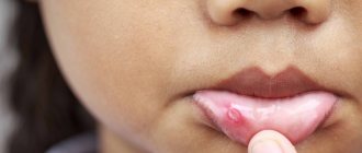
- Pseudomembranous acute candidiasis is the most common form and most often occurs in infants and the elderly. This form is characterized by the appearance of severe swelling and hyperemia (redness) of the mucous membranes. In addition, a characteristic whitish coating forms on the surface of the lips, palate, back of the tongue and the inside of the cheeks. If the plaque is scraped off, the surface of the mucous membrane underneath will be macerated (softened) or ulcerated and bleeding. In this case, patients complain of pain, burning or itching in the mouth; Eating becomes very difficult. Very often the process spreads to the esophagus and pharynx.
- Acute atrophic candidiasis of the oral mucosa usually develops due to the lack of adequate treatment. The upper part of the mucous membranes (epithelium) is exfoliated, the mucous membrane becomes thin, red or, on the contrary, swollen. The patient’s tongue and the corners of the lips also acquire a bright red color; the papillae on the tongue atrophy and smooth out. The plaque is absent or is found only in hard-to-reach places.
- Hyperplastic chronic candidiasis is characterized by the formation of a large number of papules and plaques of irregular or round shape. They are located close to each other on the mucous membrane of the tongue and cheeks and often become soldered and fused. Around each such formation there is a thin rim of reddened, inflamed tissue. It is difficult to scrape off or otherwise remove such a plaque. The oral cavity becomes dry and rough; When chewing, speaking, and even at rest, patients experience significant discomfort and pain. It should be noted that this disease most often affects men over 30 years of age.
The main cause of the chronic atrophic type is constant injury to the mucous membranes, for example due to wearing a prosthesis. Symptoms of the disease are localized in the affected area. Redness of the mucous membrane occurs (often along the contour of the lesion), plaque forms, pain and burning occur, and the membranes become dry.
What does thrush look like in the mouth - its symptoms in adults
After Candida attaches to the mucous membrane, they multiply and “grow” deep into it2, causing inflammation, swelling and redness. There is a feeling of a “scalded mouth” and discomfort when eating and swallowing. There may be a change in taste and the appearance of a metallic, sour, salty or bitter taste1.
The proliferation of the fungus leads to the appearance of small white spots on the gums, tongue, inner surface of the cheeks and palate, reminiscent of curdled milk or grains of semolina porridge. Increasing in size, the “grains” turn into plaques, which, in turn, merge to form solid white films.
If plaques and films are removed with a spatula or a cotton swab (this does not require additional effort), then a bright red inflamed, eroded mucous membrane is revealed underneath them.
Oral mycosis can spread to the red border of the lips , causing redness, dryness and peeling. Seizures appear in the corners of the mouth: the skin becomes inflamed, covered with grayish-white scales and cracks5.
If candidiasis is not treated at this stage, the fungi “spread” to the tongue and pharyngeal tonsils.
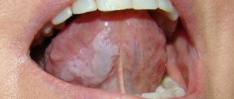
When the tongue is damaged, glossitis develops - the tongue swells, its papillae are smoothed out, a characteristic white coating appears on its back and lateral surfaces1 - and when the pharyngeal tonsils are damaged, a sore throat occurs.
A sore throat caused by fungal microflora is very different from normal. With obvious inflammation of the tonsils and the presence of white films and plugs on them, there is no temperature or pain when swallowing, and the submandibular lymph nodes remain of normal size1.
Candidiasis can spread further - affecting the respiratory tract, causing pneumonia and blood poisoning, so it is important to stop the process at the very beginning.
Up to contents
How does the disease manifest in children?
In children, the disease occurs in an acute form and is accompanied by the appearance of redness and swelling in the oral mucosa. The child sleeps poorly, may have no appetite, and becomes tearful.
The disease can occur in children for the following reasons:
- Weakening of the immune system.
- Infection during breastfeeding.
- Transmission of the fungus during childbirth.
- Infection through household items.
If the disease is not diagnosed and treated in a timely manner, a whitish coating resembling cottage cheese will soon appear in the child’s mouth, and in an advanced stage, ulcers will appear, which are accompanied by bleeding and cause severe pain in children.
Diagnostics
To make an accurate diagnosis, a combination of several methods is used - from a simple examination and questioning of the patient for complaints to laboratory methods, such as culture, microscopic examination of biomaterial, analysis of the degree of contamination of the oral cavity with fungal mycelium.
Oral candidiasis is accompanied by a number of characteristic external signs, in particular the formation of plaque, bad breath, ulceration and hyperemia of the mucous membranes. However, laboratory methods make it possible to accurately determine the type of pathogen and exclude the possibility of a secondary infection, which may affect the nature and duration of treatment.
Differential diagnosis is used to separate cases of candidiasis from aphthous stomatitis, leukoplakia, lichen ruber, streptococcal infection and other infectious pathologies of the oral cavity.
How to treat oral candidiasis?
Treatment is carried out using local and general, specific and symptomatic remedies. Among the main goals of therapy are the elimination of foci of infection in the oral cavity (sanitation), treatment of diseases that accompany candidiasis and are risk factors, and stimulation of the body's defenses. The total duration of treatment is usually at least 7-10 days.
As a means of local therapy, rinses are used - using solutions of boric acid, soda, sodium tetraborate. For a longer and more effective effect, such products can be used in the form of applications - moistening a cotton swab or bandage with the solution.
Nystatin for oral candidiasis is used to combat the main cause of the disease - a fungal infection. Treatment of candidiasis in the mouth may also include the use of other antimycotic (antifungal) drugs - for example, levorin ointment. The best effect is achieved by using several drugs, alternating them for several days.
Antifungal drugs are also prescribed for systemic therapy - in this case, medications for oral candidiasis and other infections such as Lamisil, Diflucan, Levarin, Nizoral, etc. are taken orally. In the most severe cases of the disease, the treatment regimen includes taking immunomodulatory drugs, as well as the use other agents that have a stimulating effect on the immune system and help strengthen the body’s own defenses.
An equally important task is to protect against additional fungal and bacterial infections that can join the Candida infection and complicate the course of the disease. For this purpose, rinses with antiseptic solutions - fucorcin, iodinol and others.
As an alternative, you can use ASEPTA antiseptic mouth rinse, which contains the active ingredients chlorhexidine and benzydamine. Both of these substances have broad antimicrobial effects. Regular use of ASEPTA rinses also has a pronounced anti-inflammatory effect and helps not only eliminate unpleasant symptoms, but also reduce the risk of complications.
What not to eat if you have thrush
Following your doctor’s recommendations for a healthy and nutritious diet and comprehensive treatment for oral candidiasis is the key to a speedy recovery. But there is a list of ingredients that must be excluded from your diet.
It is important to carefully ensure that they do not end up on the menu either raw or as part of any complex dishes.
So, you will have to stop using:
- Black tea. There is a possibility of the formation of invisible mold, which will have a detrimental effect on the patient’s health.
- Mushrooms of both forest and industrial origin.
- Buttermilk and sour cream. The only exception is fresh sour cream with a small percentage of fat.
- Mayonnaise, cheese.
- Dried fruits, since their surface may contain tiny mold.
- Drinks obtained on the basis of fermentation. This category includes strong alcohol, wine, beer, cider, kvass, mead, etc.
- Sprouted grains.
- Dishes containing monosodium glutamate. These are ketchups, ready-made wet condiments and soups, various dressings, all types of fast food.
- Vinegar. The only exception is apple.
- Medicines made from yeast. These are antibiotics, vitamin-mineral complexes, etc.
- Products containing refined white flour. This category of dishes includes sweet pastries, buns, cookies, pasta, muesli, etc.
- The ideal solution would be to replace animal fats with plant analogues.
- Sausages, fatty meats, various corned beef.
During the period of exacerbation of the disease, experts recommend excluding these dishes from the diet. Only after complete recovery and completion of the course of treatment should it be gradually introduced, observing the condition of the body. At the first alarming symptoms, it is necessary to contact a specialist in order to adjust the existing diet and continue treatment.
Carbohydrates are prohibited
A strict no-carbohydrate diet has worked well. It involves complete abstinence from foods such as:
- Sugar. It is important to exclude any form of it (honey, sand, condensed milk, refined sugar, syrups, etc.).
- All types of sweeteners (fructose, maltose, glucose, sorbitol).
- Any candy, as well as all types of chocolate.
- Sweet carbonated drinks, as well as packaged and bottled juices.
- Fruits and berries with a high sugar content (watermelon, raspberries, strawberries, bananas, grapes, melons, apples, etc.).
- Dairy products and milk. The only exceptions are fermented baked milk, live yogurt and fresh kefir.
- Vegetables with a high starch content. Potatoes and legumes can be consumed only a couple of times a week in the absence of alarming symptoms and persistent positive dynamics of the pathological process.
A no-carbohydrate diet requires some effort from a person. But after a few days the patient notices an improvement in his condition. By continuing treatment and adhering to the same meal plan, it is possible to obtain lasting positive dynamics.
Menu for oral candidiasis
Fungal diseases require a balanced diet. A proper diet includes a variety of foods. A strict diet does not mean eating just a few ingredients. If desired, you can develop a full menu for every taste. Let's consider possible food options for breakfast, lunch and dinner.
Breakfast for thrush
- Fresh cucumber salad, scrambled eggs, unsweetened green tea and whole grain bread;
- Sour cream and cottage cheese with a small percentage of fat, apple-carrot or carrot juice;
- Freshly squeezed orange juice, omelette;
- Tomato and herb salad, boiled eggs;
- Not sweet green tea and fruit salad in small quantities;
- Assorted nuts and oatmeal without sugar;
- Boiled fish or lean meat;
- Brown rice with boiled fish.
The main thing is to carefully monitor the absence of simple carbohydrates, and also avoid excessive consumption of dairy products.
Afternoon snack or snack
Meals for thrush should be fractional and as healthy as possible. Therefore, experts have developed several healthy snack options that can satisfy hunger and free a person from the desire to eat prohibited foods.
So, what can you snack on during the day:
- Green apple. It can be baked, steamed or fresh.
- A small handful of nuts.
- Live yogurt or kefir.
- Whole grain bread in small quantities, as well as homemade cheese.
- Orange or orange juice.
- Apple with low-fat cottage cheese.
Using the above products to satisfy hunger between main meals, the patient not only follows a diet, but also provides the body with necessary vitamins and minerals. This diet helps to activate the immune system and speed up the healing process.
Dinner
Among the allowed foods for oral thrush, experts include:
- Vegetable soups;
- Casseroles from lean meats;
- Boiled chicken breast and carrot salad;
- Tomato soup;
- Lean fish casserole;
- Pea soup;
- Buckwheat porridge or soup;
- Boiled or steamed vegetables;
- Lentil soup;
- Boiled meat;
- Steam cutlets;
- Beetroot;
- Chicken soup;
- Seafood.
Eating properly selected meals provides a complete diet, and split meals normalize metabolism and promote recovery.
Dinner dishes for oral candidiasis
Nutritionists suggest choosing several dishes for dinner as a tasty and healthy menu:
- Baked (or steamed) vegetables;
- Vegetable stew without seasoning;
- Eggplant cooked with rice;
- Potato roll with vegetables;
- Stuffed cabbage rolls;
- Zucchini or carrot fritters;
- Dumplings with cabbage or potatoes based on rye flour;
- Vegetable Salad.
To make your diet as healthy and effective as possible, you can follow a few simple rules:
- Purchase meat and fish products only from trusted places. Or on farms. Partially or completely replace pork with rabbit and lamb.
- Stop eating fried meat and switch to boiled or stewed foods.
- Introduce quail or turkey meat into your diet.
- Partially replace meat products with chicken or quail eggs.
- Reduce the amount of vegetable fats. Switch to steam or non-stick pan cooking. It is also better to reduce the consumption of animal fats.
- Increase the consumption of homemade fermented baked milk, yogurt, etc.
- Increase the amount of fiber in your diet.
Avoid eating fast food and processed foods. The manufacturer often includes components that are prohibited for you. This reduces to zero all the efforts spent on treating oral candidiasis. Strictly follow the recommendations of specialists, stick to your diet, and then the fight against thrush will be successful!
Disease prevention
Preventive measures are aimed at improving the condition of the microflora. These include:
- Proper oral hygiene.
- A thoughtful diet with a high amount of protein foods and reduced consumption of foods containing glucose.
- Quitting smoking and alcoholic beverages.
- Timely examination by the attending dentist for the prevention, diagnosis and treatment of the disease.
- Avoid taking medications, such as antibiotics, without first consulting your doctor.
- If the patient has dentures, then one of the preventive measures will be their regular treatment in a special solution.
Sources:
- The role of anti-inflammatory rinse in the treatment of periodontal diseases (L.Yu. Orekhova, A.A. Leontyev, S.B. Ulitovsky) L.Yu. OREKHOVA, Doctor of Medical Sciences, Prof., Head of Department; A.A. LEONTIEV, dentist; S.B. ULITOVSKY, Doctor of Medical Sciences, Prof. Department of Therapeutic Dentistry of St. Petersburg State Medical University named after. acad. I. P. Pavlova
- Report on clinical trials to determine/confirm the preventive properties of commercially produced personal oral hygiene products: mouth rinse "ASEPTA PARODONTAL" - Solution for irrigator." Doctor of Medical Sciences Professor, Honored Doctor of the Russian Federation, Head. Department of Preventive Dentistry S.B. Ulitovsky, doctor-researcher A.A. Leontiev First St. Petersburg State Medical University named after academician I.P. Pavlova, Department of Preventive Dentistry.
- Study of the clinical effectiveness of treatment and prophylactic agents of the Asepta line in the treatment of inflammatory periodontal diseases (A.I. Grudyanov, I.Yu. Aleksandrovskaya, V.Yu. Korzunina) A.I. GRUDYANOV, Doctor of Medical Sciences, Prof., Head of Department I.Yu. ALEXANDROVSKAYA, Ph.D. V.Yu. KORZUNINA, asp. Department of Periodontology, Central Research Institute of Dentistry and Maxillofacial Surgery, Rosmedtekhnologii, Moscow
Diet for fungus in the mouth
Antifungal Diet
- Efficacy: no data
- Terms: 3-6 months
- Cost of products: 1500-1600 rubles. in Week
In addition to complex antifungal treatment, patients are strongly recommended to follow a strict diet consisting of foods without vinegar, sugar, yeast and alcohol. in vitamins and strengthening the immune system is also recommended The menu should include bananas, onions, garlic, asparagus, chicory, olive oil, seafood, legumes, and buckwheat.