Candidiasis or thrush
is a fungal, infectious disease caused by opportunistic fungi of the genus Candida. Microorganisms are present in most healthy people. A person may encounter them for the first time in the womb, during childbirth, or during breastfeeding. Candidiasis develops against the background of reduced immunity, when the body can no longer restrain the active proliferation of the fungus.
The infection affects the mucous membranes of the mouth and genitals. It can develop on smooth skin, nails, and in the intestines, as a type of dysbacteriosis. There is visceral (systemic) candidiasis that affects internal organs. Despite the fact that Candida fungi are present in many people, and the disease itself is considered a neglected disease, it must be treated even if there are no external manifestations.
Causes of candidiasis
- Frequent damage to the skin and mucous membranes, for example, due to illness, working with aggressive chemicals, dry skin, age-related changes.
- Prolonged exposure to water, humid and warm environments. This includes a climate that is not suitable for your skin type, as well as occupational hazards.
- Mechanical damage to the skin and mucous membranes: injection drug addiction, long-term surgical interventions, catheters, shunts, endotracheal tubes.
After entering the body, the fungus may not manifest itself for a long time, since its reproduction will be suppressed by the immune system. In some cases, natural defenses weaken, leading to candidiasis (thrush). Among the factors that contribute to the proliferation of Candida:
- Chronic, endocrine and immune system diseases (diabetes, HIV).
- Metabolic disorders or taking medications that disrupt the natural microflora (antibiotics, hormonal contraceptives).
- Unbalanced diet, lack of sleep, stress and depression.
The fungus can be found in raw meat, unpasteurized dairy products, and, in more rare cases, on fresh vegetables and fruits. You can become infected with candidiasis from animals: dogs, calves, poultry.
Oral candidiasis
Manifestations of candidiasis of the oral mucosa are varied and depend on the patient’s age, the state of the immune system, the presence of concomitant diseases, medications (antibiotics, corticosteroids) and other factors.
According to the clinical course, acute and chronic forms are distinguished. Acute candidiasis can occur in the form of thrush (acute pseudomembranous candidiasis) or acute atrophic candidiasis. Chronic candidiasis also exists in two clinical forms: chronic hyperplastic and chronic atrophic. They can develop as independent forms or transform into one another.
Acute pseudomembranous candidiasis , or thrush (candidosis acuta, s. soor), is one of the most common forms of candidiasis of the oral mucosa. In infants, thrush occurs frequently and is relatively mild. In adults, acute pseudomembranous candidiasis is often accompanied by any general somatic diseases: diabetes mellitus, blood diseases, hypovitaminosis, malignant neoplasms, etc.
Most often the mucous membrane of the back of the tongue, cheeks, palate, and lips are affected. She is hyperemic and dry. Against the background of hyperemia, there is a white coating, reminiscent of curdled milk or cottage cheese, rising above the level of the mucous membrane. At the beginning of the disease, it is easily removed by scraping with a spatula, revealing a smooth, slightly swollen, hyperemic surface underneath. In severe, advanced cases, plaque becomes denser and is difficult to remove, revealing the erosive surface of the oral mucosa underneath.
Patients complain of a burning sensation in the mouth, pain when eating, especially spicy food.
Acute pseudomembranous glossitis should be differentiated from desquamative glossitis, in which areas of epithelial desquamation appear on the back of the tongue, constantly migrating along the back of the tongue and surrounded by a rim of exfoliating epithelium. Acute candidal stomatitis is differentiated from leukoplakia and lichen planus. With the latter, whitish films and nodules on the surface of the mucous membrane are formed due to hyperkeratosis, and therefore it is impossible to remove them by scraping. A differential diagnosis of candidiasis and soft leukoplakia, or white spongy nevus, is carried out, in which the lesion is localized mainly along the line of closure of the teeth and on the mucous membrane of the lips. The color of the mucous membrane in soft leukoplakia in the affected area is whitish-gray, its surface is rough, uneven, and there are multiple small superficial erosions (abrasions). The final diagnosis is made on the basis of bacterioscopic examination data.
Acute atrophic candidiasis (candidosis acuta atrophica) is characterized by significant pain, burning and dryness in the mouth. The mucous membrane is fiery red, dry. When the tongue is affected, its back becomes crimson-red, dry, shiny, and the filiform papillae are atrophied. The plaque is absent or remains in deep folds, is difficult to remove and is a conglomerate of deflated epithelium and a large number of fungi of the genus Candida in the stage of active budding (mycelium, pseudomycelium).
Acute atrophic candidiasis should be differentiated from an allergic reaction to the plastic of removable dentures. An important role in this case is played by clinical observation of the dynamics of changes in the oral mucosa after eliminating the prosthesis and conducting a bacterioscopic examination.
The general condition of patients with acute candidiasis does not suffer.
Chronic hyperplastic candidiasis (candidosis chronica hyper plastica) is characterized by the formation of a thick layer of plaque in the form of nodules or plaques on the hyperemic oral mucosa. The plaque is usually located on the back of the tongue, on the palate. The tongue is most often affected by the area typical of rhomboid glossitis.
Chronic hyperplastic candidiasis on the palate has the appearance of papillary hyperplasia. In cases of long-term, persistent disease, the plaque becomes saturated with fibrin, and yellowish-gray films are formed, tightly fused to the underlying mucous membrane. When scraped with a spatula, the plaque is removed with difficulty, revealing a hyperemic, bleeding, erosive surface underneath. Patients complain of dry mouth, burning, and, in the presence of erosions, pain. This form of candidiasis should be differentiated from leukoplakia and lichen planus.
Chronic atrophic candidiasis (candidosis chronica atrophica) is manifested by dry mouth, burning, pain when wearing a removable denture. The area of the mucous membrane corresponding to the boundaries of the prosthetic bed is hyperemic, swollen, and painful.
Chronic atrophic candidiasis in people who have been using removable lamellar dentures for a long time is most often characterized by damage to the oral mucosa under the dentures (hyperemia, erosion, papillomatosis) in combination with mycotic (yeast) infection and candidal atrophic glossitis, in which the back of the tongue is crimson-red, dry, shiny, filiform papillae atrophic. There is a small amount of whitish-gray coating only in deep folds and on the lateral surfaces of the tongue; it is difficult to remove. Under a microscope, spores and mycelium of the fungus of the genus Candida are found in the plaque. This triad (inflammation of the palate, tongue and corners of the mouth) is so characteristic of atrophic candidal stomatitis that diagnosing it is not difficult.
Risk factors for development
The likelihood of infection increases with casual sexual intercourse, uncontrolled use of antibiotics and drugs that disrupt the natural microflora. Foods with large amounts of sugar and carbohydrates create a favorable environment for fungal growth. Excessive sweating also leads to an exacerbation of candidiasis, so it is necessary to wear cotton underwear that allows the skin to breathe and moisture to evaporate.
At the same time, excessive cleanliness can also cause harm. We are talking about douching. It should not be used as a method of contraception, since it is not effective, and also as a means of hygiene, because it leads to the leaching of the protective flora. If you experience discomfort, you should first consult a doctor.
Fungi of the genus Candida that cause the development of urogenital candidiasis (UGC) include Candida albicans, the dominant causative agent of the disease (detected in 90-95% of patients with UGC), as well as representatives of Candida non-albicans species (more often - C. glabrata, C. tropicalis, C. krusei, C. parapsilosis, less often - C. lipolytica, C. rugosa, C. norvegensis, C. famata, C. zeylanoides), detected, as a rule, with recurrent UGC occurring against the background of diabetes mellitus, HIV infection, postmenopause. Candida spp. - opportunistic microorganisms that are facultative anaerobes and have a tropism for tissues rich in glycogen (for example, the vaginal mucosa).
UHK is a widespread disease, more often observed in women of reproductive age. The frequency of registration of vulvovaginal candidiasis is 30-45% in the structure of infectious lesions of the vulva and vagina. According to researchers, 70-75% of women have at least one episode of vulvovaginal candidiasis during their lives, while in 5-10% of them the disease becomes recurrent. By the age of 25, about 50% of women, and by the beginning of menopause, about 75% of women have at least one episode of the disease diagnosed by a doctor. Vulvovaginal candidiasis is rarely observed in postmenopausal women, with the exception of women receiving hormone replacement therapy.
UGK is not a sexually transmitted infection, but this does not exclude the possibility of candidiasis balanoposthitis in men who are sexual partners of women with UGK.
Endogenous risk factors for the development of UGC include endocrine diseases (diabetes mellitus, obesity, thyroid pathology, etc.), underlying gynecological diseases, disorders of local immunity; exogenous risk factors - taking antibacterial, glucocorticosteroid, cytostatic drugs, immunosuppressants, radiation therapy; wearing tight clothing, underwear made of synthetic fabrics, regular use of sanitary pads, prolonged use of intrauterine devices, vaginal diaphragms, douching, use of spermicides.
The question of the reasons for the formation of recurrent UGC has not been completely resolved, since recurrent forms of the disease also occur in women who do not have the above risk factors. The leading role in the development of recurrent forms of UGC is given to local immune disorders caused by the innate qualities of vaginal epithelial cells.
Classification of the disease
Based on the depth of damage, candidiasis is divided into superficial and systemic forms. The first appear on visible parts of the body: mucous membranes, skin and its appendages. The second, systemic, are combined forms of the disease affecting internal organs. Without proper treatment, systemic (visceral) candidiasis can lead to the development of candidal sepsis. Most often, the fungus affects:
- gastrointestinal tract (esophagus, stomach, intestines);
- urinary organs (bladder, urinary tract, kidneys);
- lower respiratory tract (trachea, bronchi, lungs).
The disease is also classified based on the rate of spread of infection and the presence of relapses. Candidiasis of the liver and spleen often takes a chronic form. The acute, generalized form is asymptomatic candidemia, candida septicemia, candida thrombophlebitis, myositis, arthritis, cerebral candidiasis and other forms.
Classification of candida lesions according to ICD-10
- Candidiasis.
- Candidal stomatitis.
- Pulmonary candidiasis.
- Candidiasis of the skin and nails.
- Candidiasis of the vulva and vagina.
- Candidiasis of other urogenital locations.
- Candidal meningitis.
- Candidal endocarditis.
- Candidal septicemia.
- Candidiasis of other locations.
- Candidiasis, unspecified.
Officially, the diagnosis is made in accordance with ICD-10. In practical healthcare, such a classification is not entirely convenient, so they use the classification of deep candidiasis, which is a deeper list. When making a diagnosis, first indicate the name and form of the infection with a description of all localization points.
Skin treatment is carried out by a pediatrician in children and by a dermatologist in adults. Therapy is also carried out by doctors of other specializations, depending on the affected organ. For example, this could be a dentist, neurologist, gynecologist, pulmonologist.
Candida
Candida
(
Candida
) is a genus of yeast-like fungi.
Yeast-like fungi of the genus Candida
are single-celled microorganisms 6–10 microns in size.
Fungi of the genus Candida
are dimorphic: under different conditions they form blastospores (bud cells) and pseudomycelia (threads of elongated cells).
Fungi of the genus Candida
are widespread in the environment.
They are found in soil, drinking water, food products, on the skin and mucous membranes of humans and animals. Favorable conditions for the growth of Candida
are considered to be a temperature of 21–37 °C and an acidity of the environment of 5.8–6.5 pH.
Prevalence of Candida fungi in healthy people
The frequency of carriage of fungi of the genus Candida
in healthy individuals reaches 25% in the oral cavity, and up to 65–80% in the intestines (Shevyakov M.A.).
According to OST 91500.11.0004-2003 “Protocol for the management of patients. Intestinal dysbiosis" in the composition of the main microflora of the colon in a healthy person, calculated per 1 g of feces, should contain no more than 104 colony-forming units (CFU) of fungi of the genus Candida
(and for children under one year old - less than 103). They are found in the vagina and genital tract in 10–17% of healthy women (as well as in 26–33% of pregnant women), on the skin of healthy people, without manifesting themselves in any way. In gastric juice, the ratio of different types of Candida fungi is approximately the following (Lazebnik L.B. et al.):
- Candida albicans
- 41% of all Candida fungi - Candida tropicalis
— 30 % - Candida glabrata
— 9 %
Diseases caused by fungi of the genus Candida (Candida)
Fungi of the genus Candida
are the most common opportunistic fungi found in humans.
Among other human candidiasis caused by fungi of the genus Candida
, in 86% of cases the infectious agent is fungi of the species
Candida albicans
, in 9% of cases -
Candida tropicalis.
Also pathogenic for humans are the following species:
Candida krusei, Candida glabrata, Candida parapsilosis, Candida guillermondii, Candida tropicalis
.
Thrush (vulvovaginal candidiasis)
manifested in the form of white cheesy discharge from the female genital organs, their itching, burning, redness, swelling.
Candidal balanitis
(inflammation of the glans penis) and
urethritis
.
Candidiasis of the gastrointestinal tract
Colonization
of the gastrointestinal tract (GIT)
Candida In healthy Europeans, Candida
are present:
- in the oral cavity - in 10–25%
- in the oropharyngeal zone - in 20–30%
- in the small intestine - in 50–54%
- in the colon - in 55–70%
- in feces - in 65–70%
- in cystic and ductal bile - in 0.8–4%
Normal biochemical, histochemical and physiological processes in the gastrointestinal tract, timely regeneration of epithelial cells, acid-enzyme barrier, and full peristalsis are also protective factors that prevent the penetration of pathogenic microorganisms. The important role of stomach acid in preventing the introduction of fungi into the mucosa has been established. In an environment with low acidity, Candida
acquire pathogenic properties, vegetative forms appear, and pseudomycelium or mycelium is formed, damaging the mucous membrane.
In HIV-infected individuals, who are characterized by achlorhydria, ingested Candida
can cause gastric candidiasis, while in people with normal immunity this localization is rare. The role of gastric acidity in the development of bacterial and fungal infections of the intestine has not been confirmed (Burova S.A.).
Candidal esophagitis
Infectious esophagitis is most often associated with fungi of the genus Candida
, among which the most common pathogen is
Candida albicans
. Infectious esophagitis is characterized by an acute onset with the appearance of symptoms such as dysphagia and odynophagia. Heartburn, chest discomfort, nausea and vomiting are possible. Sometimes abdominal pain, anorexia, weight loss and even cough are observed.
Local oropharyngeal candidiasis is a common infection, recorded mainly in children and elderly patients, people with dentures, patients receiving antibiotics, chemotherapy treatment and/or radiation therapy to the head and neck area, and in patients with AIDS. There is also an increased risk of developing candidiasis in patients using inhaled steroids. Symptoms of oropharyngeal candidiasis include loss of taste, pain when chewing and swallowing, and when trying to put on dentures. Some patients do not have any symptoms. The diagnosis is most often established during an examination of the oral cavity, during which white plaques, sometimes a cheesy plaque, are discovered, and under removable dentures there are zones of hyperemia without plaques.
Esophageal candidiasis is most often diagnosed in patients with hematological malignancies, AIDS, after organ transplantation and in those receiving steroid therapy. In this case, candidiasis of the oral cavity may also be observed, but its absence does not exclude independent candidiasis of the esophagus. Candidal esophagitis occurs in general patients in 1-2% of cases, in patients with type 1 diabetes mellitus - in 5-10%, in patients with AIDS - in 15-30%. The most characteristic symptom of esophageal candidiasis is odynophagia, i.e. pain along the esophagus when swallowing food. The diagnosis of candidal esophagitis is established, as a rule, by endoscopy, when white and whitish-yellow plaques and plaque-like plaques are detected on the mucous membrane of the esophagus.
When writing the first three paragraphs of this section, materials from the Clinical Guidelines of the Russian State Administration for the diagnosis and treatment of infectious esophagitis were used / Ivashkin V.T. and others. RZHGK, No. 6, 2015.
Fungal esophagitis is most often caused by colonization with
Candida albicans
, less commonly
with Candida glabrata, Candida tropicalis, Candida parapsilosis and Candida krusei.
Clinically manifested by pain, a burning sensation. It develops against the background of systemic chemotherapy, with immunodeficiency of various origins, with the use of corticosteroids, especially in inhaled forms, diabetes mellitus, and during chemoradiotherapy. Whitish-yellow focal deposits are difficult to remove; often, after removing the deposits, the mucous membrane of the esophagus bleeds (Pirogov S.S.).
Associations of microbial pathogens of the oropharynx of patients with GERD (Khrustaleva E.V. et al.).
Medicines active against Candida fungi
The first-line drug for the treatment of oropharyngeal candidiasis and esophageal candidiasis is fluconazole, prescribed 100–200 mg per day orally or intravenously for 2–4 weeks. Only in cases of intolerance to fluconazole or resistance of the pathogen (usually Candida krusei, Candida glabrata, Candida pseudotropicalis
) second-line drugs are indicated (also within 2-4 weeks). Second-line drugs for esophageal candidiasis are (Shevyakov M.A.):
- itraconazole in oral solution 200–400 mg per day
- ketoconazole 200–400 mg per day
- amphotericin B 0.3–0.7 mg per day per kg of patient weight
- caspofungin intravenously 70 mg per day on the first day, then 50 mg per day intravenously in one administration
- voriconazole IV 6 mg per day per kg of patient weight every 12 hours on the first day, then 4 mg per day per kg of patient weight every 12 hours
- posaconazole 400 mg (10 ml suspension) 2 times a day orally with meals.
Of the antimicrobial drugs (antibiotics) listed in this reference book, the following are active against Candida fungi: fluconazole, nifuratel (MacMirror), clotrimazole (exception: Candida guillermondii
is resistant to clotrimazole).
Antifungal activity against fluconazole-resistant strains of Candida albicans, Candida glabrata, Candida krusei, Candida parapsilosis
and
Candida tropicalis
has lactoferrin (in combination with fluconazole).
Enterol (Saccharomyces Boulardii) inhibits the growth of Candida albicans, Candida kruesei, Candida pseudotropicalis
.
Professional medical work addressing the role of Candida in diseases of the gastrointestinal tract
- Shevyakov M.A. Candidiasis of the esophagus: diagnosis and modern choice of treatment // Journal “Treating Doctor”. – 2008. – No. 9.
- Pankova L.Yu., Osipenko M.F., Vergazov V.M. Risk factors for the attachment of opportunistic fungi to defects in the gastric mucosa in peptic ulcers // RZHGGK. – No. 1. – 32–37. – 2007.
- Lazebnik L.B., Khomeriki S.G., Morozov I.A., Kasyanenko V.I., Zvenigorodskaya L.A., Khomeriki N.M., Goncharenko L.S. Yeast-like fungi in gastric mucus in acid-dependent diseases // Experimental and clinical. gastroenter. 2005. No. 4. P. 27-32.
- Khrustaleva E.V., Pedder V.V., Shishkina N.M., Lubyanskaya T.G. Relationship between the pH level of the mucous membrane of the oropharynx and the presence of fungal flora in patients with GERD // Medical Sciences. — 2013 — No. 6.
On the website GastroScan.ru in the Literature section there is a subsection “Parasitic and infectious diseases of the gastrointestinal tract”, containing articles that touch upon, among other things, the treatment of candidiasis
.
Candida in the taxonomy of biological species
The genus of fungi Candida ( Candid
a) belongs to the family Saccharomycetaceae
,
which is included in the order Saccharomycetales
,
class Saccharomycetes
,
subphylum Saccharomycotina
,
phylum
Ascomycota
, kingdom
Fungi
.
Genus Candida ( Candid
a) includes a large number of species:
C. albicans, C. ascalaphidarum, C. amphixiae, C. antarctica, C. argentea, C. atlantica, C. atmosphaerica, C. blattae, C. carpophila, C. carvajalis, C. cerambycidarum, C. chauliodes, C. corydali, C. dosseyi, C. dubliniensis, C. ergatensis, C. fermentati, C. fructus, C. glabrata, C. guilliermondii, C. haemulonii, C. insectamens, C. insectorum, C. intermedia, C. jeffresii, C. kefyr, C. krusei, C. lusitaniae, C. lyxosophila, C. maltosa, C. marina, C. membranifaciens, C. milleri, C. oleophila, C. oregonensis, C. parapsilosis, C. quercitrusa, C. rugosa, C. sake, C. shehatea, C. temnochilae, C. tenuis, C. theae, C. tropicalis, C. tsuchiyae, C. sinolaborantium, C. sojae, C. subhashii, C. viswanathii, C. utilis
.
Appendix 1. Candidiasis in ICD-10
In the International Classification of Diseases ICD-10 in “Class I. Some infectious and parasitic diseases (A00-B99)”, in block “B 3
5-B49 Mycoses” there is a heading:
B37 Candidiasis
Included:
candidiasis, moniliasis
Excluded:
neonatal candidiasis (P37.5)
B37.0 Candidal stomatitis
Thrush
B37.1 Pulmonary candidiasis
B37.2 Candidiasis of the skin and nails
Candida:
- onychia
- paronychia
Excludes
: diaper dermatitis (L22)
B37.3† Candidiasis of the vulva and vagina (N77.1*)
Candidal vulvovaginitis Monilial vulvovaginitis Vaginal thrush
B37.4† Candidiasis of other urogenital sites
Candida:
- balanitis † (N51.2*)
- urethritis † (N37.0*)
B37.5† Candidal meningitis (G02.1*)
B37.6† Candidal endocarditis (I39.8*)
B37.7 Candidal septicemia
B37.8 Candidiasis of other sites
Candida:
- cheilitis
- enteritis
B37.9 Candidiasis, unspecified
Thrush NOS
Notes. 1. An asterisk * marks optional additional codes related to the manifestation of a disease in a separate organ or area of the body, which represents an independent clinical problem. 2. The main codes of the underlying disease that must be used are marked with a cross †.
Appendix 2. Gastroduodenal candidiasis in the Kyoto global consensus
The Kyoto global consensus recommends that the following candidiasis of the gastroduodenal zone be included in the new ICD-11 categories as separate clarifying lines:
- to the new section “Fungal gastritis”: gastric candidiasis
- to the new section “Fungal duodenitis”: duodenal candidiasis
Appendix 3. Medical services aimed at identifying fungi of the genus Candida
Order of the Ministry of Health and Social Development of Russia No. 1664n dated December 27, 2011 approved the nomenclature of medical services. Section 26 of the Nomenclature provides for a large number of different medical services related to the study of Candida fungi in humans:
| Service code | Name of medical service |
| A26.01.008 | Microscopic examination of skin scrapings for fungi of the genus Candida (Candida spp.) |
| A26.01.010 | Mycological examination of skin scrapings for fungi of the genus Candida (Candida spp.) |
| A26.01.013 | Mycological examination of skin punctate (biopsy) for fungi of the genus Candida (Candida spp.) |
| A26.01.014 | Mycological examination of bedsore puncture for fungi of the genus Candida (Candida spp.) |
| A26.02.004 | Mycological examination of wound discharge for fungi of the genus Candida (Candida spp.) |
| A26.04.007 | Mycological examination of synovial fluid for fungi of the genus Candida (Candida spp.) |
| A26.05.006 | Microbiological blood test for fungi of the genus Candida (Candida spp.) |
| A26.06.014 | Determination of antibodies to fungi of the genus Candida (Candida spp.) in the blood |
| A26.07.006 | Mycological examination of oral scrapings for fungi of the genus Candida (Candida spp.) |
| A26.08.009 | Mycological study of nasopharyngeal swabs for fungi of the genus Candida (Candida spp.) |
| A26.09.022 | Microscopic examination of sputum smears for fungi of the genus Candida (Candida spp.) |
| A26.09.027 | Microscopic examination of lavage fluid for fungi of the genus Candida (Candida spp.) |
| A26.10.005 | Mycological examination of biopsy specimen for fungi of the genus Candida (Candida spp.) |
| A26.14.006 | Microscopic examination of bile for fungi of the genus Candida (Candida spp.) |
| A26.19.009 | Mycological examination of stool for fungi of the genus Candida (Candida spp.) |
| A26.20.015 | Microscopic examination of vaginal discharge for fungi of the genus Candida (Candida spp.) |
| A26.20.016 | Mycological examination of vaginal discharge for fungi of the genus Candida (Candida spp.) |
| A26.21.011 | Microscopic examination of urethral discharge for fungi of the genus Candida (Candida spp.) |
| A26.21.014 | Mycological examination of urethral discharge for fungi of the genus Candida (Candida spp.) |
| A26.23.013 | Mycological examination of cerebrospinal fluid for fungi of the genus Candida (Candida spp.) |
| A26.25.003 | Microscopic examination of ear discharge for fungi of the genus Candida (Candida spp.) |
| A26.25.004 | Mycological examination of ear discharge for fungi of the genus Candida (Candida spp.) |
| A26.26.017 | Molecular biological study of eye discharge for fungi of the genus Candida (Candida spp.) |
| A26.28.004 | Microscopic examination of urine sediment for fungi of the genus Candida (Candida spp.) |
| A26.28.007 | Mycological examination of urine sediment for fungi of the genus Candida (Candida spp.) |
| A26.30.003 | Mycological examination of peritoneal fluid for fungi of the genus Candida (Candida spp.) |
Back to section
Symptoms of candidiasis
The symptoms of the disease are directly related to the affected organ. Superficial forms have obvious signs, while systemic forms can be asymptomatic or be similar to other diseases. In the presence of chronic diseases (diabetes mellitus, HIV, AIDS, leukemia and other types of oncology), candidiasis may not be detected at all. Since its symptoms will be perceived by the patient as an unhealthy state due to the existing disease. To make matters worse, Candida susceptibility testing is not considered a routine procedure in many teaching hospitals and laboratories.
Candidiasis of the skin and nails
The infection is expressed in the form of allergic rashes (candidamycids), erythematous-squamous spots, urticarial and bullous type rashes. Nail pathologies are divided into:
- parochinia - purulent inflammation of the periungual and subungual spaces, absence of eponychium (skin at the base of the nail plate);
- onychia - purulent inflammation of the nail fold, deformation of the nail with discoloration and brittleness.
Candidal skin lesions may be accompanied by fever, inflammation and swelling of the lesions.
Candidiasis (thrush) of the oral mucosa
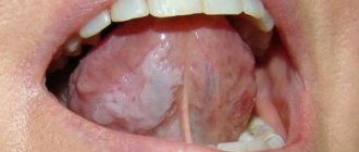
The fungus can affect both individual areas of the oral cavity and completely all mucous membranes: lips, gums, cheeks, palate, tongue, tonsils, uvula and pharynx. This is expressed in the presence of a white coating, compared to a curd mass. Swelling and redness may appear. There is often pain when swallowing, talking, or palpating. Saliva becomes viscous and bad breath appears.
Thrush may be accompanied by candidal cheilitis - damage to the lips and corners of the mouth. Painful cracks appear, covered with a white coating, and pieces of skin peel off from the lips. The disease is characterized by a long course with the possible addition of bacterial infections.
Intestinal candidiasis
Intestinal infection with fungi of the genus Candida can be an independent disease or develop as a result of damage to the oral cavity. Expressed as severe dysbacteriosis with the following symptoms:
- diarrhea;
- excessive gas formation;
- nagging pain in the rectum;
- an admixture of white flakes in the stool.
In most cases, symptoms are sluggish or completely absent. The disease is dangerous because the body does not receive enough vitamins and microelements, which is especially dangerous for a growing child.
Candidiasis of the genitourinary system (urogenital candidiasis)
Unlike sexually transmitted diseases, candidiasis of the genitourinary system is often hidden and asymptomatic. The fungus is discovered during a planned infection or against the background of other diseases. Symptoms begin to appear against the background of declining immunity, ongoing pathological processes of microflora destabilization and re-infection. In women, candidiasis manifests itself as follows:
- itching and burning of the external genitalia;
- swelling and redness;
- white, cheesy discharge;
- pain during sexual intercourse.
In men, the symptoms are similar:
- itching and burning of the head of the penis;
- white plaque and discharge:
- pain during sexual intercourse and urination.
Fungal infections of the bladder, excretory tract and kidneys are characterized by frequent urination, including false urges, pain in the suprapubic region. Candidiasis can be accompanied by bacterial infections. Complications include: cystitis, pyelonephritis, necrosis of the papillae, abscesses, formation of mycelium in the renal pelvis. Without proper treatment, against the background of chronic diseases, there is a risk of developing kidney failure.
Candidiasis
Cervical cancer
Diabetes
HIV
Thrush
19500 July 27
IMPORTANT!
The information in this section cannot be used for self-diagnosis and self-treatment.
In case of pain or other exacerbation of the disease, diagnostic tests should be prescribed only by the attending physician. To make a diagnosis and properly prescribe treatment, you should contact your doctor. Candidiasis: causes, symptoms, diagnosis and treatment methods.
Candidiasis is an infectious disease caused by yeast-like fungi of the genus Candida. It is caused by the active proliferation of fungus on the mucous membranes of the oral cavity, genital and internal organs and on the skin.
All representatives of the genus Candida belong to opportunistic microorganisms, that is, they are constantly present in the normal microflora. But with a decrease in immunity, changes in hormonal levels and for a number of other reasons, these fungi can begin to actively colonize the mucous membranes and skin.
The most common members of the genus are Candida albicans and C. tropicalis. In 90-95% of cases of urogenital candidiasis, C. albicans is the dominant pathogen.
The first contact with fungi of the genus Candida occurs during the passage of the child through the birth canal. However, the medical literature describes cases of detection of these microorganisms in amniotic fluid, which indicates the possibility of a vertical (transplacental) transmission route. Transmission of the fungus of the genus Candida also occurs through breastfeeding, skin contact between the child and the mother, and through household and food routes.
These microorganisms produce endotoxins and enzymes that cause cell death and tissue necrosis, which enhances the adhesive (attachment to cells of the mucous membranes or skin) ability of the fungus and ensures penetration into tissue.
Overproduction of these and a number of other substances determines the pathogenicity of representatives of the Candida family.
Causes of candidiasis
- Exogenous (external) factors facilitating the penetration of fungi into the body:
- occupational hazards leading to frequent skin damage;
- prolonged exposure to a warm and humid environment;
- violation of the integrity of the mucous membranes.
- Factors leading to a decrease in the body's resistance:
- presence of chronic diseases;
- long-term use of drugs that disrupt the natural microflora;
- unbalanced diet;
- frequent stress, disturbances in sleep and rest patterns.
Risk factors for developing candidiasis
- Metabolic disorders (hypovitaminosis), diseases of the immune system (HIV infection), endocrine pathologies (diabetes mellitus, etc.).
- Long-term use of certain drugs: hormonal contraceptives, systemic glucocorticosteroids, broad-spectrum antibiotics, cytostatics.
- Long stay or living in an area with high humidity and temperature, comfortable for the circulation of fungal spores in the environment.
Classification of the disease
Based on the localization of the process, the following are distinguished:
- Urogenital candidiasis.
- Candidiasis of the oral mucosa.
- Superficial candidiasis.
- Interdigital candidiasis.
- Candidiasis of periungual ridges and nails.
- Candidiasis of the gastrointestinal tract.
Symptoms of candidiasis
Urogenital candidiasis (UGC)
– a widespread disease: according to medical statistics, about 75% of women of reproductive age have registered symptoms of UGC at least once.
There are acute and chronic forms of urogenital candidiasis, candidiasis of the vulva, vagina and other urogenital localizations.
In some cases, when diagnosing, a clarification is used: complicated or uncomplicated UGC, which reflects the number of exacerbations per year and the severity of the disease. Symptoms of female urogenital candidiasis
- The appearance of white-yellow cheesy or creamy discharge from the genital tract. The intensity of discharge may increase before menstruation, which is associated with changes in hormonal levels.
- Unpleasant sensations, itching in the genital area, often aggravated by sexual intercourse or urination.
- Redness and swelling of the mucous membrane of the vulva and vagina, the presence of damage to the skin of the genital organs (cracks, microtraumas).
- In the chronic course of UGC, dryness of the mucous membranes of the genital tract develops.
Symptoms of male urogenital candidiasis
- Redness, swelling, discomfort in the genital area.
- Whitish discharge of a cheesy structure from the genital tract.
- Pain and burning during sexual intercourse and urination.
Superficial candidiasis
can be erythematous (the main symptom is reddened areas of the skin with a weeping surface) and vesicular (the formation of papules, vesicles and pustules in the affected area - inflammatory elements located in the superficial layers of the skin). The lesion begins with large folds of skin, gradually spreading to other areas of the body. In the depths of the folds, weeping occurs (separation of serous exudate through the smallest defects of the epidermis), a violation of the integrity of the skin contributes to the addition of a secondary infection.
Interdigital candidiasis
localized in the space between the fingers. In this case, redness of the skin is noted, followed by the appearance of bubbles in transparent contents. The disease spreads quickly in close groups (in kindergartens, schools, etc.).
Oral mucosal candidiasis (OCOR)
Oral candidiasis causes discomfort, especially when eating - burning, pain, dryness. Depending on the location of the process, several forms of oral candidiasis are distinguished.
Often, CSOPR and the gastrointestinal tract accompanies immunodeficiency conditions: HIV infection, acquired human immunodeficiency syndrome (AIDS) or congenital immunodeficiency (for example, with T-lymphocyte pathology). In the presence of these diseases, candidiasis occurs with the most severe symptoms, is difficult to treat, and is aggressive in nature.
The most common manifestation of CSOPR is candidal stomatitis, which mainly affects infants and adults with weakened immune systems.
With this pathology, the oral mucosa turns red, swells, and whitish films with a cheesy consistency appear on it. In the initial stages of the disease, plaque is easily removed. As the disease progresses, the films become denser, are difficult to separate, and when removed, the bleeding mucous membrane is exposed.
With candidal stomatitis, the tongue may be affected, which is manifested by redness of the back of the tongue, the appearance of plaque and desquamation of the epithelium. These symptoms are accompanied by severe pain in the affected area when talking, eating, and palpating the tongue.
Smokers, more often than other types of CSOPR, develop chronic hyperplastic candidiasis, accompanied by the formation of white, merging plaques that rise above the surface of the hyperemic mucosa.
With this pathology, the consistency of saliva changes: it becomes viscous and foaming; there is an unpleasant odor from the mouth, a gray or white coating on the mucous membrane. In 10-40% of cases, this clinical form of candidiasis becomes malignant (i.e., becomes malignant).
Older people most often develop a chronic atrophic form of oral candidiasis. The mucous membrane turns red and swells. The lesion is often localized under dentures, which causes pain.
Candidal cheilitis and candidiasis of the corners of the mouth mainly occur in children and the elderly. The lesion is usually bilateral, with the formation of red, painful cracks in the corners of the mouth, covered with an easily removable white coating or scales. With a long course of the disease, a bacterial infection may occur.
Diagnosis of candidiasis
The diagnostic search algorithm for candidiasis of any localization includes taking material from the affected area, followed by microscopy and culture to determine the type of fungus and its sensitivity to antimycotic (antifungal) drugs.
In order to diagnose conditions that lead to a decrease in immunity, a general blood test is used;
Diagnosis of candidiasis
The study is based on the isolation and identification of a species of fungus of the genus Candida. Currently, there are about 150 species, differing in morphological and biochemical properties. The most common fungus is C. albicans, accounting for up to 80% of cases of candidiasis of the digestive tract and up to 70% of genital infections. Before prescribing treatment, it is also necessary to determine the sensitivity of the isolated strain to antimycotics (antifungal medications): amphotericin B, voriconazole, itraconazole, fluconazole, flucytosine.
Microscopic diagnostics
1. Microscopy of a smear is taken from the area of the affected mucosa. Allows for comparative characterization of blastospores and pseudomycelia. During the study, fixed and native preparations that stain microorganisms are used. To increase the information content, pseudomycelia of cells are treated with dyes. The contrast in staining of microorganisms makes it easy to distinguish candida from other forms, including bacteria, under a microscope.
2. Bacterial culture allows you to identify the causative agent of infection and determine its concentration. The analysis is used to monitor the effectiveness of treatment, as well as to identify the sensitivity of candida fungi to various antimycotics.
Diagnostics by ELISA and PCP
1. Enzyme-linked immunosorbent assay (ELISA) is based on the determination of antibodies that are produced by the immune system in response to foreign substances in the blood. This technique allows you to identify the pathogen and the degree of its development, establishing whether the disease occurs in an acute or chronic stage.
2. Polymerase chain reaction (PCR) is a highly sensitive test that allows you to directly detect the infectious agent. Thanks to it, it is possible to differentiate Candida fungi with pseudomycelium from those that do not have it. These data are important for subsequent interpretation of results and deeper diagnosis.
CANDIDIASIS OF THE ESOPHAGUS
Candidiasis is an infectious disease of the mucous membranes, skin and internal organs caused by yeast-like fungi of the genus Candida. Esophageal candidiasis (OC), which is a manifestation of visceral candidiasis, occupies a prominent place among infectious lesions of the esophagus. In recent years, there has been a tendency to increase the frequency of CP, especially in patients with impaired immunity. The growth of candidiasis infection is largely due to an increase in the number of patients with HIV infection, advances in transplantation and immunosuppressive therapy, and the uncontrolled use of antibiotics. KP occurs in 0.7-1.5% of gastroenterological patients [5, 6].
The problem with severe fungal infections caused by opportunistic pathogens is that they are difficult to treat and can be fatal. The mortality rate for invasive candidal infections has been found to be 34% [16].
Etiopathogenesis. Candida species are the most common esophageal pathogen, most notably Candida albicans, with occasional occurrences of C. tropicalis, C. parapsilosis, C. glabrata, C. lusitania, and C. krusei. These microorganisms are normal components of the oral flora and their growth is inhibited by bacterial commensals. Infection with fungi such as Candida, which are widespread in the environment, occurs through endogenous or exogenous routes. Endogenous infection is associated with the activation of saprophytic fungi; exogenous infection can occur through direct contact with carriers of infection or from the environment. If the host's body is not weakened, many fungi do not exhibit their pathogenic properties. Research in recent years has shown that the source of fungal dissemination is the intestines, and candidiasis of the oral cavity, genitals, and esophagus is a manifestation of systemic candidiasis. The likelihood of developing systemic damage depends both on the properties of the microorganism itself (their number, virulence, genetic and species heterogeneity of the population), and on the state of the macroorganism, especially its immune system, nutritional status and abdominal blood flow [3, 17].
Favorable conditions for the development of the infectious process are created by various violations of the physiological, anatomical and immunological mechanisms of the body's defense. Factors that provoke the occurrence of esophageal candidiasis include the use of antibiotics, inhaled or injected corticosteroids, antacid therapy or a hypochlorhydric state, diabetes mellitus, alcoholism, the consequences of intoxication, malnutrition, old age, impaired motility of the esophagus or esophageal obstruction, organ and bone marrow transplantation , enteral and especially parenteral nutrition, etc. A weakened immune system can lead to candidiasis infection. In diabetes mellitus, elevated blood glucose levels promote fungal growth because hyperglycemia impairs granulocyte function. Hypofunction of the parathyroid glands and adrenal glands leads to disruption of calcium-phosphorus metabolism, which causes hidden spasmophilia of the esophagus, thereby reducing its local protective capabilities [9]. Impaired nutritional status due to a lack of protein in the body and low calorie food affects the state of the immune system and creates the preconditions for the development of candidiasis [3]. Risk factors for candidiasis include a decrease in the acidity of gastric juice (pH 7.4 is optimal for the growth of Candida fungi, and when the pH shifts to 4.5, fungal growth is completely inhibited) [3, 4, 7].
The pathological manifestations of KP are varied. At first, the affected areas of the esophagus have the appearance of individual whitish or yellowish lesions raised above the mucous membrane. Later, these lesions can merge, forming dense plaques with the introduction of the fungus into the submucosa or pseudomembranous deposits with the penetration of the fungus into the muscular layer and blood vessels [9]. Films that form on the esophageal mucosa in especially severe cases can almost completely close the lumen of the esophagus. Plaque consists of desquamated epithelial cells that mix with fungi, inflammatory cells and bacteria. Microscopic examination reveals uniformly colored yeast-like cells and filaments of mycelium of Candida fungi [9]. True ulceration is observed infrequently and in most cases is observed in immunosuppressed patients with granulocytopenia [29]. Sometimes necrosis of the esophageal wall occurs and phlegmonous inflammation of the esophagus and mediastinum develops, which can become one of the causes of death of the patient [1].
There is a morphological classification, according to which all cases of KP are divided into three groups depending on the severity of the process, that is, depending on the depth of damage to its wall: 1st group - individual whitish plaques with the introduction of pseudomycelium of the fungus between the epithelial cells; 2nd group - membranous plaques merging with each other and forming vast fields, while filaments of pseudomycelium grow not only the mucosa, but also the submucosa; Group 3 - pseudomembranous overlays, combined with deep changes, in which the threads of the fungus penetrate deeply into the thickness of the muscle tissue [10].
Clinical manifestations and complications. Symptoms of the disease are practically absent in 25-30% of patients suffering from KP, especially in immunocompetent individuals. However, most patients present with complaints related to damage to the gastrointestinal tract. The most typical clinical manifestations of KP are dysphagia and, somewhat less commonly, odynophagia. The severity of esophageal symptoms ranges from moderate difficulty swallowing to severe pain, resulting in the inability to eat and the development of secondary dehydration. In severe odynophagia, there may be other causes or co-infection, especially in patients with AIDS. Much less frequently, patients may complain of chest pain not associated with swallowing, heartburn, nausea, sometimes vomiting with the release of films (pseudomembranes), decreased appetite and weight, and the appearance of loose stools with mucus (see figure) [4, 9, 29].
| Symptoms of candidal esophagitis (RS Orlando, 1996) |
Physical examination may be helpful in KP. Approximately two thirds of patients with
AIDS and esophageal candidiasis have candidal stomatitis. KP is observed in patients with chronic mucocutaneous candidiasis, which is a severe form of candidal infection and is more often observed with dysfunction of the adrenal glands and parathyroid glands [29].
Complications of esophageal candidiasis are rare. Esophageal bleeding can be observed in severe cases of the disease, accompanied by the formation of erosions, ulcers, and be associated with coagulopathy; perforation may develop. Secondary obstruction of the lumen by mycetoma has been described. Necrosis rarely occurs with the development of phlegmonous inflammation of the esophagus and mediastinum [1]. In severe cases, specific esophagitis can be complicated by the development of candidiasis sepsis [6].
Diagnostics. Suspicion of esophageal candidiasis should arise in any patient if there are risk factors for the development of esophageal infection and complaints of dysphagia and odynophagia. The presence of candidal stomatitis confirms this diagnosis, but in its absence, damage to the esophagus is also not excluded.
Barium x-ray of the esophagus is usually used for initial evaluation before endoscopy. However, in the early stages of candidal esophagitis, X-ray examination of the esophagus does not have much diagnostic value, since it reflects only nonspecific changes common to all esophagitis [2]. Classic radiographic signs of acute esophagitis caused by Candida spp. are linear or irregular filling defects with clear edges. In severe cases of candidal esophagitis, fusion of lesions occurs, which is why large filling defects sometimes form clusters in the form of bunches of grapes [2]. In this case, the esophagus acquires a “shaggy” (“hairy”) appearance, simulating ulceration [25]. The presence of large, well-circumscribed ulcers is not a sign of candidal esophagitis. Impaired motility and narrowing of the lumen of the esophagus due to pseudomembranes may occur. It should be remembered that a normal barium radiograph of the esophagus does not exclude esophageal candidiasis. Due to severe odynophagia, the patient will not be able to drink barium, which makes X-rays of the esophagus difficult [29].
The double contrast radiological method is considered more informative for the diagnosis of candidal esophagitis, the effectiveness of which reaches 70% [26].
A cytology brush and balloon catheter are used to quickly diagnose esophageal infections without endoscopy. These instruments can be easily inserted through the nasal passages or the mouth through a protective probe that prevents contamination. The material obtained from the protected brush or balloon catheter after it is removed from the esophagus is evaluated cytologically and culturally. The technique using protected brushes has a sensitivity of 88% and a specificity of almost 100% [26].
The cytological method involves staining impression smears or swab sediment from a cytological brush in search of active forms of Candida - budding yeast cells, pseudomycelium and mycelium. The cultural method involves placing the test material on Sabouraud's glucose-enriched medium or other media, in order to then judge the etiology of the infectious process in the esophagus by the nature of the colonies formed.
Endoscopic examination of the esophagus is the most sensitive and specific method for diagnosing esophageal candidiasis. The endoscopic picture of KP is most often characterized by the presence of easily removable fibrinous loose overlays of white or yellow color, under which easily wounded and/or edematous mucosa is found. Catarrhal and erosive-ulcerative esophagitis are less common [19]. Candida spp. rarely causes true ulceration. The presence of an ulcer in candidal esophagitis is often a sign of an additional pathological process in the esophagus [29]. There are various endoscopic classifications of esophageal candidiasis (Tables 1 and 2).
During endoscopy, affected areas of the mucosa may be subjected to brush biopsy for cytological examination or biopsy for histological diagnosis. When ulcers are identified endoscopically, repeated biopsies help rule out the presence of coexisting pathological processes. Cytological examination of brush biopsy material has a higher sensitivity level than histological examination of biopsy specimens for mild superficial candidiasis because microorganisms may be washed off the tissue surface during processing of the biopsy material [19]. In rare cases, positive cytology in the presence of negative histology indicates colonization rather than infection. For more severe candidiasis of the esophagus, the greatest diagnostic value is histological examination of mucosal biopsies using special staining for neutral mucopolysaccharides according to Schiff PAS (CHIK reaction) or according to Gomori with silver hexamethylenetetramine. Only histological examination demonstrates invasion of the mycelium or pseudomycelium of the fungus deep into the tissue of the esophagus.
Skin testing and serological tests are not very informative for diagnosing esophageal candidiasis.
Treatment. There are many oral and intravenous medications that are used to treat candidiasis esophagitis. Despite the relatively wide choice of drugs, the treatment of KP is an urgent problem, since some drugs are not effective enough, others have serious side effects; In addition, there is currently an increase in resistance to antifungal drugs.
When treating KP, oral therapy should initially be prescribed; intravenous administration is used only in case of refractory disease or if there are contraindications to oral use of medications. Patients with moderate severity of the disease and minimal immunocompromise require a short course of therapy using systemically absorbed drugs such as oral azole. Immunocompromised transplant patients and AIDS patients with KP are best treated with longer courses of azole. In patients with granulocytopenia, when there is a significant risk of dissemination of Candida infection, the use of intravenous systemic drugs (azoles, amphotericin B) is justified [29].
The arsenal of modern antifungal agents is quite wide. Antifungal drugs of several groups are used to treat esophageal candidiasis. The most effective drugs are from the azole group. Non-absorbable azoles (clotrimazole, miconazole) are used orally; however, systemic drugs from this group (ketoconazole, fluconazole and itraconazole) are more effective. These drugs, like others in the azole group, alter fungal cell membrane permeability through cytochrome P450 (CYP)-dependent interference with ergosterol biosynthesis, resulting in fungal cell damage and death. New triazoles (itraconazole and fluconazole) have higher affinity similarity than imidazoles (miconazole and ketoconazole) for fungal CYP enzymes [14]. Although other drugs, such as miconazole, clotrimazole, and nystatin, can be used to treat candidal stomatitis, as well as to prevent esophageal lesions, these drugs are less effective as the main group of drugs for the treatment of KP [24].
Clotrimazole and miconazole are imidazole drugs. Clotrimazole tablets and miconazole for oral use are currently available. However, they are not absorbed from the gastrointestinal tract. These drugs can be used for mild candidiasis of the esophagus in people without immunodeficiency.
Ketoconazole (nizoral, oronazole) is an imidazole derivative and, when taken daily in a dose of 200 to 400 mg, gives a good effect in the treatment of esophageal candidiasis. In AIDS patients who usually require higher doses of ketoconazole, the daily dose can be increased, if nausea does not occur, to the maximum (800 mg). Ketoconazole penetrates well into various organs and tissues, but poorly through the blood-brain barrier. The drug is well absorbed from the gastrointestinal tract, but requires an acidic environment for optimal absorption. With gastric hypochlorhydria and the use of antacids, its bioavailability decreases. To improve absorption, ketoconazole should be taken 2 hours before taking antiulcer medications. Approximately 10-25% of AIDS patients experience decreased gastric acid secretion. Ketoconazole can cause a temporary blockade of the synthesis of testosterone and cortisol [6, 8, 29].
Itraconazole (Sporanox) belongs to the group of triazoles, like ketoconazole, and is prescribed at a dose of 200 mg per day. Further increases in the dose lengthen the half-life of the drug and increase its effectiveness. The absorption of intraconazole decreases when the pH of gastric juice decreases [23]. Ketoconazole and itraconazole are metabolized in the liver and excreted in the bile. The half-lives of these two drugs are 7 to 10 hours and 24 to 42 hours, respectively [14]. No dose adjustment is required in patients with renal failure.
Fluconazole (Diflucan, Diflazon, Forkan, Flucostat - domestic fluconazole) is a water-soluble triazole and is prescribed at a dose of 100 mg per day. Fluconazole is a drug whose absorption is independent of gastric pH and is significantly more effective in the treatment of esophageal candidiasis in AIDS than ketoconazole (200 mg daily) [21]. Fluconazole is available for oral and intravenous use. It is minimally metabolized and excreted unchanged in the urine. Fluconazole has a high tissue tropism and does not affect the synthesis of androgens and penetrates well through the blood-brain barrier. Unlike ketoconazole and intraconazole, it is highly soluble in water and minimally protein bound. The drug has a long half-life (approximately 30 hours, unless renal function is impaired and the presence of food or hypochlorhydria does not alter absorption), allowing it to be taken once daily. It has been shown that the administration of fluconazole improves immune parameters in the T- and B-systems [18]. Both fluconazole and itraconazole can be taken orally as solutions. These forms may be more effective than tablets because they enhance the local effect and improve absorption.
Adverse effects of ketoconazole, fluconazole and itraconazole are primarily dose dependent and include nausea, hepatotoxicity, decreased steroid production and cyclosporine metabolism [14]. In rare cases, ketoconazole can cause fatal hepatitis [12]. A slight increase in aminotransferases is a common side effect of all three drugs, but this should not be used as an excuse to discontinue them. The effect on steroidogenesis is most pronounced with ketoconazole. Reversible inhibition of gonadal and adrenal steroid synthesis by ketoconazole may occur when the dose exceeds 400 mg per day [27]. At recommended doses, fluconazole and itraconazole do not affect steroidogenesis. As a result of their effects on hepatic microsomal enzymes, all three azoles inhibit the metabolism of cyclosporine, which leads to an increase in the level of cyclosporine in the blood; this effect is most pronounced with ketoconazole [14].
Another main group of antifungal agents is polyene antibiotics, represented by amphotericin and nystatin. These drugs irreversibly bind to sterols in fungal cell membranes, thereby altering the permeability properties of the membrane, disrupting its barrier function and causing cell death. Nystatin (anticandin, mycostatin, fungicidin) is practically not absorbed from the gastrointestinal tract. It is used to treat candidal stomatitis, but is less effective in cases of esophageal candidiasis. In addition, the effectiveness, safety and ease of use of azole derivatives make it possible to consider nystatin as a second-line therapy. Amphotericin B (amphostat, fungizone) is the only polyene antibiotic for parenteral administration. It is not absorbed in the gastrointestinal tract, is used intravenously, penetrates well into various organs and tissues, and is excreted from the body by the kidneys. The half-life is 24-48 hours, but with systematic use it can increase to 15 days due to accumulation in tissues [8]. Although amphotericin B is the most effective drug used to treat systemic mycoses, its use in the treatment of KP is limited due to serious side effects. Side effects of amphotericin include neurotoxicity, hematoxicity, nephrotoxicity, local irritation (phlebitis), allergic reactions, dyspeptic disorders, fever, etc. [8]. The most adverse side effect resulting from long-term use of amphotericin is nephrotoxicity, which is usually reversible. This medication is now available as an oral solution and lozenges. In patients with KP who are refractory to treatment with fluconazole or other azoles, low doses of intravenous amphotericin B (10 to 20 mg daily) may be effective. The total dose of the drug for the treatment of esophageal candidiasis ranges from 100 to 200 mg [29].
Flucytosine is a drug with a narrow spectrum of antifungal activity that works by interfering with RNA translation. It is incorporated into fungal cells, where it is converted into 5-fluorouracil and inhibits thymidylate synthetase. This oral drug, which is given at a dose of 50 to 150 mg/kg per day every 6 hours, can be used in combination with amphotericin B, but it should not be used as monotherapy because fungi quickly become resistant to it. In addition, flucytosine monotherapy appears to be only moderately effective [12].
The newest class of antifungal drugs are candins, which interfere with the synthesis of the fungal wall. They are effective against most Candida species, including C. krusei. The first studies showed that capsofungin, which represents this group of drugs, was as effective in KP as amphotericin B [16].
When treating patients with KP, one should take into account the presence of resistance, which has now increased significantly due to the widespread use of azoles. If resistance develops, it is often useful to increase the azole dose. If this is not enough, switch to another drug from this group or use an oral solution of itraconazole [13], which must be prescribed in higher doses due to frequently observed cross-resistance. When a high dose (i.e. 400 mg daily) of fluconazole is not enough, switch to intravenous amphotericin B, and the result is achieved in 90% of cases. Resistance to amphotericin is rare [29].
In table 3 presents the treatment of candidal esophagitis depending on the function of lymphocytes and granulocytes.
In the treatment of candidal esophagitis in patients with AIDS, the first-line drugs are ketoconazole and fluconazole, with fluconazole being preferred. Due to better tolerability, it is primarily indicated for patients at an advanced stage of the disease who have many comorbidities. If swallowing is impaired, parenteral forms of fluconazole can be used. If first-line drugs are ineffective, drugs from the reserve group (amphotericin B, itraconazole), which are more toxic and/or more expensive, are used. Etiotropic therapy of esophageal candidiasis, in addition to the main course of treatment, requires maintenance treatment, which can be lifelong (Table 4) [4].
Treatment of candidiasis against the background of severe immunodeficiency and leukopenia is a difficult task. Along with antifungal therapy, it is important to restore the pool of neutrophil leukocytes and their functional activity, since neutrophil leukocytes are one of the main links in the defense mechanism against Candida spp. It is proposed as an additional agent in the treatment of candidal infection against the background of neutropenia to use granulocyte colony-stimulating factor, which reduces the deficiency of myeloperoxidase in neutrophil leukocytes and enhances their oxygen-dependent anti-candidal activity [7]. A good effect has been obtained from the endoscopic administration of granulocyte concentrate and high-intensity pulsed laser radiation to patients with KP, which improves immune functions [5].
Thus, to achieve success in patients with severe fungal infections, including candidiasis, an integrated approach to diagnosis and treatment is advisable. Increased survival will be facilitated by prompt diagnosis followed by the selection of effective specific antifungal therapy and therapeutic measures aimed at increasing the number of granulocytes and stimulating phagocytosis [16].
For questions about literature, please contact the editor
Interpretation of results
If there are clear signs of candidiasis (thrush), and during a laboratory test blastospores and pseudomycelia of the fungus were identified, the study ends here. A diagnosis is made and treatment begins.
If a microscopic examination gives a negative result, this does not indicate the absence of infection. The disease can occur in a latent chronic form. It is necessary to carry out a number of other tests, for example, microscopic examination of scrapings, determination of Candida DNA in scrapings, urine, and prostate secretions. Also prescribed:
- Clinical blood test.
- Test for HIV infection.
- Determination of trace element reserves in the body.
- Test for glucose and carbohydrate metabolism metabolites.
These laboratory tests can help identify conditions that may be causing the fungus to grow.
Advantages of taking tests at JSC "SZDCM"
- Own laboratory with the latest diagnostic equipment.
- Convenient location of terminals within transport accessibility from anywhere in the city.
- Qualified laboratory technicians and friendly staff.
- Fast analysis and several options for obtaining results. Choose the one that is most convenient for you.
Medical centers and laboratory terminals of the North-Western Center for Evidence-Based Medicine are located in St. Petersburg, Leningrad region, Veliky Novgorod, Okulovka, Kaliningrad and Pskov.
Analyzes
- Bacteriological study for opportunistic pathogenic flora (OPF)
- NC yeasts of the genus Candida: C.albicans, C.krusei, C.glabrata
- Study of the biocenosis of the urogenital tract in women (“Femoflor 13 - screening”)
- Candida albicans
- Mycoses: identification of clinically significant fungi with determination of sensitivity to antimycotic drugs (only for fungi of the genus Candida and Cryptococcus neoformans)
- Specific immunoglobulin E - Candida albicans
go to analyzes