Oral leukoplakia is a disease that affects the mucous membrane and is characterized by its visual and structural changes.
In the initial stages, the pathology is manifested by keratinization and a whitish coating of the mucous membrane of the palate, gums, tongue, and the inside of the cheeks. In the future, the disease can “degenerate” into a malignant tumor formation.
Causes
Experts identify several main factors that provoke the development of oral leukoplakia. Most often, the cause of the disease is thermal damage or chemical irritation of the mucous membrane. A similar effect is caused by smoking (smoker's leukoplakia), drinking alcohol, and eating disorders (excess spicy, excessively hot, sour foods).
The most dangerous is the so-called smoker's leukoplakia, which is common among adult patients. Smoking provokes the occurrence of several forms of pathology at once and increases the risk of cancer.
Other factors that provoke the development of leukoplakia include:
- Mechanical injury with damage to the epithelial layer. This may include a bruise, gum burn, tongue biting, malocclusion, chipped teeth, incorrectly placed correction systems, for example, braces.
- Formation in the oral cavity of an environment favorable for the development of the disease. This is the galvanic environment that occurs in the presence of metal crowns. Metal oxidation provokes persistent irritation of the mucous membrane and degeneration of the epithelial layer.
- Leukoplakia can result from the use of a number of medications, a lack of vitamin A, and iron.
- The pathology is provoked by a number of diseases of the internal organs: diseases of the digestive tract, pathologies of the endocrine system (hormonal imbalances, diabetes). Hereditary factors also influence the development of the disease.
Hairy leukoplakia - symptoms and treatment
First of all, non-drug treatment , which involves complete sanitation of the oral cavity (treatment of teeth, gums, mucous membranes) in the clinic and normalization of personal oral hygiene.
During hygiene lessons, the patient is taught the correct technique for brushing teeth at home:
- Brushing your teeth should be done every day, 2 times a day: in the morning after breakfast and in the evening before bed.
- The duration of cleaning should be 2-3 minutes.
- The brush must be held in your hand so that the bristles are directed at an angle of 45° to the surface of the teeth. This improves the quality of cleaning.
- The brush should be chosen with atraumatic bristles, ideally polyester bristles.
- The toothpaste chosen is medicinal for the mucous membrane. It may contain extracts of medicinal herbs and antiseptics.
- To improve the quality of teeth cleaning, it is necessary to use floss (dental floss), dental brushes and irrigators (devices that, by exposure to a stream of water or a medicinal product, wash away bacteria and food debris even from hard-to-reach areas of the oral cavity).
Professional rehabilitation is necessary to stop the impact of etiological factors that cause diseases of the mucous membrane. Routine examinations are carried out to monitor the condition of the oral cavity and routine professional hygiene, during which hard (supragingival and subgingival tartar) and soft dental plaque are removed. Pigmented plaque is removed. The surface of the teeth is ground and polished.
It is necessary to replace amalgam fillings with composite ones, since amalgam fillings contain heavy metals, including mercury, and can be an additional irritant and contribute to the development of mucosal pathologies. Teeth that cannot be treated must be removed, as they are a source of chronic infection and cause a weakening of the general and local immunity of the oral cavity. Replace orthopedic structures containing different metals to avoid galvanism.
Galvanism is the occurrence of weak electrical currents in the oral cavity due to the presence of metals in orthopedic structures or amalgam fillings that can interact with each other. Galvanic syndrome occurs when using structures made of alloys of nickel, copper, zinc, and gold. It is accompanied by a burning sensation, dryness, and an unpleasant aftertaste that is not associated with food consumption. Against the background of galvanic syndrome, tastes are often distorted (sweet begins to taste bitter, salty seems bland). Swelling of the mucous membrane and tongue appears, keratinization processes are disrupted, and a grayish coating appears in the mouth. As the pathology progresses, it provokes headaches, decreased immunity, and increased fatigue. Galvanism can become a provoking factor in the occurrence of hairy leukoplakia against the background of existing immunodeficiency or aggravate its course.
During orthopedic treatment, the patient’s allergic status must be taken into account; temporary crowns are made from hypoallergenic materials. The occurrence of local allergic reactions leads to constant irritation of the mucous membrane and worsening of the clinical picture in hairy leukoplakia. Orthopedic structures are made from precious metals or biocompatible modern materials. For the manufacture of removable plate structures (prostheses), colorless plastic is used. If a partially removable plate structure has clasps (means of fastening removable dentures), they must be placed taking into account the location of the focus of hairy leukoplakia to avoid injury. Partial removable and complete plate dentures in patients with hairy leukoplakia should be thoroughly polished. Any injury to the mucous membrane by a clasp or the sharp edge of a prosthesis, especially chronic, contributes to a more pronounced clinical manifestation of hairy leukoplakia, an increase in complaints and aggravation of the process. Microbes and fungi are fixed in the pores of poorly polished plastic dentures, which will contribute to the addition of microbial or fungal pathology and complicate the course of the disease.
It is necessary to carry out constant careful care of the oral mucosa. For rinsing, antiseptic solutions and decoctions based on chamomile and linden flowers are used. For intraoral (intraoral) local use, retinol (vitamin A) and tocopherol acetate (vitamin E) are prescribed. It is recommended to keep oil solutions of these drugs in the mouth for several minutes before swallowing. B vitamins (riboflavin, etc.) take 0.25 g orally 2 times a day, use general strengthening agents, biogenic stimulants. The use of cauterizing agents is strictly prohibited, as they irritate the oral mucosa and contribute to the transition of the disease to a malignant form.
Treatment must be comprehensive; consultation with an infectious disease specialist, oncologist, endocrinologist, therapist and gastroenterologist is necessary.
Drug therapy includes the prescription of antiviral drugs, which, although they do not eliminate the virus, do reduce the rate of productive replication of EBV. The drugs that were previously used required more frequent use and larger dosages, which led to more frequent side effects with significantly more severe clinical symptoms. New drugs are more effective and, accordingly, have a smaller frequency of administration. Widely prescribed antiviral drugs include acyclovir, valacyclovir, and famciclovir.
The use of local keratolytics (drugs that soften the stratum corneum and promote its faster rejection) has a good effect: “Podokon-25”, “Podofin”, etc. It is necessary to use these drugs twice with an interval of a week. Vitamins and drugs that improve tissue trophism are also prescribed. During treatment, the patient may experience complaints in the form of local local pain, discomfort and taste disturbances.
Local therapy is also carried out with retinoic acid preparations. They regulate the processes of proliferation (reproduction), differentiation and function of target cell populations. In addition, they normalize nucleic acid, glycoprotein and lipid metabolism in mucosal tissues. They have a pronounced effect on the synthesis of carbohydrate complexes. Thus, they have a positive effect on the functioning of the immune system, on the growth and maturation of epithelial cells.
If necessary, use painkillers, desensitizing (reducing sensitivity) drugs, and microelements.
Physiotherapeutic methods used in the local treatment of hairy leukoplakia include diathermocoagulation and cryodestruction, which are carried out to remove areas of hyperkeratosis.
Diathermocoagulation is carried out with high-precision electric current obtained from diathermy devices. The process of cauterization of pathological tissues to a significant depth occurs without causing bleeding. Diathermocoagulation is carried out intermittently. Complete healing of the wound occurs in 5-10 days.
Cryodestruction is a method of removing altered, pathological mucosal tissues by exposure to low temperatures using liquid nitrogen. At low temperatures, ice crystals form inside pathological tissues, which contribute to the destruction of damaged tissues. In seriously ill patients with concomitant somatic pathologies and in hard-to-reach areas of the mucosa where surgical intervention is difficult, this is the method of choice. This technique has virtually no contraindications. Within the actual mucous membrane of the oral cavity, contact freezing is carried out for 1.0-1.5 minutes at a temperature of -160... -190 °C. It takes 2-3 minutes to defrost. The healing process of the mucosa after cryodestruction takes 6-10 days.
Surgical methods are also used in the treatment of hairy leukoplakia . This is an excision method, which has become widespread, and laser ablation is a more modern type of treatment. The essence of the laser ablation technique is to use a low-power laser pulse, under the influence of which substances from the surface undergo evaporation, or sublimation of the substance occurs (transition from a solid state immediately to a gaseous state). The substance breaks down into free atoms, molecules or ions.
The work and rest regime for patients with hairy leukoplakia do not have specific features and requirements. Treatment and rehabilitation of such patients also takes place according to generally accepted standards without any special individual requirements. After completing the course of treatment, you must undergo professional examinations of the oral cavity at least 2 times a year and carry out preventive procedures to prevent relapses. There are no special care requirements. Such patients are also not required to undergo auxiliary procedures.
The diet of patients with hairy leukoplakia should not contain spicy, sour and salty foods. It is recommended to cook food without the use of spices; it is not advisable to eat hot food, as it causes a feeling of pain and discomfort. It is preferable to consume foods containing vitamins A, B, C and microelements [3][5][7][8][11][17].
Symptoms of oral leukoplakia
Symptoms of oral leukoplakia vary depending on the degree of development of the pathology. General symptoms are expressed in the following manifestations:
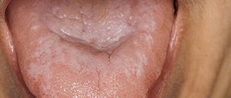
- The initial stage is characterized by the appearance of a gray-white or whitish coating, which is formed from dead epithelial cells. Small numbers of lesions are concentrated on the tongue, gums, palate, cheeks from the inside, lips, and in the sublingual area. On palpation, keratinization of the mucous membrane and compaction in the area of plaques are felt.
- The development of pathology is accompanied by the growth of lesions, their size increases in comparison with healthy epithelium. Over time, keratinized plaques become more sensitive and painful.
- The growth of spots is accompanied by a feeling of tightness, dryness and hard roughness. The sensations intensify under the influence of any irritant.
The uncontrolled course of leukoplakia leads to its persistent progression and the appearance of painful ulcerative and erosive lesions.
Leukoplakia in adults: what is it and what does it look like?
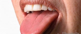
Symptoms of leukoplakia do not appear immediately. At first, minor swelling areas may appear, usually on the inside of the cheeks. The affected area can be one or several, sometimes their number increases. The patient feels roughness when touching the affected areas with the tongue; they can cause severe discomfort (itching and burning). Quite often, despite the presence of painful symptoms, patients are extremely dismissive of the manifestations of leukoplakia, turning to them only in the later stages of the disease.
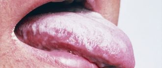
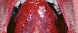
As it progresses, the affected areas become covered with a hard white coating, which is easily removed, but appears again after a while. They do not cause painful sensations. It is noted that the pathology is often accompanied by fungal infection; and a tenth of cases become harbingers of cancer. In this case, leukoplakia of the oral mucosa usually remains at the stage of benign tumors, but leukoplakia of the tongue (pictured) or located in the lower part of the mouth is much more dangerous.
The main symptoms of the disease include:
- the appearance of papillary-shaped neoplasms;
- thickening of the area where the inflammation is located;
- rapid increase in the affected area;
- frequent injury and bleeding of diseased areas;
- development of erosions, ulcers, white crust,
- significant enlargement of lymph nodes.
Types and forms of oral leukoplakia
The main forms of oral leukoplakia:
- Flat.
- Verrucous.
- Erosive.
- Tappeiner's leukoplakia.
- Soft.
Oral flat leukoplakia
The most common type of disease is flat or simple oral leukoplakia. It is often characterized as the initial stage of a pathological condition. At this stage of development, the pathology does not manifest itself as severe pain.
Photo: leukoplakia of the tongue
The initial phase of the disease is accompanied by the appearance of whitish or gray-white plaques, irregularly shaped stripes located on the tongue, palate, inner surface of the cheeks, and in the corners of the mouth. The plaques are dry and rough, without pronounced elevation or hardening. You often feel tightness, burning, and excessive dryness in the area of the damaged areas.
Simple leukoplakia often affects the tongue, resulting in a decrease in its sensitivity and taste perception.
Inflammatory processes do not occur at this stage of development of the disease. In most cases, this type of pathology occurs without deterioration in general well-being or complications and can accompany a person throughout life.
Verrucous form
The uncontrolled course of leukoplakia with constant traumatic effects on the mucous membrane - irritation from chipped teeth, incorrect dentures, consumption of excessively hot food and liquid - provokes its development and transformation of the simple form of the disease into verrucous.
The dominant sign of the transformation of simple leukoplakia into verrucous is pronounced keratinization and elevation of the upper layer of epithelial cells of the affected area. At this stage of the disease, the plaques noticeably rise above the healthy epithelium; upon palpation, compaction is clearly felt.
Verrucous leukoplakia of the oral cavity is accompanied by a pronounced feeling of tightness and dryness, and the occurrence of painful sensations during eating.
According to external signs, leukoplakia is of two types:
- Plaque. Plaque areas appear on the oral mucosa. Plaques are characterized by pronounced roughness, density, and elevation above the healthy epithelium. Their color varies from milky to bluish.
- Warty. Lesions similar to warts appear on the mucous membrane. Compared to the plaque form of the disease, the wart is characterized by the appearance of more keratinized, lumpy compactions. The formations rise 2–3 mm above healthy epithelial cells and most often have a gray-white tint.
The verrucous form of the disease more often than others turns into a malignant lesion of the mucosal epithelium.
Erosive stage
The erosive form of the disease occurs with its progression and prolonged exposure to negative factors on epithelial tissues. This stage of oral leukoplakia is accompanied by a number of symptoms:
- Severe pain occurs, which intensifies when any type of irritant comes into contact with the lesion. Eating food, alcohol and hot drinks provokes pain.
- Externally, the disease is manifested by the appearance of cracks and ulcers and erosive lesions in the affected areas. Inflammatory foci are localized around erosive lesions, often accompanied by bleeding.
- Unpleasant sensations persist even at rest. There is a burning sensation, excessive dryness and roughness. The development of a lesion at the base of the tongue causes a sore throat and speech impairment (coughing occurs).
This type of pathological condition has the highest risk of relapse. The erosive stage of the disease is very difficult to treat; ulcerative lesions take a very long time to heal.
Soft oral leukoplakia
This type of pathology is a benign neoplasm. The main prerequisites for the development of soft leukoplakia of the oral cavity are hormonal imbalances, disturbances in the psycho-emotional background, and chronic fatigue.
Visually, a slight elevation of white plaques above the healthy epithelium is noted. The formations are rough and painless. There is slight compaction and dryness of the affected tissue. When the lateral surfaces of the tongue are affected, sensitivity to temperature influences decreases, and taste perception is impaired.
The mild form of pathology is divided into several more varieties:
- Partial (focal), manifested by the appearance of affected areas in the form of foci localized in certain places.
- A diffuse variety, characterized by the “spreading” of plaques over the entire surface of the mucosa. The formations have a spongy, porous surface, loose structure, and increased size. The lesions are painless, but interfere with talking and eating, creating the feeling of “extra” tissue.
Tappeiner's (smoker's) leukoplakia
Adult patients, especially long-time smokers, are most susceptible to this form of the disease. Tappeiner's leukoplakia is provoked by tobacco decay products and chemical irritation of the mucous membrane by tobacco smoke.
In most cases, the formations affect the palate and the gum tissue located next to it. Large folded plaques of brown or grayish color appear in the oral cavity. The affected areas are painless.
The appearance of formations leads to blockage of the salivary glands and the appearance of reddish nodules. This condition causes excessive dry mouth and “thick” saliva.
It is not difficult to treat this type of leukoplakia in the oral cavity. It is necessary to give up smoking by first reducing your consumption and then stopping smoking completely. Getting rid of a negative habit leads to the complete disappearance of the pathological condition.
Treatment methods for leukoplakia
Despite the fact that leukoplakia should not be treated, the patient should still take a number of measures that can help get rid of the disease as quickly as possible. If the disease could be caused by trauma to the oral cavity, bite correction, dentures, or uneven edges of the teeth should be corrected. In case of leukoplakia, smoking should be limited or (preferably) completely abandoned. If the doctor has diagnosed the presence of systemic diseases, be sure to direct all efforts to cure them. In mild forms, you should restore damaged tissues using rosehip oil, sea buckthorn and take vitamin A.
Treatment of leukoplakia in a specialized clinic usually involves surgery, but another effective method can be cryodestruction (freezing out pathological formations with liquid nitrogen), subsequent chemotherapy and the use of anti-tumor drugs.
The staff of the AlfaDent dental clinic in Orenburg are ready to provide professional medical assistance in restoring oral health and getting rid of leukoplakia at any age.
"AlfaDent" is a professional approach to complex tasks.
Diagnosis of the disease
The diagnosis is made based on examination and questioning of the patient. Mandatory diagnostic studies confirming the diagnosis include:
- Cytology is the collection of epithelial cells to identify cancerous lesions.
- Histology is taken to perform a biopsy, which allows examination of the affected epithelial tissue to detect cancer cells.
- Blood from a vein.
Additional clarifying studies make it possible to distinguish leukoplakia from other pathologies.
Classic treatment of oral leukoplakia
The choice of treatment method for oral leukoplakia depends on the symptoms, the form of the pathology, and its severity. Initially, it is necessary to eliminate the negative factors that provoke the onset of the disease: giving up bad habits, changing your diet.
Photodynamic therapy
A number of physical procedures can be used to effectively treat the disease in a hospital. Such events include:
- Diathermocoagulation intervention to remove keratinized plaques. After a course of procedures, healing of the mucous membrane occurs within 10 days.
- Freezing the affected epithelial layer of the oral cavity (cryodestruction) for 1 minute. The course of treatment is 1 week.
- Carrying out photodynamic therapy (PDT). Pathogenic cells are destroyed by irradiation with a light wave.
The treatment regimen includes taking medications aimed at stopping infectious processes and inflammation. Local treatment of the mucous membrane is mandatory.
Features of treatment of leukoplakia of the tongue
When the disease is localized in the tongue, sanitation of the oral cavity is mandatory. If there are incorrectly installed dentures, fillings, or chips on the teeth, the fillings, dentures are polished (or replaced), and chipped teeth are restored. The affected areas are treated with antiseptics and wound healing solutions.
The flat form of the disease is easily treated. Sometimes it is possible to eliminate pathological plaque through laser or radio wave therapy. But if a patient is diagnosed with malignant tumors, treatment will be carried out in two stages - first, surgery is performed, then long-term radiotherapy is performed.
Upon completion of the treatment course, the patient should remain under outpatient observation, since leukoplakia of the tongue is characterized by a sluggish course, and symptoms of the disease may appear over several years.
Causes of the disease
The exact mechanisms of the appearance of oral leukoplakia are not yet known, but there are confirmed studies that the main influence is heredity and the presence of certain types of papillomavirus in the body. However, the factors that provoke this disease have been fairly reliably established.
Long-term (chronic) mechanical damage to the mucous membrane. For example, dentures, braces, chipped teeth.
Long period of smoking.
Long-term and constant intake of strong alcoholic drinks.
Deficiency of vitamins in the body (especially groups A and B).
Pathologies and anemia (iron deficiency).
Diabetes.
Pathologies of the gastrointestinal tract (gastrointestinal tract).
Consequences and chronic bacterial and fungal infections, especially in the oral cavity.
But in some cases, the disease occurs for no apparent reason and without provoking factors (so-called idiopathic leukoplakia of the oral mucosa ).
Treatment of oral leukoplakia at home
At home, treatment of leukoplakia of the oral cavity and tongue must begin with proper hygiene procedures. You need to use toothbrushes with soft bristles. Brushing your teeth and tongue should be done smoothly, without excessive intensity. After each meal, rinses and applications with anti-inflammatory and restorative properties are used. Particularly careful hygiene is required when treating leukoplakia of the tongue.
The affected areas must be treated with antiseptics: Chlorhexidine, Furacilin. Irrigation with antiseptic solutions is carried out up to 5 times a day. Taking vitamin complexes containing vitamins A and B groups helps to cure the pathological condition at home.
Folk remedies can be used as additional treatment. Decoctions of sage, chamomile, and calendula, which have an anti-inflammatory and calming effect, are considered particularly effective. A positive result is achieved by treating the affected areas with sea buckthorn oil and Kalanchoe juice.
Adult patients are recommended to consume natural juices, which have a general strengthening effect that increases immunity. Similar drinks include juices from apples, carrots, and cabbage. It is recommended to dilute freshly squeezed drinks with water.
Oral leukoplakia is a pathological condition that should not be left to chance. When the first signs of pathology appear, you must consult a specialist; self-medication is fraught with the development of serious complications and consequences.
Causes of leukoplakia in the oral cavity
To date, scientists have not reached a consensus on why the disease occurs. There are factors that can lead to the development of excess keratinization:
- Constant exposure to irritating factors: smoking, regular burns from hot food, strong alcohol, etc.
- Work in industries where there are chemicals, alkalis, acids.
- Living in a hot, dry climate.
- Taking drugs that reduce immunity.
- Local traumatic factors: failed fillings, sharp edges of dentures and damaged teeth, poor-quality braces.
- Endocrine pathologies: diabetes mellitus, hyperthyroidism, hypothyroidism.
- Reduced immunity: HIV, AIDS, immunosuppressive state. Oral hairy leukoplakia is often observed in patients with HIV and AIDS.