What is sinusitis?
Sinusitis is an inflammation of the maxillary sinuses. It usually develops as a complication of a runny nose. The most characteristic signs:
- temperature rises to 37 - 37.5 C;
- nose is stuffed;
- yellow-green nasal discharge appears;
- the bridge of the nose, cheeks near the wings of the nose and temples hurt;
- headache worsens in the evening;
- the sense of smell disappears;
- the voice becomes nasal;
- feeling of general weakness.
When the maxillary sinuses are inflamed, a person quickly gets tired, it is difficult for him to concentrate, and his memory weakens. It is very difficult to work with sinusitis, so it is better to take sick leave.
Sinusitis can be acute or chronic.
Causes of sinusitis
The disease occurs more often in the cold season in people with weakened immune systems, but this is not the only reason. Inflammation of the maxillary sinus can develop due to:
- complications of a runny nose;
- curvature of the nasal septum;
- damage to the nasopharynx;
- adenoiditis;
- infections and bacteria;
- allergies;
- too dry indoor air;
- work in hazardous industries;
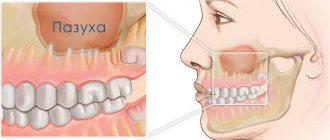
- poor condition of the upper teeth;
- trauma to the mucous membrane of the maxillary sinus.
Causes of inflammation of the trigeminal nerve on the face
Usually the disease is caused by infection or bacteria. List of reasons why inflammation of the facial nerve may occur:
- Temporomandibular joint injuries
- Tumors (benign and malignant) of the brain and facial area
- Anomalies of skull development
- Skull injuries - birth, fracture, base, damage to the face or jaw
- Polio
- Pulmonary tuberculosis
- Otitis
- Sinusitis
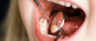
- Chronic caries
- Inflammation after tooth extraction or treatment
- Hypertension
- HIV and AIDS
- Poisoning
- Inflammation of the middle ear
- Severe hypothermia of the head
- Changes in hormonal levels in women
- Gum inflammation
- Ramsay Hunt syndrome
- Stroke
- Bell's palsy
Causes range from minor to life-threatening illnesses. Each of the reasons determines the further treatment of the patient. In some cases, special tests are performed for diagnosis - auditory, lacrimal, infectious, salivary or gustatory. In this way, the functioning of the receptors and sensory organs is checked.
Diagnosis of sinusitis
Treatment of sinusitis will be effective if the patient contacts an ENT specialist in time, the specialist will diagnose and prescribe treatment. The examination includes a number of procedures:
- examination of the nasal cavity;
- palpation or feeling the area around the nose and under the eyes;
- clinical blood test;
- X-ray of the paranasal (paranasal) sinuses - according to indications;
- computed tomography of the sinuses.
X-rays can detect the accumulation of mucus in the sinuses and swelling of the mucous membrane of the sinuses. With sinusitis, a darkening will be visible in the picture, since the mucus and swelling of the tissues in the cavities do not allow x-rays to pass through.
CT scan of the paranasal sinuses is necessary to identify polyps, cysts and anatomical changes. The examination is painless and lasts no more than 5 minutes.
If you feel heaviness when you tilt your head down, and the discomfort subsides some time after you raise your head, contact an ENT specialist. Self-medication with folk remedies, randomly selected antibiotics and nasal drops causes serious complications. Sinusitis can become chronic and cause more severe diseases: otitis media, meningitis, brain abscess, orbital phlegmon (inflammation of the tissue around the eyeball).
Causes of nasal congestion with headache
The causes of headaches and nasal congestion can be inflammatory diseases of various structures. The nose is an organ of the respiratory system with a complex anatomical structure. It is involved in the passage of air through the respiratory tract, its purification from harmful impurities and microorganisms, as well as in the capture of odors. These functions are carried out due to the specific anatomy of the nose. In its structure, there are a number of structures, disruption of the functioning of each of which can lead to the development of headaches and runny nose:
- the outer nose, formed by bones and cartilage, as well as a muscle layer;
- nasal cavity - a space for the passage of air, which gradually narrows and passes into the nasopharynx;
- mucous membrane - lines the nasal cavity and passages through which air moves;
- paranasal (paranasal) sinuses, or sinuses - voids filled with air;
- vessels and nerves.
The entire length of the airways is lined with mucous membrane. Its structure differs in different areas, depending on the function performed. So, in the thickness of the mucosa there are nerves and blood vessels, and in the initial areas there are olfactory receptors. Inflammatory processes begin on the mucous layer and then spread to other parts. Many diseases manifest themselves with similar symptoms, but the cause lies in damage to different parts of the respiratory system.
Sinusitis
Sinusitis is an inflammation of the mucous membrane of the paranasal sinuses. They are paired cavities formed by the bones of the skull. Normally they are aseptic, filled with air. However, inflammatory processes lead to swelling of the walls, secretion of mucus or pus, which leads to the development of the characteristic picture of sinusitis. The disease begins like a common cold and can be a complication of it. The patient experiences nasal congestion, fever, and general deterioration in health. Nasal discharge and headache appear. During diagnosis, it is necessary to determine in which area the inflammatory process occurs - this will allow choosing effective treatment.
- The maxillary (maxillary) sinuses are located to the right and left of the nasal cavity and have a common septum with it. They connect to it in the area of the middle nasal passages. Inflammation of the maxillary sinuses is called sinusitis and is the most common form of sinusitis.
- The frontal sinuses are located deep within the frontal bone. They are paired, separated by one common partition. The sinuses are superficial, their superficial walls are directed towards the face, the posterior ones - towards the internal structures of the skull, and the lower ones - towards the eye sockets. They are connected to the nasal cavity through the frontonasal canal. Frontal sinusitis, inflammation of the frontal sinuses, is also a common manifestation of sinusitis.
- Sphenoid - belongs to the posterior paranasal sinuses. They are located in the thickness of the sphenoid bone and connect to the nasal cavity in the area of the upper nasal passages. The lower wall of the sinuses is the upper vault of the internal nose. Some brain structures (frontal lobe of the brain, pituitary gland) are adjacent to the sphenoid sinuses. Sphenoiditis (inflammation of the mucous membrane of the sphenoid sinus) is rare due to the location of the sinus, but occurs in a complicated form.
- Ethmoid - located in the thickness of the ethmoid bone and formed by its cells. Their number may vary and range from 8 to 10 pieces. The anterior and middle sinuses connect to the middle nasal passages, the posterior ones to the upper ones. Inflammation of the mucous membrane of the ethmoid sinuses is called ethmoiditis. This is a rare disease that in most cases develops simultaneously with other forms of sinusitis.
Diseases can occur in acute or chronic forms. The course is considered chronic if the patient experiences 3 or more cases of exacerbation of inflammation per year. If sinusitis is caused by a viral infection, nasal rinsing and over-the-counter antiviral medications are sufficient to treat it. However, if a bacterial infection occurs, antibiotics will be required. Sinusitis may require surgical treatment. It consists of puncturing the contents of the maxillary sinuses and washing them with antibacterial solutions.
Flu and ARVI
Headache, nasal congestion, cough and fever are the first symptoms of respiratory viral diseases. They often occur during the off-season and are a sign of decreased immune defense. The infection enters the nasal mucosa, where it lingers and begins to multiply. This leads to an acute inflammatory reaction, the formation of exudate, swelling of the walls of the nasal cavity and paranasal sinuses. The patient experiences typical cold and flu symptoms:
- runny nose, nasal congestion;
- headache;
- cough - can be dry or wet, with sputum production, depending on the degree of spread of infection along the respiratory tract;
- muscle pain, aches;
- elevated temperature.
Viral diseases can be easily treated at home, but after mandatory consultation with a doctor . For this purpose, over-the-counter antiviral drugs, cough and cold remedies are used. It is also important to maintain bed rest and drink plenty of fluids regularly. However, a simple cold can cause dangerous complications in some patients if the natural immune system is weakened and cannot cope with the infection. For prevention, as well as for the treatment of bronchitis, pneumonia, sinusitis and other complications, the doctor will select antibiotics.
Respiratory syncytial virus
Respiratory syncytial virus (RSV) belongs to the group of pneumoviruses. It causes cold symptoms of varying severity and often occurs in children. According to statistics, most children become ill with RSV by the age of 4 years. Outbreaks are diagnosed in children's groups during the cold season. They are associated with a seasonal decrease in immunity, as well as vitamin deficiency. Viral particles penetrate the nasal mucosa, from where they spread further through the respiratory system.
The disease manifests itself with characteristic symptoms:
- stuffy nose, poor sense of smell;
- sore throat, redness of the mucous membrane;
- headache, general weakness;
- increased body temperature;
- dizziness, pain and muscle aches.
In most patients, the disease proceeds without complications and resolves on its own within 1–2 weeks. It causes typical symptoms of a cold, so the virus is rarely differentiated. Treatment is symptomatic, aimed at facilitating breathing and reducing pain. Bed rest, nasal rinsing, and drinking fluids in increased quantities are recommended. Complications are rare, and changes in the respiratory system caused by the virus quickly return to normal.
In some children, respiratory syncytial virus can cause capillary bronchitis (bronchiolitis). This is a pathological condition in which the spread of a viral infection to the bronchial mucosa is observed. This leads to necrosis (death) of cells, atelectasis (sticking together) of individual alveoli. During listening, wheezing and sounds of crepitus are clearly detected. The prognosis is favorable, but symptoms of respiratory failure may appear for some time after recovery.
Allergic reactions
One of the reasons why a stuffy nose and a headache is allergic reactions . They occur in the case of an individual increased reaction of the immune system to certain irritants. Normally, its action is aimed at combating viruses and bacteria that daily enter the mucous membranes from the external environment. In response to them, cellular and humoral defense mechanisms are launched, which identify the pathogen, stop its reproduction and destroy the infection. These processes are accompanied by inflammatory reactions, swelling of the mucous membranes, and increased body temperature.
An allergy is a malfunction of the immune system. If normally the mechanisms for recognizing normal and foreign proteins work, then in allergic patients they do not function correctly. The immune system perceives various substances that cannot harm the body as dangerous factors, so it begins to fight them. These proteins are called allergens and can be of different nature. When they enter the body, a typical set of symptoms appears:
- runny nose, swelling of the mucous membrane of the nasal cavity and paranasal sinuses;
- sharp pain in the head, dizziness;
- labored breathing;
- inflammatory reactions.
The main reason why allergic reactions occur is the release of immunoglobulin E into the blood. This is a protective protein of the body that stimulates the formation of other substances, including histamine and mast cells. Chemical reactions occur at the cellular level, but clinical manifestations are noticeable already in the first stages. They may differ depending on the way allergens enter the body. If they are in the inhaled air, the first thing that occurs is swelling of the nasal mucosa and difficulty breathing. The most dangerous manifestation of allergies is anaphylactic shock. This is an immediate type of reaction that occurs upon repeated contact with the stimulus. Symptoms increase sharply, and swelling of the mucous membranes can interfere with normal breathing. The prognosis in this case is favorable only in case of urgent medical care.
To treat this condition, it is necessary to eliminate contact with confirmed allergens as much as possible. However, for some patients this is not possible: during the flowering period of plants, in conditions of constant work with chemicals and a combination of other factors. Antihistamines help reduce allergies and relieve nasal congestion. The doctor will also select vasoconstrictor drops that will help reduce the level of swelling of the mucous membranes of the nasal cavity. If signs of an allergy develop rapidly, you should immediately take an antihistamine and consult a doctor.
Middle ear diseases
One of the causes of headaches and nasal congestion is ear disease. This organ not only performs the auditory function, but also provides balance. It has a complex structure, which includes several sections. Each of them performs its own functions, but is connected to other areas.
- The outer ear is represented by the auricle, auditory canals and eardrum. This organ picks up sound signals and transmits them further.
- The middle ear is a cavity filled with air. It contains small bones (the malleus, incus and stirrup) that not only transmit sound waves, but also amplify their power.
- The inner ear is also called the labyrinth. At its center is the organ of Corti, which picks up sound vibrations and transmits them to the cerebral cortex. The semicircular canals are also located here - this organ is responsible for balance and coordination of movements in space.
Otitis media is a dangerous disease that can occur independently or as a complication of a cold. Common pathogens are rhinoviruses and respiratory syncytial virus. They get on the nasal mucosa, but do not stay there, but move further to the nasopharynx and the organ of hearing. The disease can also be of bacterial origin, and its causative agent is streptococci or influenza bacillus.
Depending on the type of inflammation, acute and chronic, aseptic and purulent otitis are distinguished. It differs in the nature of its course, symptoms and the possibility of causing complications. Thus, acute inflammation is manifested by the following symptoms:
- severe attacks of headache, which can be concentrated in only one part of the head;
- discharge from an inflamed ear - they may have a yellowish tint, admixtures of pus or blood, and an unpleasant odor;
- nasal congestion, runny nose, deterioration of sense of smell;
- temperature rise, sometimes to critical levels.
Treatment of otitis media should begin immediately after the first symptoms appear. This is a dangerous disease that can become chronic and cause complications. The internal environment of the hearing organ is a favorable environment for the development of pathogenic microflora. Bacteria quickly multiply under conditions of oxygen deficiency and high temperature, causing purulent inflammation. This further leads to purulent melting of soft tissues, hearing impairment, and the appearance of discharge with a characteristic unpleasant odor.
Migraine
Primary headaches that develop without an infectious agent are called migraine. They may first appear in childhood or adulthood. Doctors distinguish several possible causes of migraines, proven through laboratory tests. The first of them is vascular pathologies, which cause a sharp spasm. This occurs in response to external stimuli, changes in atmospheric pressure and other factors. Some doctors are of the opinion that migraine occurs due to pathologies of the nervous system. If brain cells become too excitable and overreact to stimuli, they transmit impulses to neighboring areas. At the same time, the areas responsible for pain are excited, which is accompanied by attacks of pain in the head.
A characteristic symptom of migraine is its aura. During the course of this disease, there are 4 main phases, which can sequentially replace each other or be absent. Regardless of the presence of all the signs, the doctor will be able to diagnose migraine based on the clinical picture.
- Prodromal phase – occurs frequently (in more than half of patients). It lasts from several hours to a day before the onset of the aura or the headache itself. Only those patients who are experiencing attacks not for the first time will be able to note the symptoms and associate them with migraine. During the prodromal phase, frequent mood swings, disruption of the gastrointestinal tract, general weakness and fatigue are observed.
- Aura is a neurological disorder that can occur before or during a headache. The attack lasts from several minutes to an hour, rarely longer. It cannot be confused with other diseases. In most cases, visual disturbances occur. Flickering effects appear in the field of view, spreading from the center to the periphery. In addition, the patient may develop sensory symptoms such as tingling or numbness in the skin of the hands or face. In acute cases, there is a lack of coordination of movements, auditory hallucinations, and confusion. The aura may be absent or occur without headache in some patients.
- A headache attack can last from 2-3 to 72 hours; in children it rarely lasts more than an hour. The pain is characterized as acute, pulsating, it is often one-sided and affects only half of the head. However, in almost half of patients it is bilateral, accompanied by discomfort and pain in the neck. Pain also causes additional symptoms: nausea and vomiting, confusion, mood swings, nasal congestion, and digestive tract disorders. Each case is individual, so attacks even in one patient may differ.
- The postdromal phase is a period that continues for several days after the headache ends. At this time, patients pay attention to confusion, weakness and dizziness, symptoms of deterioration of the digestive system.
Migraines most often occur in women over 25 years of age. It is considered chronic if it occurs with a frequency of at least 15 times a month within three months. Regardless of the frequency and conditions of occurrence, a typical migraine requires examination by a doctor. Patients who experience attacks frequently can identify the relationship between migraine and its triggers. They cite changes in weather conditions, consumption of certain foods and other reasons.
At the first symptoms of a migraine, you need to lie down in a well-ventilated area, without harsh natural or artificial light. This will help slightly reduce the duration of the attack and provide relief during an exacerbation of migraine. Initially, doctors recommend simple over-the-counter analgesics to reduce headaches. However, migraine headaches are often so severe that they cannot be treated with these drugs. In this case, specific drugs are prescribed for the treatment of migraine (triptans, ergotamines). They can be purchased at a pharmacy, but you will need a prescription from your doctor. In the period between attacks, it is recommended to pay attention to the prevention of migraines, especially if it is not possible to exclude trigger factors. For this purpose, drugs such as sodium valproate, timolol, metoprolol, propranolol, topiramate and others are used.
Neoplasms on the nose and nasal passages
A common cause of nasal congestion and headaches in both children and adults is the appearance of tumors in various parts of the nasal passages. They block passages and prevent the normal passage of air. As a result, there is an insufficient supply of oxygen to the lower parts of the respiratory system and a low concentration of this element in the blood. This leads to ischemia of organs and tissues, oxygen starvation of the nerve cells that form the brain. There are several conditions in which neoplasms appear in the nasal cavity and passages and require urgent surgical treatment.
- A furuncle is a purulent inflammation of the sebaceous gland or hair follicle. The process is manifested by the formation of a dense, painful swelling, which is often located at the tip of the nose. In most patients, the boil resolves without complications, but further infection can cause it to increase in size.
- A carbuncle is a similar formation that is larger in size. Several hair follicles are simultaneously involved in the formation of the carbuncle. They form a large, painful elevation above the skin with several heads from which pus is released. Carbuncles are painful and can cause fever, nasal congestion and shortness of breath, as well as other factors. Their contents must be removed surgically, since the presence of bacterial microflora causes rapid suppuration of the carbuncle.
- Erysipelas of the nose is clearly visible on the surface of the skin. The redness is pronounced and can be separated from healthy skin with a small roller. The disease is acute, caused by a specific microorganism - hemolytic streptococcus. Treatment is selected individually by the doctor, taking into account general health and the results of laboratory tests.
- Hematoma of the nasal cavity - can appear after bruises or blows, as well as as a result of weakness of the walls of blood vessels. This neoplasm is an accumulation of blood surrounded by a membrane. Hematomas are often found close to the nasal septum. This is a painful internal growth that is at risk of further rupture and infection. After some time, the hematoma goes away on its own, but large formations and those located deep in the nasal cavity may require surgical intervention.
- Abscess of the nasal cavity - develops as a complication of purulent bacterial processes. It is a large cavity filled with pus, with strong walls. Abscesses are formed as a result of infection of wounds, hematomas, and they can also be caused by improper structure of the nose. These tumors cause chronic pain and nosebleeds. Treatment is surgical - the abscess is removed, followed by sanitation of the wound and the use of antibiotics.
- Entry of foreign objects into the nasal cavity. This situation often occurs in children, and in some of them it is discovered only during the examination. The foreign object must be removed mechanically so that it does not move further along the respiratory tract. They often turn out to be small parts of toys, plant seeds, buttons, berries and other objects. They can damage the nasal mucosa with sharp walls and also clog the respiratory passages.
Various neoplasms and foreign objects are easily detected as a result of examination. Treatment will vary depending on the type of lesion, its size and other factors. Thus, foreign objects can be easily removed from the nasal passages. If they injure the mucous membrane, it is necessary to treat it with disinfectants. Most neoplasms, including abscesses and hematomas, must be opened. The surgeon makes an incision in the side wall through which purulent or bloody contents can be removed, and then cleans the resulting cavity with antibacterial drugs.
Other reasons
Headache and nasal congestion are typical manifestations of many diseases. These symptoms signal inflammatory processes, impaired nasal breathing for any reason. It can be difficult to differentiate between the two at home, so you should see a doctor for an examination. As a result, the following disorders and conditions may be detected:
- runny nose and headache during pregnancy - manifests itself as a result of a decrease in the rate of metabolic processes, swelling and deterioration in the removal of fluid from the body;
- herpes - during the period of exacerbation it can manifest itself, including in the nasal cavity, leaving behind ulcers that do not heal for a long time;
- injuries – as a result of a fall or blow, damage to external or internal structures occurs, which is accompanied by headache and swelling;
- being at a high altitude above sea level - the lack of oxygen in the inhaled air significantly affects well-being.
If headaches are accompanied by difficulty breathing, nasal congestion, and the appearance of discharge, in most cases this indicates the initial stages of colds. However, similar signs can also signal dangerous disorders that significantly affect well-being and can occur with complications.
Treatment of sinusitis
Depending on the patient’s condition and the course of the disease, the doctor prescribes therapy and selects antibacterial, decongestant and anti-inflammatory drugs. Treatment can be conservative or surgical.
Conservative treatment
For mild sinusitis, in addition to drug treatment, according to indications, nasal cavity lavage according to Proetz, or “cuckoo”, is performed. A special solution is injected into the nose of the patient lying on his back. It passes through the nasal cavity, sinuses, after which it is pumped out with a vacuum pump. “Cuckoo” is carried out in a course of 2 to 10 procedures.
For moderate and severe cases of the disease, antibiotics are prescribed in the form of tablets, solution for injection or inhalation.
Surgical treatment (puncture of the maxillary sinus)
In case of severe purulent sinusitis or lack of effect from conservative therapy, a puncture is used. This allows you to quickly remove pus from the sinus, clean it and treat it with an anti-inflammatory drug. Sometimes several similar manipulations are required. Punctures are performed under local anesthesia.
When sinusitis cannot be treated or is in an advanced stage, surgery is performed under general anesthesia. The period of complete recovery after surgery can take up to two weeks.
What are the dangers of abusing decongestants?
Uncontrolled use of vasoconstrictor drops leads to the fact that the capillaries are constantly in spasm. They stop functioning normally, blood flow is disrupted, and nearby tissues suffer because they do not receive the required amount of oxygen and nutritional components.
Systematic drying of the mucous membranes and disruption of its restoration threatens the development of malnutrition and the formation of drug-induced rhinitis. In an effort to get rid of seasonal runny nose as quickly as possible, a person acquires an illness that requires serious treatment.
The next danger is psychological addiction. A reflex is formed in the brain that normal breathing can only be restored with the help of medications. Using vasoconstrictor drops makes a person feel better; he does not think about the alarming consequences for his own health. Improper use of decongestant drops harms the entire body. Violation of the dosage regimen is especially dangerous in childhood. There are cases when a child has to be hospitalized because the mother intensively instilled drops into his nose. The danger is that vasoconstrictor drugs enter the general bloodstream and negatively affect all tissues and organs. Arterial hypertension may develop, shortness of breath, headache, insomnia, etc. may appear.
Most often, dependence on nasal drops is formed due to illiteracy of the population. Not everyone knows that harmless medications can cause intoxication of the entire body. If dangerous symptoms are detected, it is important to immediately seek medical help and receive timely treatment.
Prevention of sinusitis
To avoid sinusitis you should:
- spend enough time outdoors;
- strengthen immunity;
- Healthy food;
- take vitamins;
- dress according to the weather;
- seek medical help in a timely manner.
If you have discovered the first symptoms of sinusitis and are looking for a qualified specialist who will diagnose and help you cope with the disease, call the phone number listed on the website or leave a request in the feedback form. Medical staff will answer your questions and conduct all necessary examinations within one business day. Let's take care of your health together!
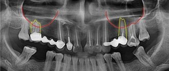
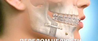
How to prevent perforation
If patients with anatomical features of the location of the roots in the sinus are treated according to the standard protocol, the threat of rupture of the membrane is very high
The doctor must have the necessary information about the anatomical and topographical features of the patient and be prepared for an emergency situation.
- To prevent perforation of the sinus membrane, computed tomography is required. After studying the CT image, the doctor selects the optimal treatment tactics. But not every equipment allows you to identify all the nuances. In our Center, diagnostics are carried out using a modern 3D tomograph Sirona Galileos with ENT mode settings. Provides high quality images, allows you to evaluate the structural features of the upper jaw and sinus, calculate the height of the septum, find out the location of the roots, their anatomy.
- When working with the canals of teeth located in the sinus, all manipulations must be extremely careful. The dental canals are very thin, sometimes tortuous, the doctor must control every action. Endodontic dental treatment in our Center is carried out using a Seiler dental microscope. Multiple magnification expands the view, allows you to examine the entire length of the dental canal in great detail, and prevent the filling material from leaving the root.
- The experience of a specialist is of great importance so that he can give an adequate assessment from a CT image, choose tactics, and calculate efforts. Our Center employs highly qualified endodontists, with excellent clinical thinking and honed manual skills. Qualifications and skills allow us to prevent complications and act promptly in the event of an emergency situation.
We have minimized the likelihood of complications after dental treatment
. Advanced CT diagnostics in ENT mode and work using a dental microscope allow us to select competent tactics to prevent perforation of the bottom of the maxillary sinus.
Levin Dmitry Valerievich Chief physician and founder of the Doctor Levin center
After endodontic treatment, control CT diagnostics is mandatory! This preventive measure allows you to timely identify perforation (even if the doctor did not notice it during treatment) and take measures to eliminate the consequences.
If a piece of instrument or filling material gets into the sinus, removal of the foreign body must be prompt and urgent ! Otherwise, there is a high risk of infection, development of inflammatory processes and neoplasms.