1024
Occlusion defects and their treatment are the most difficult issues in dentistry.
To a large extent, because there is still no consensus among experts about what is most important in this problem - the condition of the maxillary joint, the relationship of the chewing teeth with each other, or the condition of the muscles and ligaments of the dentofacial apparatus.
General overview
Dr. Kois offers his perspective and approach to solving occlusion problems.
He developed a device called a deprogrammer, which, according to the developer, makes it possible to eliminate occlusion dysfunction relatively quickly and easily.
But before talking about this device, it is necessary to say a few words about one of the main parameters of the dentofacial apparatus - the central ratio (CR) of the TMJ.
The CS is the position of the MF in which the condyle is relative to the glenoid cavity with optimal occlusion.
A feature of the temporal mandibular joint is that the articular head can be in different positions. This provides the lower jaw with the ability to move in different directions - open, move back and forth and left and right.
The main question: what position should be taken as the initial one, ensuring correct occlusion when closing the jaws? For a long time, experts disagreed about what the position of the condyle should be during CS. Today it is believed that the optimal position of the head of the joint is the anterosuperior position.
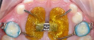
The Kois device is a removable orthodontic device designed to automatically install the movable jaw in the center position. Or, in other words, a device that facilitates the registration of a CA.
Structurally, the device is a plastic base with a vestibular arch embedded in it, running outside the dentition and serving as a retainer. The base is made of acrylic, the vestibular arch is made of thin but durable wire.
The device is placed on the upper jaw. The plastic base has a platform (stop) that serves for contact with the incisors of the lower jaw. It is this platform, in contact with the “units” of the lower jaw, that ensures the installation of the dentofacial apparatus in the CS.
When wearing the deprogrammer for a long time, the masticatory muscles are programmed in such a way that installation in the centric relation, and, consequently, correct occlusion occurs automatically.
In essence, the device performs the same function as occlusal splints (splints).
Orthodontic methods of closing gaps in the absence of incisors and other techniques used in modern dentistry.
Visit here to learn more about the purpose of the HIP Plane Analyzer.
At this address https://www.vash-dentist.ru/ortodontiya/prikus/vyityazheniya-zuba.html we will talk about the need for teeth traction and how to carry out the procedure.
Duration of wearing and confirmation of results
You need to wear the Kois device until muscle deprogramming is completed, that is, until the old muscle memory is replaced by a new one. This usually takes two to four weeks.
The duration of wearing the device per day varies and mainly depends on the complexity of the malocclusion. Thus, some patients are advised to wear it for 6-8 hours, others – around the clock with the possibility of removal only for oral hygiene and eating.
For the reliability of the result, the initial contact point should always be marked when closing the jaws on a strip of articulation paper. If the initial contact is confirmed while closing the mouth, then the person has been deprogrammed.
Indications and contraindications
The device is indicated in the following cases:
- The need to reprogram the masticatory muscles to ensure correct occlusion.
- For diagnostic purposes, when it is necessary to establish the dental system in centric relation.
- With increased tooth abrasion. The device allows you to determine the type and cause of the pathology - occlusal dysfunction, bruxism or other parafunction.
- To control the height of the bite.
- If necessary, avoid premature contact of teeth.
Contraindications to using the device are:
- periodontitis of the lower incisors, manifested by their mobility (before using the device, mobility must be eliminated);
- gag reflex (to relieve it, you can try to reduce the area of contact of the acrylic base with the palate);
- the presence of inflammation in the TMJ (arthritis, arthrosis, etc.).
Important. Bruxism can be caused by neurological disorders. In these cases the device is useless. Patients with neurological deficits are advised to wear a mouth guard to prevent tooth wear.
Aesthetic rehabilitation of a smile in an unmotivated patient with pathological abrasion
Summary : The patient had previously contacted the clinic with functional complaints, but was not motivated for aesthetic rehabilitation. Clinicians, taking into account the individual characteristics of the patient, can simultaneously ensure aesthetics and function with minimal risks to the dentition. In this clinical case, the patient was interested in solving the problem of pathological wear of the upper anterior teeth in the most effective and predictable way. After a thorough diagnosis, the risks are assessed and the treatment prognosis is determined. To demonstrate to the patient the aesthetic and functional benefits of the proposed treatment, a mock-up was made. During treatment, it was necessary to reduce the chewing load and eliminate the influence of parafunction using an occlusal splint. The occlusion height was restored using minimally invasive techniques. The length of the incisors is increased through prosthetic construction and correction of the gum contour.
The patient complained of wear of the upper frontal teeth; he was not interested in aesthetics issues. The patient's interest is important for the doctor - the treatment should completely change the appearance of the smile. It was necessary to correct the function, achieve dense, simultaneous, bilateral contact of the chewing teeth and reduce the load on the anterior teeth. To motivate the patient and better understand the clinical situation, a mock-up and a Kois deprogrammer were used.
Analysis of a clinical case
The patient, 25 years old, complained of wear of the upper frontal teeth. He was also concerned about the decrease in the length of the crowns of the upper teeth, thinning of the lingual surfaces and changes in the smile (Fig. 1-3) .
Fig.1 Initial situation
Fig. 2 Initial situation: patient’s lips at rest
Fig. 3 Initial situation: teeth with maximum fissure-tubercle contact of antagonist teeth
Initially, the patient did not want any aesthetic changes to his smile. However, he managed to get interested not only in functional rehabilitation, but also in the aesthetic restoration of his smile.
Anamnesis
The general medical history was unremarkable; the patient was not taking any medications. There were no concomitant diseases, no contraindications to treatment were identified.
No periodontal pathology was identified. Previously, the patient rarely sought dental care. Proximal caries was treated 3 years ago. The patient notes reduction of the crowns of the upper teeth and their thinning on the lingual surface.
There was no contact between the lateral teeth and appeared only when the teeth were clenched and the lower jaw was displaced posteriorly. After discussing this with the patient, he noted that he often clenches his teeth when working out in the gym.
Diagnosis, risk assessment and prognosis of periodontal tissue: probing depth was less than 3 mm, bleeding was not detected during probing. The bone level was 1-2 mm below the cemento-enamel border; there were no intraosseous defects. No recession detected.
Risk: low.
Prognosis: favorable.
Biomechanics: As noted above, the patient has been treated for proximal caries over the past 3 years. Upon examination, this restoration was found in tooth 4.6 (Fig. 4) .
Fig. 4 Lower dental arch before treatment. The consistency of the restoration of tooth 4.6 is questionable
On the vestibular surface of teeth 3.6-3.3, 4.3-4.6, erosion and minimal signs of abrasion were detected. On the occlusal surface of teeth 1.4, 2.6, 3.7-3.5 and 4.5-4.7 there is a minimal and medium degree of erosion and signs of abrasion. Teeth 1.1 and 2.1 with severe wear.
Risk: moderate; for teeth 1.1 and 2.1 high.
Prognosis: unfavorable.
Functional assessment: The mouth opens to its full extent (44 mm). Movements in the TMJ are symmetrical, without clicking or crunching. The TMJ stress test is negative. When performing a test with immobilization of the lower jaw, minor discomfort was detected. Severe abrasion was detected on the lingual surface and cutting edge of teeth 1.1 and 2.1; mild and moderate abrasion was detected on teeth 1.3, 1.2, 2.2 and 2.3 (Fig. 5) .
Fig. 5 Initial situation: upper dental arch
The average degree of pathological abrasion was determined on the lower incisors. An occlusal injury was detected with slight mobility of teeth 1.1 and 2.1.
Based on the diagnostics and dental history, it was revealed that the movements of the lower jaw were blocked: in order to close the lateral teeth, the patient had to move the lower jaw to a forced posterior position. This pathology was facilitated by parafunction: the patient clenched his teeth during training. In the presence of blocking of the movement of the lower jaw, the risk is usually defined as average, but in this case, taking into account the parafunction and pronounced pathological abrasion of teeth 1.1 and 2.1, the risk increases.
Risk: high.
Prognosis: unfavorable.
Examination of the maxillofacial area: The patient had a gummy smile, the contours of the gums were asymmetrical in the area of the front teeth of the upper jaw (Fig. 1) . At rest, the teeth of the upper jaw were hidden under the gum, the canines were located 2 mm above the lip (Fig. 2) . Anomalies in the spatial position of the teeth of the upper jaw were noted, and minor tremors were identified between the teeth of the lower jaw. Based on the criteria described above, the risk of aesthetic restoration was determined to be high.
Risk: High.
Prognosis: Unfavorable, especially if we take into account the patient’s initial refusal to make any changes in his smile.
Treatment Goals
1). It was necessary to correct the function, achieve dense, simultaneous, bilateral contact of the chewing teeth and minimize the pathological abrasion of the anterior teeth. The patient must be interested in creating a new smile: aesthetic aspects will certainly be affected during treatment. To motivate the patient and better understand the clinical situation, a mock-up and a Kois deprogrammer were used.
2). It was necessary to take preventive measures to preserve the patient's health. To minimize functional risk, it is necessary to eliminate the blocking of movements of the lower jaw and parafunction - for this purpose, it is necessary to make a protective mouthguard for sports activities. The bite height must be restored using minimally invasive methods: most of the patient's posterior teeth are intact.
3). The length of the incisors must be restored using restoration and changes in the contour of the gums.
Treatment plan
Treatment options were discussed with the patient, incl. possibility of orthodontic intervention. Orthodontic treatment allows you to eliminate anomalies in the position of individual teeth and correct the shape of the dental arches. In this clinical situation, the shape of the dental arches was within normal limits, there were no anomalies in the position of individual teeth (Fig. 3-5) . During orthodontic treatment, restoration would be necessary to restore the height of the tooth crowns. Instead, it was decided to fabricate a ceramic restoration to restore the height of the bite and the length of the anterior teeth. The patient was interested in the most predictable and effective treatment.
Stage 1: Motivation, production of the Kois deprogrammer
Previously, the patient was informed about the causes of pathological abrasion, and attention was drawn to the importance of maintaining optimal pH. For this purpose, the patient was recommended to use a paste and rinse that increase and maintain neutral pH. A diet excluding low pH drinks was also recommended. During follow-up visits, it is recommended to coat the teeth with fluoride varnish. To perform treatment, it was necessary to determine the position of the TMJ at the “true” centric relation. For this purpose, a Kois deprogrammer was used.
Stage 2: Diagnostic wax -up
After 2 weeks of wearing the device, the patient noted that every time before it was removed, the teeth were in contact. At the same visit, occlusion fixators were made (the deprogrammer was in the oral cavity). Photographs were taken with the “true” centric relation of the jaws fixed. Impressions were made of the upper and lower jaws (vinylpolysiloxane). Next, the models are plastered into the articulator in a previously determined central ratio (Fig. 6) .
Fig.6 Models installed in the articulator
When evaluating the photographs obtained, taking into account the position of the anterior teeth, a decision was made to lengthen the incisors by 2 mm. When planning the restoration, much attention was paid to the upper canines: they were 2 mm above the lip. Also, to obtain an aesthetic result and the required crown length in the area of teeth 3-8, it was necessary to perform mucogingival surgery with gum reduction from 1 to 2 mm. A wax-up was modeled (Fig. 7) , and a silicone key for mock-up was obtained based on it.
Fig.7 Diagnostic wax-up
Stage 3: Mock -up
At the next visit, a mock-up was made to demonstrate the treatment result (Fig. 8) . This allowed the patient to evaluate the expected positive changes in aesthetics and function.
Fig.8 Mock-up to evaluate aesthetics and function
Stage 4: Gingivoplasty and osteoplasty
Using wax-up, a surgical template was made for gingivoplasty, on which the planned gum height for teeth 3-14 was marked (Fig. 9) .
Fig.9 Gingivotomy, surgical template installed
Next, the mucoperiosteal flap was detached to perform osteoplasty and create an optimal level of the alveolar ridge and harmony of white and pink aesthetics after tissue healing (Fig. 10) .
Fig. 10 View after reposition of the mucoperiosteal flap
There were no complications during tissue healing (Fig. 11) .
Fig. 11 Patient’s smile 3 months after gingivoplasty and osteoplasty
Stage 5: Fabrication of temporary restorations
For 3 weeks before the manufacture of the orthopedic structure (healing period after surgery), the patient wore a Koys deprogrammer to maintain the functional stability of the TMJ. At the next visit, a composite aesthetic mock-up was made as a template for preparation. In order to maintain deprogramming, a small bite block was created on the palatal surface to visualize the “true” centric relation during preparation between teeth 1.1 and 2.1 (Fig. 12) .
Fig. 12 Bite area between teeth 1.1 and 2.1; the contact of the lateral teeth is as close as possible
A minimally invasive preparation was carried out through a mock-up of the vestibular and occlusal surfaces of the teeth in the upper and lower jaw. Next, occlusion recorders were made (the lower incisors touched the platform at the initial point) (Fig. 13) .
Fig. 13 Determination of the central ratio
The last procedure was carried out after preparing the chewing group of teeth for working with models in the laboratory. On the lower jaw, the incisors were prepared for veneers. For the upper frontal teeth, to restore tissues that had been subjected to pathological abrasion, it was necessary to make crowns. Gum retraction was performed using the 2-strand technique, and vinyl polysloxane impressions were obtained from both dental arches. Provisional restorations are made on a mock-up basis from cold-cured plastic and fixed with cement for temporary structures.
Next, all-ceramic E.max® restorations were made: overlay occlusal-vestibular inlays for 12 lateral teeth, veneers for the frontal teeth of the lower jaw, crowns for the upper frontal teeth. Four second molars were planned to be restored using direct composite restoration.
Stage 6: Final restoration
The provisional restorations were removed and the final restorations were fitted. Veneers and overlay inlays are fixed using an adhesive protocol with dual-curing composite cement; crowns are fixed with self-adhesive universal cement. To achieve the required occlusal contacts, direct composite restorations of the second molars were fabricated. Next, the occlusal relationships were checked and minimal adjustments were made. After this, impressions are taken to make a protective mouthguard. The final result was above the patient's expectations (Fig. 14-19) .
Fig. 14 Lower dentition after treatment
Fig. 15 Upper dentition after treatment
Fig. 16 Final result with the jaws positioned in centric relation
Fig. 17 Final result: lips at rest
Fig. 18 Patient's appearance after treatment
Fig. 19 Control image (OPTG)
conclusions
Analysis of the patient's individual risk factors allows us to create the most predictable treatment plan. In this clinical case, biomechanical risk was minimized: almost all preparation was carried out within the enamel. Pathological abrasion led to blocking of the movements of the lower jaw, which contributed to the progression of the pathology. Increasing the bite height eliminated this problem. Despite this, the patient was recommended to wear a mouth guard to prevent damage to the restorations during sports as a result of parafunction. Follow-up visits were scheduled every 4 months. The possibility of complications only from the restoration side was assumed - chipping of the ceramics. The prognosis of periodontal tissue is favorable. The prognosis for the functional state of the maxillofacial region has improved.
Source: AEGIS Dental Network
Translation from English by Elena Kovshik for the portal
Preparatory activities
Preparatory operations performed during treatment with a deprogrammer are largely determined by the individuality of the patient.
However, all the main parameters on which the patient’s occlusal status depends must be determined.
Risk assessment
Dr. Kois argues that for the successful treatment of dental anomalies, a correct approach to diagnosis is necessary, which consists of assessing the risks in all 4 areas that determine the functioning of the dentofacial apparatus - the biomechanical system, dentofacial, functional or periodontal.
The greatest difficulty is the assessment of functional (occlusal) parameters, since changes in the vertical position of the lower jaw cause its horizontal displacement.
Important parameters
The human dentofacial apparatus consists of 3 main components:
- joints (VNSJ);
- teeth;
- muscles and ligaments that move the LF.
In accordance with this, Dr. Kois suggests considering 3 positions when diagnosing occlusion dysfunction - P1, P2 and P3.
P1 determines the position of the joint, namely the condyle. Since its location is not strictly defined due to the structural features of the TMJ, there are difficulties in establishing the CS.
This may be MIP, myocentric or habitual occlusion if functionality is not impaired.
P2 affects the occlusion of teeth, P3 affects muscles and ligaments.
Questions for the patient
To determine the degree of occlusal risk a patient is exposed to, it is necessary to test them by asking 5 questions.
Depending on his answers, a conclusion is made about the presence or absence of occlusion dysfunction:
- Do you have trouble chewing gum? A positive answer (“yes”) may indicate limited chewing amplitude.
- Do you have difficulty chewing dry foods (such as bagels) that require a lot of chewing? The answer “yes” indicates occlusal dysfunction.
- Have you noticed any changes in the functioning of your dentition over the last 5 years? Increased wear of teeth, their mobility, formation of teeth, and weakness of the masticatory apparatus should be considered as changes.
It should be borne in mind that normal wear of enamel is 11 microns per year. That is, normally, 100 years should pass before a 1 mm thick layer of enamel wears out.
- Do you need to bite into your food more than once? An affirmative answer means dysfunction of the dentofacial apparatus.
- Do you have trouble sleeping? This refers to throwing off the blanket at night, movement disorders or restless legs. If the answer is positive, the presence of parafunction or neurological deficit can be assumed.
A positive answer to any of the questions indicates a certain degree of dysfunction. When conducting the test, it is important that the answers are made based on the normal state of the dentofacial apparatus, without taking into account the patient’s adaptation to a negative situation regarding teeth.
If he has adapted to pathological changes in the functioning of his dental apparatus (for example, he began to eat only soft foods) and answered “no” to the first 2 questions, this does not at all indicate the absence of pathology.
How the device for distalization of molars works and the timing of its wearing.
In this publication we offer characteristics of the permanent dentition.
Here https://www.vash-dentist.ru/ortodontiya/prikus/mehanizm-stanovleniya-molochnogo.html we will discuss methods for correcting primary occlusion.
Questions for the patient
Violation of occlusion is accompanied by a number of symptoms that the patient experiences when eating, chewing gum, sleeping, and talking.
In order for the doctor to be able to determine the nature of occlusal dysfunction, he must know what the patient experiences during the listed actions. This can be found out using a small test consisting of 5 questions.
Depending on how the patient answers these questions, a conclusion is made about the presence or absence of occlusion:
- Do you have problems sleeping - throwing off the blanket at night, restless legs, etc.? A positive answer suggests the presence of parafunction.
- How are things going with chewing gum? Are there any difficulties or not? If the patient answers “yes,” this may indicate a limited chewing range.
- Do you have any difficulty chewing dry foods such as cookies? A “yes” answer indicates some occlusion problems.
- Do you have to bite into your food more than once to get the right piece? An affirmative answer indicates dysfunction of the dental system.
- Have there been any negative changes in the functioning of your dental system in the last five years that you noticed? In this case, the patient needs to explain that this means such negative phenomena as increased wear and mobility of teeth, weakness of the masticatory muscles, and the formation of teeth.
An affirmative answer to at least one of these questions may indicate dysfunction of the dentofacial apparatus.
Important. The patient should be warned that answers should be given based on a normal lifestyle, without refusal, for example, from regular solid food, without taking sleeping pills, etc.
Stages of formation of permanent occlusion and causes of abnormal phenomena.
In this publication, we will consider methods for determining central occlusion.
Here https://zubovv.ru/ortodontiya/prikus/kak-opredelyaetsya-vyisota.html read how to correctly determine the height of the bite.
Manufacturing stages
Manufacturing a Kois deprogrammer includes the following operations:
- Obtaining alginate impressions from both jaws.
- Casting from plaster model impressions.
- Installation of models into the articulator with complete coincidence of fissures and tubercles.
- Making a vestibular arch from wire and installing it in a model of the upper jaw.
- Pouring the acrylic base. In this case, the vestibular arch is imprinted into the base.
- Trimming the base to form a platform (stop) for the mandibular incisors. Its width should be 3 mm, and its height should provide a bite gap in the range of 1-1.5 mm.
- The final stage is polishing the deprogrammer.
Preparatory stage
In preparation for the procedure, an individual approach is used, determined by the peculiarities of the anatomical structure of the patient’s jaw. The quality of the correction depends on the accuracy of the readings obtained, so the doctor must competently assess potential risks and pay attention to diagnostic procedures. Dr. Kois's theory identifies five treatment risk conditions, ranging from acceptable functionality to neurological impairment and bruxism. Among the parameters identified during the research, three indicators were identified:
- Joint position;
- Chewing unit ratio;
- Specifics of the operation of the chewing system guide.
To make a diagnosis, a standard questionnaire is used, which includes five points and allows one to assess the severity of changes, as well as draw a conclusion about the presence of signs of dysfunction. In addition, establishing a dialogue between the doctor and the patient contributes to a better understanding of the need to follow recommendations for oral care and calculate the permissible load, which ultimately allows the latter to get rid of constant discomfort.
Manufacturing Features
Preparing a deprogrammer is a multi-stage process, which is determined by the high demands placed on the accuracy of the product. To develop the correct apparatus, it is necessary to take impressions from the upper and lower rows, and then place the plaster models in the articulator. To make a vestibular arch, a wire is used, the configuration of which eliminates contact with the surface of the units. The horseshoe-shaped base is created from polymer and provides a platform for the lower incisors. The finished structure is processed and polished, after which the doctor checks the compliance of the dimensions and tries the device on the dentition. It is worth emphasizing that the contact area should have a narrowed area and affect the midline of one of the frontal elements of the lower section.
The device works the entire time it is in the mouth. The principle of operation is based on registering the occlusion in the correct position and changing the contacts of the teeth. With the help of this device, neuromuscular coordination changes, joint and muscle pain goes away, the functioning of the TMJ improves, and the chewing load is distributed evenly. After two to three weeks of wearing, the discomfort caused by the abnormal development of the TMJ is eliminated, as well as the normalization of sleep and the restoration of the natural state of the jaw region during wakefulness.
Principle of use
Before using the device, be sure to check the width of the platform. If it exceeds 3 mm, it must be corrected by abrasive treatment.
The device is placed on the upper jaw, and if everything is done correctly, it should be securely fixed on it, thanks to the shape of the base and the tight-fitting vestibular arch.
The patient is asked to take a comfortable position in the chair and press the articulation paper with his teeth, moving the jaw left and right, back and forth.
It is necessary to check the bite separation, which should be up to 1.5 mm. The contact of the platform with the mandibular incisors should be along their midline. If these conditions are not met, the device is corrected by removing excess material .
If necessary, occlusion correction is carried out by creating correct fissure-tubercle contacts of the chewing teeth, holding the LF in centric relation.
In the video, a specialist talks about the design and use of the Kois deprogrammer.
Muscle deprogrammer AQUALIZER
AQUALIZER is a special dental splint in which water is placed. The design is used to achieve relaxation of the jaw muscles. The design is one of a kind, so dentists recommend it if a problem develops in the form of involuntary teeth grinding.
The advantage of the system is its versatility. This is achieved in a simple way - the water that gets into the splint after the device is installed on a person, itself adapts to the structural features of the patient’s jaw. In this case, the person’s discomfort and pain go away almost immediately.
Your doctor will help you buy the AQUALIZER deprogrammer. The appointment is made after consultation. The price of an equalizer depends on the model.
The tire is different:
- effective pain relief;
- muscle relaxing effect;
- performing their tasks with an uncertain diagnosis.