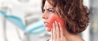
Orthodontics is a branch of dentistry aimed at correcting defects in the development of teeth and the maxillofacial skeleton. The result of orthodontic treatment largely depends on the correct diagnosis. Modern diagnostics in orthodontics
performed using special devices and equipment.
In this article we will consider popular methods of orthodontic diagnostics and answer frequently asked questions about orthodontics
.
Diagnostic methods in orthodontics
The treatment of defects of the masticatory-speech apparatus is carried out by a specialized orthodontist. After a visual examination of the patient’s oral cavity, the specialist decides on the need to conduct additional research procedures using modern diagnostic methods. Based on the analysis obtained, it is possible to establish an accurate diagnosis and select the correct treatment to achieve a successful result.
Diagnostic methods in orthodontics:
1. Radiography.
2. Tomography of the temporomandibular joint.
3. Orthopantomography.
4. Examination using diagnostic models (taking impressions of the patient’s jaw with subsequent production of working models).
5. Electromyography.
6. Mask model (determining facial asymmetry taking into account three planes).
7. Simon's photostat.
8. Teleradiography.
The most popular diagnostic methods in orthodontics are dental radiography and orthopantogram, which allow identifying various deviations of the dentofacial apparatus. In difficult cases, additional research methods are used to clarify the identified pathologies.
3D cephalometric analysis of a CT image (a modern alternative to two-dimensional TRG images)
Standard TRG analysis includes an assessment of the position of the teeth already in the space of the skull and how the teeth stand relative to the jaws, how the jaws are located relative to the skull, and what size they are.
3D diagnostics is a much larger amount of information compared to what we see from TRG. Additionally, a CT scan evaluates the position of each tooth in the bone tissue, the therapeutic condition of the teeth, and the condition of the root canals of pulpless teeth. It is only possible to accurately assess the causes of malocclusion pathology and understand what is causing it - a decrease in the size of the jaw or its displacement - only with the help of 3D diagnostics.
Without 3D computed tomography, a full diagnosis is impossible today - this one image replaces and unites everything.
Unfortunately, not all orthodontists have tomographs and are able to analyze 3D images. Now this is the gold standard for all diagnostics and a huge plus for the patient, because not only the orthodontist, but also any other doctors involved in the treatment can take a comprehensive approach to a single, verified treatment plan using just one CT image.
Questions about orthodontics
Many patients have different questions about orthodontics. Let's look at the most popular ones.
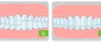
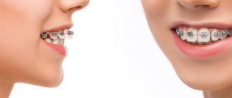
Is it possible to correct dental defects without braces?
The vestibular brace system is by far the most effective way to correct bite defects. But as an alternative, the patient may be offered aligners. A set of mouth guards is made from impressions taken from the patient’s jaw. While wearing them, you must maintain careful oral hygiene. The orthodontic structure is changed once a month. The patient needs to regularly visit an orthodontist to monitor the treatment process.
Cons of aligners:
- Not suitable for everyone;
- not used in complex cases;
- are more expensive than braces.
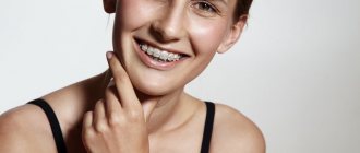
How quickly do teeth straighten with braces?
On average, teeth move into an even arch after 3-4 months. These periods can be shortened or increased, depending on the type of braces chosen and the complexity of the particular case.
What is the function of orthodontic screws?
Screws in orthodontics
are used to stabilize the brace system in the absence of some teeth in a row, as well as in case of bone tissue atrophy. They eliminate the movement of supporting teeth and speed up the process of dental correction.
How do teeth move?
In some cases, it becomes necessary to move one or more teeth. For this purpose, orthodontists use removable or non-removable structures. Fixed bracket systems include bracket systems that ensure the movement of teeth in the desired direction, under the supervision of a specialist.
Types of tooth movement in orthodontics
:
1. Corpus technique, which involves moving the tooth crown and root in one direction.
2. Oblique-rotational – involves moving the crown and tooth root to different distances.
In orthodontic treatment, it is possible to move not only one tooth, but also a whole series. This technique is successfully used in the treatment of occlusion with a distal position of the lower jaw.
When are orthodontic implants installed?
When treating complex defects of the dental system, there is often a need to install orthodontic implants aimed at providing the necessary support for fixed structures that correct bite defects.
Indications for installation of orthodontic implants:
- crowded dentition;
- strong protrusion of molars or front incisors;
- distal bite;
- severe curvature of teeth;
- if it is necessary to change the size or position of the jaw;
- to ensure the required degree of impact on the dentition (if braces do not cope with this task);
- lack of positive results after wearing braces for a long time.
Implants in orthodontics
differ from standard designs used to restore lost teeth. They are smaller in size and thickness, are installed temporarily, and the implantation method itself is minimally invasive and does not require cutting the gums or suturing.
Photo protocol, assessment of smile and facial harmony
Plays an important role in monitoring changes and quality of treatment. At the consultation stage, photographs are necessary to visualize the patient's problem. During the diagnosis, intraoral photographs are taken, photographs of teeth in a closed, open state, from different angles of the face and smile - including the so-called “Emma test”, which shows the patient how his teeth are usually visible during speech. The younger the person, the more the upper incisors are visible when speaking and the lower ones are not visible. With age, soft tissues sink, lose tone, and the upper teeth become less and less visible and the lower teeth become increasingly visible. If a patient comes for orthodontic treatment, we pay attention to this because we can change this and “rejuvenate” him in terms of perception during speech.
The patient takes up to 20 photographs of teeth and face from different angles
What is being assessed?
- Facial aesthetics
- Profile
- Bite and condition of teeth in their positions
- Incisor centerline relationship
- Width and arc of the smile (visibility and parallelism of the lip line)
- Condition of tooth enamel
The movement of the dentition can affect the position of the lips, so the orthodontist can reduce the worsening of wrinkles by increasing the height of the bite. A decrease in bite height leads to a greater likelihood of wrinkles and folds, which the doctor must take into account in the treatment plan.
More details about the photo protocol
Orthodontic treatment of bite defects in
Experienced orthodontists who see patients at the specialized dental clinic “Aesthetics” are ready to answer all your questions about orthodontics. The center is equipped with modern diagnostic equipment that gives highly accurate results, which guarantees a quick correct diagnosis and the correct choice of treatment protocol.
Orthodontic treatment uses high-quality lingual braces, mouth guards, plates and other structures that can effectively eliminate various malocclusion defects, even in the most difficult cases. You can make an appointment with an orthodontist by phone. The first consultation appointment is free.
All patients who leave reviews on our website are guaranteed to receive an additional 3% discount on everything dental (the discount is added to the client’s main discount). Share your opinion and experience of orthodontic treatment in our clinic, do not miss the opportunity to save your own money on the services of an experienced orthodontist!
Taking dental impressions
Nowadays, few people do calculations using models; they are created more formally. In general, all this can be done using computed tomography, provided the orthodontist has sufficient knowledge and skills. Using plaster models, the orthodontist calculates the size of the teeth, the amount of space required to move them, looks at the proportionality of the teeth, whether restoration will be required, and how they will fit together after treatment.
Why do we continue to make impressions at the Confidential Clinic?
At the Confidence clinic, we do not treat the patient so that he has straight teeth and everything is fine only according to calculations. We treat so that your smile is beautiful in real life.
The first and most important reason is the need for models for indirect fixation. The second reason is that they provide an opportunity to once again see some real object live, because CT and TRG are virtual things, and models make it possible to visually see the patient’s bite.
It happens that an orthodontist, during the treatment process, encounters some unexpected effect for him and is lost. At the Confidence clinic, we calculate everything “on the shore” in order to continue strictly according to the treatment plan, which we drew up even before fixing the braces.
All patients want to know how much orthodontic treatment will cost.
A complete diagnosis makes it possible to accurately announce to the patient the final amount of treatment, since the orthodontist understands the situation in detail and is confident in the treatment plan, and therefore in its cost. If additional medical procedures are required, such as implantation, prosthetics, restorations, this will be discussed immediately. High-quality preparation for orthodontic treatment allows you to minimize the likelihood of pitfalls.
And all this is not only told to the patient, but also clearly shown on the computer.
Diagnostic presentation.
After a comprehensive analysis, the orthodontist prepares a detailed diagnostic presentation. It can consist of up to 100 or more slides with cut-ups of images and a step-by-step architecture for building the result of orthodontic treatment. By opening it, any doctor from any other clinic will be able to assess the patient’s clinical situation.
A diagnostic presentation is a detailed, ready-made treatment plan that the patient can use even if he moves to another city and continues treatment with another doctor. In Russia, only a few orthodontic clinics practice this approach.
All this makes it possible to plan treatment, predict its timing and complexity. We do all the necessary preparation in advance for a clear understanding of the treatment process at the start, and not at its stages.
For what reasons do pathologies arise?
- The most common reason is breathing through the mouth when the nose is not breathing. If ENT treatment has been carried out, the child may continue to breathe through his mouth out of habit. If this is not compensated for in any way, it remains as a bad habit. As a result, there may be a narrowing of the upper jaw, and we begin to develop malocclusion pathology, lack of space for teeth, deep bite, distal bite.
- The second reason is that when a patient breathes through his mouth, his tongue is usually located in the lower floor, the tongue can push the lower jaw forward and excessive growth of the lower jaw occurs and a medial bite, open bite, cross bite is formed - this is also a pathology. Sometimes children suck their fingers. Sometimes they bite their lip. Which lip is bitten by the upper or lower one - this also affects what the pathology will be.
- Genetic factors. If a patient comes with a medial bite and the jaw is forward. It is advisable for the orthodontist to look at the parents, if one of them has the same jaw - this is genetics. If a patient has supernumerary teeth erupting, then it is possible that one of his relatives had this.
- Congenital pathologies - this could be, for example, cleft lip and palate, but the orthodontist does not work with such complex issues - special institutions deal with this.
Pathological mobility of teeth, caused by inflammatory-destructive changes in periodontal tissues, is one of the leading symptoms of periodontitis. Many authors believe that splinting is indicated at the very first signs of pathological tooth mobility [1, 2, 6]. However, clinically it can be difficult to detect incipient tooth mobility, so splinting is often used at later stages of the disease [6]. At the same time, increasing tooth mobility can contribute to the progression of the disease. Previous studies have shown that without stabilizing mobile teeth with splinting structures, it is impossible to achieve positive results and prevent relapse of the disease [1, 3, 7]. Therefore, splinting teeth is an important stage in the treatment of periodontitis, especially moderate and severe cases [4, 7].
Assessing the condition of the periodontium today is possible using periotestometry, implemented by the electronic-mechanical device “Periotest S” (Siemens, Germany). The microcomputer of the “Periotest S” device registers the response to a push applied to the tooth crown, calculates the characteristics of the damping properties of periodontal tissue for 16 blows, and monitors the accuracy of the results. The “Periotest S” values are shown on the display and at the same time accompanied by audio information. The question of assessing the effectiveness of various types of splinting for periodontitis using periotestometry remains open, since the available studies are still few [1, 5].
The purpose of the study was to evaluate, using periotestometry, the effectiveness of splinting the anterior group of teeth in a patient with severe periodontitis.
Material and methods
Complex periodontal treatment was performed for patient Shch.A. A 60-year-old man who complained of tooth mobility, especially 4.2 (Fig. 1-A). According to the patient, while fishing in the winter, he began to tighten the knot of the fishing line with his teeth, as a result of which the tooth “jumped out of the gums.” He put it back in.
A comprehensive periodontal examination was carried out, supplemented by intraoral and panoramic radiography, as well as periotestometry of the teeth (three times for each tooth). Measurements were made according to the standard method specified by the manufacturer: the patient sits upright, the teeth of the upper and lower jaws do not contact, the tongue does not touch the teeth. The position of the tip is perpendicular to the tooth axis (± 200), the percussion point is in the center of the vestibular surface of the anatomical crown. During the measurement, the tip sleeve did not touch the tooth, but was located at a distance of 0.7–2.0 mm from its surface. Periotestometry was carried out before and after splinting teeth. Based on the results of the examination, a diagnosis was made, treatment was planned and implemented.
Results and discussion
Examination of the patient showed the following. Objectively: the mucous membrane is pale pink. Green-Vermillion hygienic index – 3.0, Muhlemann bleeding index – 2.5 points.
Teeth mobility according to Miller as modified by Flesar: 4.2-II, 4.1-I, 3.1-I, 3.2-II. On a panoramic radiograph: resorption of bone tissue of the alveolar processes of the jaws by 2/3 of the length of the root. On the intraoral radiograph: a horizontal fracture of root 4.2 in the middle third, its root canal is sclerotic, the lumen of the canal can be traced only in the lower third of the root (Fig. 2-A).
Diagnosis: chronic generalized severe periodontitis, horizontal fracture 4.2 in the middle third of the root, chronic apical periodontitis 3.3.
Treatment: 4.2 is temporarily splinted with 4.1 and 4.3 using Filtek Flow (3M ESPE) flowable composite in the area of the contact surfaces. Root canal 4.2 is passed to the fracture line. Next, using a Piezon Master 400 (EMS) ultrasonic device, the composite was removed from contact surfaces 4.1, 4.2, 4.3. Comparison of fragments, passage of the apical part of the root canal, expansion. The entire length of the canal is prepared for a titanium pin; after fitting, the pin is removed from the root canal, and the canal is temporarily sealed with Metapex (Biomed).
At the next visit, an “AH+” sealer (Dentsply) was inserted into the fracture line, the root canal was sealed with “AH+” at the apex, a titanium pin and a “Valux” filling (3M ESPE) were fixed in it (Fig. 2-A).
In parallel with treatment 4.2, local anti-inflammatory therapy was carried out: applications with a mixture of metronidazole with a 0.2% chlorhexidine solution, hard dental plaque was removed using an ultrasonic scaler "Piezon Master 400" and soft plaque using an "Air Flow" (EMS) device.
When choosing a splinting technique, we were guided by the clinical degree of tooth mobility. With degrees I–II of mobility, one could expect good long-term results using a conventional set of adhesive technologies. But in this situation, when mobility 3.2 was defined as II according to Miller, and based on periotestometry indicators (48 c.u.) it turned out that it was much more pronounced and corresponded to grade III, it was decided that it was necessary to create additional conditions for fixing the structure on the teeth. For this purpose, groove creation technology was used. According to the literature, no fundamental differences in the therapeutic effectiveness of fiberglass and polyethylene adhesive splints have been identified [1, 2]. When choosing a specific fiber system, with all other relatively similar parameters and capabilities, the determining criteria for a doctor are most often ease of use and price.
Using GlassSpan fiberglass tape, 4.3, 4.2, 4.1, 3.1, 3.2, 3.3 were splinted using an invasive technique: a cut was made along the lingual surface of 4.3, 4.2, 4.1, 3.1, 3.2, shaped like a reverse cone, to the thickness of the GlasSpan tape. , with a distance from the cutting edge of 2 mm. Chemical adhesion of the composite to tooth tissue is achieved through the “Single bond” bonding system; and a 38% etching gel was used to remove the dentin smear layer, increase surface area and micromechanical retention.
Since 3.3 was located under the crown as part of the bridge, it was decided to splint the tooth through the hole in the crown. Glass fiber tape "GlasSpan" was laid in the cut, covered with a fluid composite "Filtek Flow" + "Valux", the tire was ground and polished (Fig. 2-B).
A.N. Ryakhovsky et al. (2007) indicate that the “Periotest S” readings are representative for a splinting structure weight of no more than 12.71 ± 0.81 g. The results of periotestometry before and after teeth splinting are presented in the table.
When comparing the reference values and those we obtained before splinting, it turned out that they were greater than the normal values. After splinting the teeth, periotestometry indicators 4.3 and 4.2 were within the normal range, and 4.1, 3.1, 3.2 were less than normal.
After splinting the teeth, functional selective grinding according to Jenkelson was performed. A month after splinting, periotestometry indicators for this group of teeth decreased: for 4.1 – by 2.0. e.; for 4.2 and 3.1 – by 1; for 4.3 the indicators remained at the same level; and for 3.2 they increased by 1.00. e. We believe that the change in periotestometry indicators one month after splinting is associated with the adaptation of periodontal tissues to the redistribution of the load associated both directly with the combination of teeth into a splint and with the elimination of supracontacts during selective grinding. Before splinting, periodontium 3.1, 4.1, and 4.2 (due to fracture) had a deficiency of chewing load, and after splinting, the ligamentous apparatus of the teeth was not ready for the load applied to it, demonstrating its rigidity.
The direction of pathological mobility of teeth depends on their location in the dental arch, the state of occlusion, and it is always natural. From our point of view, the best result when splinting is achieved by combining teeth whose mobility vectors lie in intersecting planes. For incisors and canines, the mobility vectors lie in planes located at an angle to each other. To immobilize the frontal group of teeth, we used a structure that combines incisors and canines (anterior immobilization).
The glass fiber tape “GlasSpan” filled with filling material, used in this clinical situation for splinting teeth, becomes rigid after polymerization and does not have elastic properties. Therefore, the fixed attachment of the teeth to each other and, as a result, the absence of a minimal, but physiologically necessary excursion of the teeth in relation to each other, to the dentition and the alveolar process are a significant drawback of this method of splinting (Fig. 1-B)
The main negative result when using this method of splinting is the rapid destruction of the periodontal supporting structures, due to which the teeth are stabilized as part of the structure.
As a result, traumatic tangential stresses arise on the supporting teeth of the splinting structure, similar to those for cantilever dentures [7].
conclusions
- Periotestometry is a fairly informative modern method for assessing the condition of the periodontium, allowing one to assess the degree of elasticity of the ligamentous apparatus of the teeth. An increase in periotestometry values relative to reference values is an early sign of a violation of the elastic properties of the periodontium, which requires temporary splinting of teeth (for the period of active treatment of periodontitis or longer).
- The presented clinical case clearly shows that teeth whose mobility vectors lie in intersecting planes must be combined into a splint.
- Negative periotestometry indicators after applying a splint made of glass fiber tape "GlasSpan" indicate that this method of dental immobilization is unphysiological.
Indications for wearing removable appliances
It is better, of course, to take your child to the orthodontist on a preventive basis once every six months, at least to understand whether he needs treatment or not. The classic indicator is a small jaw. Let's say a patient was at the dentist and was told that his jaw was small and his teeth would erupt crookedly. Or an incorrect bite - the upper jaw overlaps the lower jaw too much. Or the child has a bad habit - mouth breathing, which stems from problems with the ENT organs. The ENT doctor also refers you to an orthodontist with this problem.