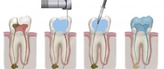
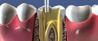
What are root canals
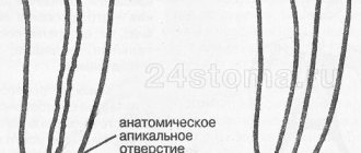
The dental nerve, or pulp, is located not only inside the tooth, its crown, but also inside the roots. Root canals are elongated cavities located inside the roots of teeth. This is where the dental nerve passes within the root. The number of root canals in one root may vary. There may be two or more channels in one root. The root canal can be very thin, thinner than a human hair, so high-quality treatment can take quite a long time. The difficulty is that even with the use of modern microscopes, the doctor cannot see absolutely all parts of the root canal. Work in the deepest part of the root is carried out almost blindly, which requires experience and skill from the doctor.
Endodontics under a microscope at Dentistry on Smolenskaya
Treatment of root canals of teeth under a microscope allows you to control the process of tooth treatment at any stage. Firstly, it is initially important to determine how many canals there are in the tooth so as not to miss any. High-precision expert equipment with ZOOM magnification at Dentistry on Smolenskaya gives a clear result, and the doctor can confidently say what condition the tooth tissue is in.
Secondly, during treatment under a microscope, any perforations of the root walls, violation of the integrity or incomplete filling of the canal are clearly visible. If the canals were initially sealed, a microscope will make it possible to qualitatively unseal the cavity. In addition, the microscope will help remove the remainder of a previously broken instrument, regardless of when it was broken off in the canal.
Dentistry on Smolenskaya employs doctors who are members of the expert community of the Russian Dental Association. Our specialists not only treat teeth, but also write books about trends in modern dentistry and its development. Attend symposia and conferences at the global and European level. They share their original developments in dental practice with colleagues.
Dental treatment should be trusted to professionals! At the Dentistry on Smolenskaya clinic, you are guaranteed to receive the best medical care, provided by doctors recognized for their services in the field of dentistry in Russia and in other countries of the world. To clarify prices for root canal treatment, please contact our specialists.
Root canal treatment
Root canal treatment takes place in several stages. Before treatment, X-rays or computed tomography must be taken. Using CT or X-ray images, the doctor will determine the approximate length of the root canals, shape, and the presence of obstacles to their cleaning.
Preparation for root canal treatment involves administering anesthesia and isolating the tooth from saliva using a rubber dam. If a tooth is severely damaged, the doctor can first restore its walls using temporary filling material.
Factors leading to the appearance of obliteration
- Heredity. With Stanton-Capdepont syndrome or marble disease (osteopetrosis), the formation of hard dental tissues is disrupted due to the characteristics of dentinogenesis imperfecta (the process of dentin formation).
- Diseases of the tooth and surrounding tissues. For example, pulpitis, caries, periodontitis, etc.
- Long-term and systemic exposure to chemicals and factors.
- Malocclusion. Chewing overload occurs and there is a risk of developing obliteration.
- Inactivity of the tooth or decrease in its functionality.
- Injuries, bruises, tooth dislocations.
- The presence of diseases of the endocrine system and metabolic disorders.
- Age and changes in the body associated with it.
In most cases of detection, obliteration occurs asymptomatically for the patient and is detected only during an intraoral radiograph.
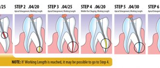
Cleaning and expansion of root canals
Root canal cleaning begins with measuring its length. A special instrument that resembles a needle in appearance is inserted into the root canal. A special apparatus is attached to this instrument - an apex locator. When the root apex is reached, the device beeps and the distance to the apex appears on its display.
After measuring the length, the doctor begins to clean the canal from the remnants of the nerve and expands it. Root canals can be expanded using hand instruments that the doctor holds in his hands or using machine or rotary instruments. Rotary instruments are inserted into a special tip - an endomotor. Development with machine tools is more modern and more expensive. It speeds up the process and provides higher quality processing. In order for a doctor to achieve a similar effect using hand instruments, the doctor will need much more time. After each insertion of each instrument, the doctor rinses the root canal with a chlorine-containing antiseptic.
After processing and drying the root canal, it is filled.
Search for the mouths of heavily sclerotic canals
Clinical case of endodontic treatment of upper incisors with heavily sclerotic canals. How to navigate a situation where there are “no mouths.”
Today I would like to share a clinical case that will demonstrate how a seemingly trivial situation can lead to serious complications, as well as how to avoid these complications.
The reference patient was referred by a colleague with a request to help carry out endodontic treatment before prosthetics in teeth 12 and 21. The doctor made an independent attempt at treatment, but unexpectedly the problem of severe obliteration of the orifices in both teeth was revealed, which did not allow the doctor to find the entrances to the root canals. The referring dentist only had binoculars in his arsenal, which, alas, did not allow him to confidently navigate inside the tooth.
This is how these teeth came to me. What do we see?
Firstly, we see that the doctor is great for realizing in time the risk of searching for the mouths “blindly”, without magnification. Since in both cases there were a couple of strokes of the tip left before the root wall was perforated.
Secondly, the search vector was shifted to the palatal side in both cases.
Thirdly, the obliteration really did not leave even a hint of the mouth where it should have already appeared. The situation in tooth 21 turned out to be especially difficult.
The green line shows the outline of the actual root canal. And here you can clearly see how the search for the mouth was carried out away from the true course of the root canal, threatening to end in perforation.
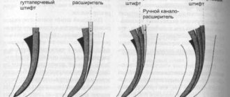
How to find the entrance to the canal when even the thinnest file (No. 6, No. 8) does not “catch” anything? It is necessary to remove sclerotic dentin. This can be done with a rotating instrument on a long stem or with an ultrasonic nozzle. But the main thing to remember is 2 important rules:
1. Any actions with aggressive instruments inside the tooth (canal) can ONLY be carried out under visual control. Ideally, under the control of armed vision, i.e. good lighting and magnification. We predictably can only do what we see with our eyes. If we work virtually, by touch, “by experience” - predictably, we can only do perforation, which will significantly worsen the future prospects of the tooth and the confidence of your patients.
2. Use practical laws of orientation in the pulp chamber when searching for orifices. One of the most important is the color of the fabrics...
This helps to understand where to “dig” to find the entrance. By removing a certain amount of sclerotic dentin under a microscope, we obtained a point that was barely noticeable at high magnification.
However, this turned out to be quite enough to make sure that this was the entrance to the root canal. Further work on processing and expanding channels in this situation was quite simple. The working length was determined using manual K-files No. 10, No. 15. Further instrumentation of the channels was carried out with the BioRaCe system to sizes No. 50.04.
Filling of the canal in the 12th tooth was carried out only in the apical third using hot gutta-percha (for subsequent restoration of the tooth using a pin structure). In 21 teeth, cold hydraulic obturation of gutta-percha with a bioceramic coating and a bioceramic sealer was used.
Progress of treatment of both teeth according to radiographs:
The patient was returned to the referring doctor for continued treatment and prosthetics.
Conclusions from this case, especially for young doctors:
- Inside the channels, do only what you see, do not work by touch!
- If you encounter a problem, stop on time! There is no need to be a hero if you do not have enough equipment, skills, or experience to cope with the situation. Refer the patient to more experienced and equipped colleagues, or at least warn the patient of the significant risk to him if he continues. This will be better for both of you and will save you from unnecessary conflict situations.
- use the rules of orientation in the pulp chamber! They are very effective, there are only a few of them, we talked about one of them today.
In subsequent articles and cases, I will tell you about other rules of orientation inside the tooth, which will help you not to make any mistakes. Stay tuned!
Measles canal filling
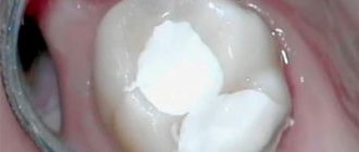
The procedure for filling root canals is carried out in various ways, for example: by the method of cold lateral (side) condensation of gutta-percha pins (reliable and affordable). Pins that resemble gutta-percha sticks in appearance are inserted into the root canal. After insertion, these sticks are compacted against one of the walls of the root canal. Between the gutta-percha there is a special paste for filling the canals.
The most modern method is filling the root canals using the vertical condensation method of hot gutta-percha. With this method, the doctor seals the top of the root canal using a heated gutta-percha pin. After this, the remaining volume is filled with hot liquid gutta-percha. This method is more expensive.
After the filling, if everything is done well, the doctor removes excess gutta-percha and sealer, and then installs a temporary filling. X-ray control is required. Permanent tooth filling in one visit is carried out only in exceptional cases, since it is necessary for the material to cool completely.
After root canal filling, discomfort may persist for some time. The tooth may be sensitive when biting. Within a few days the pain goes away.
Cost of endodontics
| Name of service | Cost, rub. |
| Intracanal procedure for the treatment of pulpitis | |
| with root canal filling with pastes (for one canal) | 2500 |
| with root canal filling with gutta-percha (for one canal) | 2750 |
| Mechanical and medicinal treatment of one canal | 1320 |
| with root canal filling with thermoplastic gutta-percha Beefil, Termofil (for one root canal) | 2750 |
| passage of the obliterated canal | 2000 |
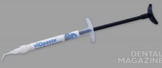
| use of the drug "Metapex" for 1 channel | 1650 |
| laser processing of 1 channel | 1200 |
| EDI (determination of pulp viability) | 400 |
| Removing a foreign body from the canal | 4150 |
| Intracanal procedure for the treatment of periodontitis | |
| with root canal filling with pastes (for one canal) | 2500 |
| with root canal filling with gutta-percha (for one canal) | 2750 |
| Mechanical and medicinal treatment of one canal | 1320 |
| with root canal filling with Beefil thermoplastic gutta-percha (for one root canal) | 2750 |
| Passage of the obliterated canal | 2000 |
| Use of the drug "Metapex" | 1650 |
| Laser treatment of one channel. | 1200 |
How to understand whether the root canal is filled properly
Signs of a properly treated canal are visible on x-ray: filling to the tops of the root, dense filling of the canal without any excess or voids. It is not desirable to have material residues outside the root canal. If the paste does not dissolve, an inflammatory process may develop in the future. But the quality of work depends not only on the attending physician: the patient himself also plays an important role. By following all the dentist’s recommendations, observing the rules of oral hygiene, as well as a careful approach to the health of your teeth, this is the only way you can achieve the desired results.
Root canal treatment and filling should always be trusted only to experienced specialists. An experienced dentist in Minsk is waiting for you at the Family Dentistry Center. Comfortable treatment, modern equipment of European quality, highly qualified specialists and an individual approach to each patient. Thanks to the use of drug sedation, all procedures can be performed without stress and pain, and children's fears of the dental office will go away on their own.