What is endodontics
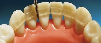
Endodontics is the treatment of the root canals of teeth. It is one of the most difficult in dentistry, because it requires experience and precision. The process involves cleaning the dental canals with diseased or dead pulp. The painstaking work of a dentist is aimed at preserving the functional characteristics of teeth and their aesthetically attractive appearance.
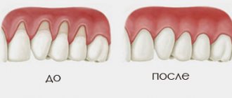
The need for treatment is due to the fact that inflammation occurs in the soft tissues of the tooth and severe pain appears. Over time, the process develops into a chronic stage. The patient no longer feels pain, but the inflammation is becoming increasingly widespread, penetrating deeply into the nerve and jawbone.
Features of the anatomy and topography of all groups of teeth
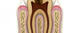
The structure of the root canal system differs from each other in different groups of teeth. They have additional, lateral channels, which can branch off from the main one at any level and have different configurations - from simple to complex. In structure, root canals can be compared to the crown of trees. Currently, there are 9 types of root canal structure. Type I - there is one root canal, which begins at the bottom of the pulp chamber and continues, gradually narrowing to the apex of the root. This type of root canal is most often found on the upper central incisors. Treatment of teeth with this type of canal structure is not difficult. Type II - the structure is represented by two root canals, which open at the bottom of the pulp chamber separately from each other, and near the root apex they merge into a common lumen, which ends in one apical opening. The root systems of the lower incisors and upper premolars most often have this structure. Type III - one orifice and one root canal open at the bottom of the pulp chamber; along the course of the root, the common canal bifurcates in the lower third of the root into two independent canals, which then unite again at the apical part and open with a common opening. This configuration is observed in the teeth of the lateral groups.
Type IV - characterized by the presence of two independently running root canals in one root, which in the area of the root apex open with two independent apical openings. The lower molars, premolars and lower incisors have this type of root canal. This is a fairly simple anatomical situation for endodontic treatment. Type V is characterized by the presence of one canal inside one root, but near the root apex the common canal is divided into two ending with independent apical foramina. This type is more often observed in the lower premolars. In some clinical situations, it can be quite difficult to treat both branches to the apical foramen. Type VI - two canals open at the bottom of the pulp chamber of the tooth, which unite into a common canal approximately in the middle of the root length, and then again divide into two independent passages and open with two apical openings. This structure of the canal system is difficult to process and fully clean the entire lumen of the root canal.
Type VII has the following structure: one root canal begins at the bottom of the pulp chamber of the tooth, then narrows towards the middle of the root like an hourglass, then it is divided into two independent canals, which in the apical part again unite into a common canal and immediately at the apex of the root they re-branch and open with two apical openings. This type is very complex and is observed in the teeth of the lateral groups of the lower jaw. Type VIII - 3 independently running root canals in one root. This type of structure is very simple and is found in various morphological groups of teeth. However, the incidence of this type of root canal structure is not very high. Type IX - the presence of 3 root canals along the entire length, which are then combined into one canal. This type is found in wisdom teeth.
It is necessary to highlight some structural features of the root canals of teeth of different groups. Endodontic treatment of the central upper incisors is not difficult, but sometimes there are clinical situations when there may be 2 canals at the root of the central upper incisor. During instrumentation of the lateral incisor canal, problems such as perforation or apical displacement can sometimes occur, especially when using rigid or large files, since about 70% of the upper lateral incisors have a pronounced distal bend. The lateral incisors, just like the central ones, can have 2 canals in one root. Endodontic treatment of the upper canines can be problematic during root canal treatment, since their structure has a buccal curve at the root apex (last 2-3 mm). There is also evidence of the presence of a canine structure with 2 canals. The most difficult is endodontic treatment of the lower incisors. In 40% of cases there are 2 channels. To create conditions for straight-line access of endodontic instruments, the tooth cavity is expanded in the vestibular-lingual direction, and the width of access must correspond to the width of the pulp chamber. Treatment of the lower canines can also cause problems with endodontic treatment, since in 10% of cases the lower canines may have 2 canals. An atypical form with the presence of 2 roots may also occur. In this case, the main difficulty in treating the root canal is the passage of both branches. However, this task will require pre-bending of the tools. The difficulty in treating upper premolars is that the apical part of the root canal ends in very narrow and curved apices. Therefore, these channels should be treated with care and precision. Often, in the area of the bottom of the pulp chamber, you can see a common mouth and a division into 2 independent canals, which runs significantly below the neck of the tooth, which makes it difficult to access the pulp chamber. In addition, the upper premolars may have 3 canals, 2 buccal and one palatal. When treating lower premolars, the dentist must remember that these teeth have a rather complex canal structure system; in 20% of cases there are 2 canals, buccal and lingual. In most cases, the buccal canal is located more directly, and the additional, second canal is located lingually. In some cases, lower premolars may have 3 canals. The lower second premolars may also have 2 roots. When forming a tooth cavity, the walls of the burr hole of the lower premolars should smoothly transition into the wall of the pulp chamber and reach the tops of the cusps. It is also important to take into account the anatomical location of the mental foramen and the vessels and nerves passing through it. Due to the proximity of these structures, an acute inflammatory process in the area of the lower premolars can cause temporary paresthesia. Exacerbation of the pathological process in this area is more pronounced and more difficult to treat conservatively. The upper first molars have a complex anatomical and morphological system. Therefore, during root canal treatment of these teeth, the highest percentage of endodontic errors and complications is observed. There is evidence that in 95% of cases there are 2 canals in the mesial-buccal root, which looks like spread out, flattened, and this determines the presence of 2 canals. The additional canal is identified on the line connecting the main mesiobuccal canal and the mouth of the palatine canal. The orifice of the main mesial-buccal canal, as a rule, is located almost under the apex of the mesial-buccal cusp of the crown, and the mesial-palatal orifice is located closer to the center relative to the apex of the mesial-palatal cusp. In dentistry, there are clinical cases where the upper first molars may have 3 canals in the palatal root and even 2 palatal roots. Therefore, the dentist must carefully examine the bottom of the tooth cavity to identify all existing canal mouths. The upper second molars are characterized by significant diversity in structure. For example, they have 2 mesiobuccal, 2 distobuccal, 2 palatal roots and canals. There may be one root and one canal, 3 roots and 3 canals, there may be cases of a two-root structure with 2 canals and even the presence of 4 roots, sometimes 3 roots fused together. In lower molars, in 30% of cases, 4 canals can be found: 2 canals in the distal root and 2 in the mesial root. These teeth can have 3 roots. The third root is distal-lingual. It is necessary to identify and treat all existing root canals, since “skipping” any of them will subsequently lead to the development of an inflammatory process in the periodontium. Along with knowing the percentage of the number of root canals, knowledge of the average (calculated) length of the root canal is also important, which helps to initially set the working length. However, we should not forget about the individual characteristics of the length of the root canals. For example, the average length of a lower molar ranges from 20 to 21 mm. In clinical practice, there are canals of this group of teeth up to 26 mm long.
The most important condition for achieving positive results in endodontic treatment is the formation of correct access to the root canal. Basic requirements for the formed tooth cavity: - there should be no overhanging edges or undercuts; — the entrance to the cavity should be straight and smooth. For all teeth of the frontal group, the entrance to the pulp chamber in 95% of cases passes through the cutting edge or near the cutting edge. The endodontic instrument must enter the root canal freely and not bend, this is especially important when using rotating nickel-titanium instruments. When forming a dental cavity in the chewing group, access must fully correspond to the volume of the roof of the pulp chamber. In some cases, it is necessary to excise part of the wall of the tooth crown. However, in this case, one must be guided by the principles of biological expediency, since the removal of an excessive amount of hard tooth tissue (removal of part of the tooth) leads to a decrease in the tooth’s resistance to stress.
Who is an endodontist and what does he do?
This specialist treats root canals and cysts neurosurgically, using an operating microscope. This equipment makes it possible to diagnose the condition of the canals and carry out treatment, while ensuring extreme accuracy.
A dentist-endodontist (or endodontist) is a rather narrow specialization, so a dentist-therapist has the right to perform his functions. But for this he must have the necessary qualifications. Compliance with this requirement is of great importance, since the endodontist must be able to carry out various manipulations with the pulp and root canals. Also, this specialist must have an impressive theoretical base and be extremely attentive. Any mistake during the procedure can lead to tissue infection and tooth loss.
Good to know. To become a specialist in endodontics, you must obtain an additional three-year education at a medical university.
Introduction
In recent years, thanks to the introduction of new technologies, instruments and materials into endodontic practice, there have been positive trends in increasing the effectiveness of endodontic dental treatment, but this does not mean that in clinical dentistry the number of unsuccessful treatment outcomes for complicated caries has currently decreased. The success of primary endodontic treatment in Russia remains at a fairly low level of 32%. The main errors that occur during endodontic treatment are:
- tooth perforation;
- breakage of an endodontic instrument or pin in the root canal;
- poor-quality passage and expansion of the root canal;
- inadequate antiseptic treatment of the root canal;
- poor quality filling (obturation) of the root canal.
The effectiveness of endodontic treatment is guaranteed by three components: cleaning, sterilization and obturation of the root canal system. And at each stage of endodontic treatment there is a significant number of errors and, as a consequence, the development of complications. Thus, according to an X-ray study, it was found that only in 13.4% of cases the root canals were filled satisfactorily. Even with high-quality obturation, inflammation in the periodontium is observed in 5-8% of cases.
When to start treatment
The patient should contact a specialist if the following symptoms occur:
- severe aching pain in the tooth;
- there were difficulties with chewing;
- teeth become sensitive to heat and cold;
- tooth color has changed;
- swelling of the gums occurred.
It is important to know. The use of modern techniques and drugs makes it possible to treat impassable canals and save teeth that need to be removed.
Patients must undergo treatment:
- with pulpitis, deep caries;
- periodontitis, periostitis (flux);
- the presence of inflammation in the root region;
- preparation for prosthetics;
- replacing old fillings;
- injuries that lead to tooth damage and the formation of large cavities.
You can make an appointment with an endodontist at the Saint-Dent Clinic in Moscow. The clinic has modern equipment; innovative techniques are used in the provision of dental services. You can find out the cost of treatment here, the clinic’s contacts are posted here.
A number of clinical cases
In complex clinical situations, it is necessary to conduct a thorough analysis of the clinical case in advance and draw up an appropriate treatment plan, otherwise there is a risk of underestimating the problem. This can lead to failure not only of the endodontic treatment itself, but also of subsequent direct or indirect restorations. In fact, a clinical case was reported in a patient with pulp inflammation due to a carious lesion on the 3.3 tooth, required as a support for a removable denture and accordingly restored with a fiberglass pin and a metal-ceramic crown on a gold alloy. At the end of treatment, which lasted approximately six months, the patient continued to complain of discomfort in the canine during chewing. This was initially thought to be due to occlusal problems, but a digital periapical x-ray revealed that the tooth had another lingual root that had not been seen previously and was therefore not treated (Figure 2). Once the problem was identified, the situation was easily resolved, and after treating the diseased root, the symptoms disappeared (Figure 3). A similar clinical case involved an upper premolar with three roots (two buccal and one palatal) that had previously been treated and restored with a single crown (Fig. 4). Also in this situation, careful examination of the x-ray revealed an unexpected anatomical feature, and retreatment eliminated the pain (Figure 5). In both cases, despite their diagnostic complexity, treatment was not particularly difficult. However, in some situations, shape abnormalities are more problematic, especially during the surgical phase (17). This was the case with a patient who reported pain symptoms in the left lower quadrant. Careful examination of the orthopantomogram (Fig. 6) revealed a deeply located filling with recurrent caries on the mesial surface of the lower left second molar, fused with the adjacent third molar (Fig. 7). Root canal treatment was difficult due to the peculiar anatomy, especially at the stage of forming endodontic access and identifying the canal orifices. In particular, 4 canals were found: large - mesial and distal (Fig. and two smaller intermediate ones (Fig. 9); the mesial canal was located in the mesial root, while all three others were found in the distal (Fig. 10). Pronounced Curves can also cause some difficulties during endodontic treatment (Fig. 11).In the past, they were treated using pre-bent stainless steel files, the operation took considerable time, and could lead to straightening of the canal. However, today, thanks to modern nickel-titanium files and using a combined technique (stainless steel - nickel-titanium), more extensive treatment can be performed, and the original curvature is preserved without unnecessary time investment. In the following case, the patient presented with serious pain in the fourth quadrant. Clinical examination and OPTG revealed a bridge that was incongruent at that time prosthesis for 4 teeth (from 4.5 to 4.8), the distal abutment tooth was affected by secondary caries and pulpitis (Fig. 12). The patient did not want to undergo implantation, and therefore the only option, without resorting to removable prosthetics, was the treatment and restoration of tooth 4.8. The anatomy of the third molar is known to be complex and unusual, and this case was no exception; it was elongated in the mesio-distal direction, narrow in the buccolingual direction and consisting of two roots with two canals with a pronounced distal curvature. Despite the significant difficulties present in this case, it was successfully resolved using a hybrid technique using stainless steel hand files and rotating nickel titanium files; the tooth was then restored with two fiberglass posts and processed as the distal abutment of a five-tooth fixed prosthesis, thereby restoring the patient's fourth quadrant (Figure 13). Another difficult case to treat is the so-called “MB2” or fourth canal (2 in the mesiobuccal root) of the upper molars. It should definitely be looked for as it is present in a significant percentage of patients. In such situations (Fig. 14), the canal is often very narrow and curved and requires searching in the mesiobuccal root (Fig. 15), located palatally and slightly mesially to the main mesiobuccal canal (Fig. 16).
Rice. 2. X-ray of the left lower canine with two roots and two canals (one not treated).
Rice. 3. Control radiograph.
Rice. 4. X-ray of the right upper premolar with three roots: two buccal and one palatal.
Rice. 5. Control radiograph.
Rice. 6. Orthopantomogram showing a deep filling with secondary caries on the mesial surface of the lower left second molar.
Rice. 7. Left lower second molar fused with adjacent third molar.
Rice. 8. X-ray to determine the working length.
Rice. 9. X-ray to determine the working length.
Rice. 10. Control radiograph.
Rice. 11. Control X-ray with adequate filling of the canals.
Rice. 12. Orthopantomogram demonstrating an incongruent bridge for 4 teeth (from 4.5 to 4.8), the distal abutment tooth of which is affected by secondary caries.
Rice. 13. Control X-ray with adequate filling of the canals and restoration of the tooth stump using two fiberglass pins.
Rice. 14. Diagnostic radiograph of the left upper first molar.
Rice. 15. Control X-ray with adequate filling of the canals and confirmation of the presence of the MB2 canal.
Rice. 16. Control X-ray with adequate filling of the canals and confirmation of the presence of the MB2 canal.
Preparation for endodontic treatment
After examining the diseased tooth, the endodontist will prescribe an x-ray. The next stage is endodontic preparation for the upcoming procedure. It consists of filling carious cavities that are not endodontic entrances and treating periodontitis. The specialist must find out from the patient whether he has an allergy to anesthetics and other contraindications.
The preparation process also involves ensuring asepsis of the root canal during the preparation process. To do this, a rubber dam is installed and the working field is disinfected.
Sequence of endodontic dental treatment
- Introduction of anesthesia. Additionally, you can use a special spray for local treatment.
- Drilling out the cavity.
- Depulpation (nerve removal). It is usually removed in one procedure. But if the patient has contraindications for anesthesia, the doctor applies a devitalizing arsenic paste and places a temporary filling, which needs to be treated for several days. During this time, the nerve dies and only after this is it removed.
- Treatment and cleaning of canals, removal of pulp residues. If there is an inflammatory process, medicine is placed inside the canals and a temporary filling is placed. After 3-7 days, you need to re-visit the endodontist and, if necessary, repeat the canal treatment.
- Canal filling with gutta-percha.
- Restoration of the coronal part using filling material or prosthetic structure.
Complications during endodontic treatment
The appearance of pain and inflammation
is a common side effect of endodontic treatment.
Most often, complications in endodontics are caused by infection.
Patients should be warned about possible adverse post-endodontic events and told that this does not mean treatment failure. The best prevention of exacerbations of endodontic treatment is a thorough diagnosis.
The causative tooth must be accurately identified, and all factors simulating toothache must be eliminated. Complete endodontic treatment protocol (including results of pulp viability tests) - in the treatment of possible complications.
In case of infection -
conduct a prophylactic course of antibiotics, and then begin treatment. The correct technique is extremely important: opening access, detecting the mouths of all canals, their expansion, irrigation, preliminary bending of the instrument, formation of canals, step-down protocol.
Proper treatment of the canals prevents the infection from spreading beyond the apex. The precise working length of the instrument allows the apical third of the canal to be completely cleared. For safe irrigation, the use of endodontic needles is mandatory.
Irrigation
necessary for effective antibacterial treatment of canals, and especially areas inaccessible to endodontic instruments.
After cleaning, the canal should be dried with paper points. And if the canal is formed and passable, the next stage begins - obturation
.
If exudate is released from the canal, then the canal is subject to temporary filling with calcium hydroxide. This will help avoid re-infection.
If a patient complains of post-endodontic pain, all diagnostic procedures must be repeated.
Inflammation can be caused by many factors, most often by infection. The occlusion should be checked for premature contact.
Then check the endodontic treatment protocol and review the images. If in doubt, it is necessary to treat the tooth.
If the doctor is sure that the treatment was correct, then it is necessary to explain to the patient that postoperative pain is not uncommon, and it will soon subside.
A course of antibiotics and analgesics is prescribed, as well as a repeat appointment (to monitor treatment). For inflammation, intraoral injections of corticosteroids are performed. Surgical treatment can be used
.
An extreme method of treating periapical inflammation is perforation of the cortical plate
adjacent to the apex of the causative tooth. The exudate is released, then a drain is installed to drain the exudate. But this creates another pathway for infection and can lead to subsequent tooth extraction.
Contact only trusted and well-proven specialists!
Possible complications
- swelling of the gums in the area of the diseased tooth;
- feeling of discomfort, severe pain;
- perforation of the tooth root wall;
- poor canal filling;
- root fracture.
The main cause of complications is poor-quality filling and the inability to monitor the intermediate results of treatment. The attitude of patients towards their health is also of great importance; if they do not follow the doctor’s recommendations, the risk of complications increases. In modern clinics, microscopes are used during endodontic treatment, thanks to which it is possible to significantly improve the quality and predictability of treatment.
Dentists - attention: diagnostics
Already at the diagnostic stage, as well as in the subsequent process of endodontic treatment, errors can be made. Examination of patients is of great importance for making an accurate diagnosis:
- careful history taking;
- analysis of patient complaints;
- analysis of the results of an oral cavity examination.
Dentistry and statistics show: a large number of diagnostic errors are due to insufficient frequency of X-ray examinations. According to literature data, in private clinics, diagnostic X-rays for inflammation of the dental pulp are performed in 19% of cases, and in municipal institutions - only in 8%. Neglect of the mandatory X-ray examination procedure before the start of endodontic treatment, which makes it possible to study the anatomical features (the number and configuration of roots and canals, the size and structure of the pulp chamber), the angle and radius of curvature of the root canals, as well as to objectify the criteria for the degree of root formation, leads to the occurrence of perforations of the cavity bottom tooth, root canal walls, over-expansion of the root canal, apex transportation, under-detection of the main and additional root canals, etc. During the process of endodontic treatment, the doctor, using an x-ray method, controls the working length and the degree of mechanical treatment of the root canals, and also determines the quality of obturation and long-term results of treatment. However, when conducting an x-ray examination, it is important to observe patient safety in order to avoid excessive radiation exposure. For this purpose, at certain stages of treatment, the electrometric method using an apex locator is used. An important problem is also the lack of knowledge by practicing doctors of the anatomical and morphological features of the structure of root canals and the percentage of their number.
Advice and recommendations from a specialist after treatment
- Immediately after the procedure, it is not recommended to eat hard and tough foods, too hot and cold dishes.
- During the first days, while brushing your teeth, the treated area should not be touched.
- Moderate toothache may continue for 48 hours; painkillers must be taken to relieve it. If after 48 hours the pain in the tooth does not subside or swelling appears, you must come for a consultation with an endodontist.
- In order to keep your teeth healthy for as long as possible, you need to visit a dental hygienist 2-4 times a year to remove tartar and plaque.
Prevention and treatment of pain symptoms in endodontics
The article presents the results of clinical observation of 20 patients after endodontic treatment of 26 teeth in accordance with standards and protocols. To prevent and treat pain symptoms, 10 patients were prescribed a non-steroidal anti-inflammatory drug of the nimesulide group for 5 days immediately after dental filling. Comparison of observation results in two groups of patients led to the conclusion that the administration of nimesulide increases the effectiveness and safety of endodontic treatment.
Prevention and treatment of pain symptoms in endodontics
The article presents the results of clinic management of 20 patients after endodontic treatment of 26 teeth in accordance with standards and protocols. For the prevention and treatment of pain symptoms, ten patients right after tooth filling were prescribed a nonsteroidal anti-inflammatory drug of a nimesulide group for 5 days. By comparison of results of clinic management of two groups, a conclusion was made that nimesulide prescription improves the efficiencyand safety of endodontic treatment.
Pain is a very common symptom both in diseases of the maxillofacial region and in the provision of dental care, which is determined by the rich mixed (somatic and autonomic) innervation of this area. Tooth extraction, dental implantation, surgical interventions on periodontal and soft tissues of the maxillofacial area, endodontic treatment can be a source of pain. Therefore, the doctor’s task in the process of dental intervention is, among other things, to prevent and/or eliminate pain symptoms.
Unpleasant sensations and even the appearance of pain, especially when biting on a tooth, are a normal condition for the patient after endodontic treatment, because The causative tooth is essentially subjected to surgical treatment, which can lead to an inflammatory process even simply due to the removal of endodont tissue. Pain in this case is due to the high content of nociceptors in the periapical tissues [1].
However, such pain should not be confused with the occurrence of an exacerbation during or after endodontic intervention associated with an error in treatment. In order for the mechanism of development of apical periodontitis to start, the presence of microorganisms in any part of the root canal system is necessary (bacteria and nutrient substrate from the root canal’s own anatomy). Therefore, the main errors in endodontic treatment, leading to an exacerbation of the process and, as a consequence, the appearance of pain, are:
- root canals not identified and/or missed during processing;
- insufficiently cleaned root canals (mechanically and chemically, including using ineffective irrigants);
- removal of infected sawdust and aggressive antiseptics from the apical foramen;
- leaving the tooth open during treatment, which contradicts the principles of etiotropic therapy of pulp and periodontal diseases, which should be aimed at eliminating microorganisms in infected canals. In the root canal system of teeth left open at different stages of treatment, microorganisms of the genus Enterococcus are then more often identified, which subsequently determine resistance to traditional treatment.
To prevent and relieve pain not associated with exacerbation of the inflammatory process after endodontic treatment carried out without following proper protocols and standards, drugs of various groups are used, but non-steroidal anti-inflammatory drugs (NSAIDs) are most often used in dental practice. The presence of combined analgesic and anti-inflammatory effects in NSAIDs makes them the main group of drugs used in outpatient dental practice, not only in the complex treatment of inflammatory processes in the maxillofacial area, but also as part of drug treatment for the consequences of traumatic interventions. However, the severity of the analgesic effect does not always coincide with the anti-inflammatory activity of NSAIDs [2].
Purpose of the study
Selection of the most effective and safe NSAID for the prevention and relief of pain symptoms resulting from completed endodontic treatment carried out in accordance with standards and protocols.
Material and research methods
Two groups of patients, 10 people each (distribution into groups was carried out randomly), who had completed endodontic treatment of at least one tooth at the beginning of the study, were observed for 7 days. Endodontic treatment was carried out for orthopedic indications (depulpation of teeth) and for various forms of pulpitis (without radiological changes in the periapical tissues). Patients of the main group (14 teeth were treated) immediately after root canal filling and sealed permanent restoration of the tooth crown were prescribed an NSAID - the drug nimesulide in tablets at a dosage of 100 mg 2 times a day for 5 days. Patients in the comparison group (12 teeth treated) were not prescribed drug therapy after completion of endodontic treatment. Repeated examinations were carried out on the third and seventh days after treatment.
The choice of nimesulide is due to the fact that it belongs to the group of selective NSAIDs, and with the same anti-inflammatory and analgesic efficacy, it has the advantage of greater safety compared to the use of non-selective NSAIDs (diclofenac, ibuprofen, indomethacin, etc.) [3]. Side effects of using non-selective NSAIDs, even for a short time, are dyspepsia, gastropathy and an effect on blood pressure. In addition, the choice of drug was determined by the rapid onset and long duration of the effect, as well as the affordable cost.
Research results and discussion.
All patients of the main group during the entire observation period did not complain of pain in the treated teeth. During an objective examination of patients in this group, in 21.4% of cases (3 teeth in 3 patients), on the 3rd day after root canal filling, slight pain remained during vertical percussion of the treated teeth, which disappeared by the seventh day. The state of the transitional fold remained calm throughout the observation period.
The following results were recorded in the comparison group. During the first follow-up examination, 4 patients (40.0%) complained of pain when biting in endodontically treated teeth. In total, pain during percussion during an objective examination was observed in 7 people (9 teeth - 75.0%). On the seventh day, 2 patients (20.0%) continued to experience pain when biting, which was the reason they were prescribed physiotherapeutic procedures (magnetolaser No. 3) and the drug nimesulide. During an objective examination, pain during percussion persisted in 3 patients (4 teeth - 33.3%). The transitional fold in all patients in the area of the treated teeth remained normal.
conclusions
The correct choice of NSAIDs, taking into account the pharmacodynamics and pharmacokinetics of the drug, the ratio of expected benefits and the risk of side effects, can increase the efficiency and safety of endodontic treatment. Nimesulide can be recommended as the drug of choice for prescription after endodontic treatment for the prevention and treatment of pain symptoms associated with manipulations in the root canal. However, even safe selective NSAIDs, which include nimesulide, should be prescribed with caution to patients who have risk factors for the development of complications. These are patients over the age of 60 years with a history of peptic ulcer disease and diagnosed Helicobacter pylori
, taking corticosteroids, aspirin or anticoagulants, long-term use of other NSAIDs (mostly non-selective). Such patients are advised to prescribe selective NSAIDs in combination with proton pump inhibitors.
E.A. Tyo, I.A. Those
Kemerovo State Medical Academy
Elena Aleksandrovna Tyo - Doctor of Medical Sciences, Professor, Head of the Department of Therapeutic Dentistry
Literature:
1. Hülsman M., Sheffer E. // Problems of endodontics. Prevention, detection, elimination. - M.: Azbuka, 2009. - 586 p.
2. Nasonov E.L. Non-steroidal anti-inflammatory drugs (Prospects for use in medicine). - M.: Anko, 2000. - 143 p.
3. Balabanova R.M. Nimesulide is an anti-inflammatory drug with selective inhibition of COX-2 // Russian Medical Journal. - 2001. - No. 9. - P. 291-292.