Endodontics of temporary teeth
Author: FSL Wong
Endodontic treatment of primary teeth is challenging due to the morphology of the root canal system, difficulties in correct diagnosis, and problems in managing the behavior of young patients. Vital, uninfected pulp should be preserved whenever possible. As with any endodontic treatment, elimination of infection is critical to a successful outcome. The abandonment of formocresol has led pediatric dentists to advocate the use of alternative medications. Although endodontic treatment of infected primary teeth is often the best treatment option, it can be difficult or unfeasible. Sometimes a referral to a specialist may be required for endodontic treatment of primary teeth.
The main goals of endodontic treatment of primary teeth are to preserve teeth to maintain space until the permanent teeth erupt; preventing or treating an infection that may cause permanent tooth malformation; and relief of symptoms, including pain. It is generally accepted that primary teeth, especially molars, should be retained until they fall out to avoid loss of space and crowding of permanent teeth.
Although the response of primary teeth to infection is similar to that of permanent teeth, there are morphological and physiological differences between them, which leads to different methods of endodontic treatment. In addition, extreme care must always be taken when performing endodontic treatment, especially on primary incisors. Parents should be advised of the risks and benefits of treatment and the potential for damage to the underlying permanent tooth. Endodontic treatment of primary teeth may require referral to a specialist pediatric clinic.
When deciding whether to undergo endodontic treatment or remove primary teeth, the following factors should be considered.
Patient-related factors
- Patient Cooperation – If a child is uncooperative or unable to cope with treatment in the dental chair, the potential benefits and risks of endodontic treatment under general anesthesia must be carefully considered. Although endodontic treatment has a high success rate, results cannot be guaranteed in the presence of exacerbating factors. If unsuccessful, a second general anesthetic may be needed just to remove the tooth.
- Medical history – Some medical conditions determine whether endodontic treatment should be performed or not, for example in children who are immunocompromised after chemotherapy or those at risk of infective endocarditis; There are concerns about a potential source of infection in the primary tooth. Endodontic treatment has previously been suggested for children with bleeding disorders to avoid the risk of bleeding associated with extraction. However, it should only be performed if the clotting disorder is mild; stopping bleeding is necessary during extractions, but not during endodontic treatment. For those who require bleeding control when a local anesthetic is administered, it may be appropriate to have the tooth extracted rather than risk another case requiring an infusion of clotting factors, such as in patients with severe hemophilia. For any child with a relevant medical history, specialist advice should be sought before deciding on the best treatment option.
- Irregular visits and dismissive
Stages of endodontic treatment.
Infection of the dental pulp.
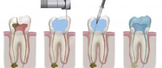
Inflammation of the tooth pulp occurs due to the penetration of infection from the carious cavity. Stage No. 1: Opening the tooth cavity. Using a drill machine, carious tissues are removed from the carious cavity, and access to the equine canals is opened. The nerve of the tooth or its remains are removed.
Stage No. 2: Mechanical and medicinal treatment of the root canals of the tooth. The infected tooth pulp or what is left of it is removed, the canal walls are thoroughly cleaned and disinfected with strong antiseptics.
Stage No. 3: Obturation of the root canals. The treated sterile tooth canals are obturated (filled) with a special material based on gutta-percha.
Stage No. 4: Placement of the filling. Endodontic treatment ends with closing the tooth cavity and placing a filling. If necessary, the tooth is covered with a crown.
Stage No. 5: Installing a crown on the tooth. If a tooth is more than 50% damaged, it is recommended to restore the tooth with a crown rather than a filling. The crown allows you to create a stronger and more durable structure.
Anesthesia
High-quality anesthesia is very important to ensure comfort and painlessness of the procedures.
Creating conditions for quality treatment.
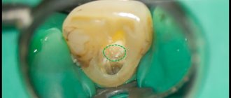
Photo: dental treatment with a rubber dam The tooth is isolated from the oral cavity, from saliva and tongue using a rubber dam. It is convenient for both the doctor and the patient, as it allows the patient to partially cover their mouth to rest during treatment without compromising quality.
The main goal of endodontic treatment is high-quality treatment and disinfection of the root canals of the tooth. During work, the ingress of saliva or other liquids can nullify all the doctor’s efforts. To isolate the tooth from the oral cavity, a rubber dam is used, a special rubber curtain installed on the tooth, which allows you to create ideal sterile conditions. A rubber dam (rubber dam, etc.) also eliminates the possibility of concentrated solutions getting into the oral mucosa.
The rubber dam greatly facilitates the doctor’s work and allows the patient to rest during the work. The patient can close his mouth, rest, and the doctor, at the same time, may not worry that something (infection) will get into the tooth cavity, etc.
Stage No. 1: Opening the tooth cavity
Using a drilling machine, damaged and carious tooth tissues are removed and the tooth cavity is opened to gain access to the canal openings.
Stage No. 2: Mechanical and medicinal treatment of the root canals of the tooth.
Before starting canal treatment, the inflamed remains of the tooth nerve are removed. To treat root canals, special instruments (files) and medications are used. In appearance, the files resemble small files, which are used to “scrape out” necrotic and infected tissue from the canal walls. The task of this stage is to completely remove the infection from the canals and microtubule system and clean the tooth cavity from decay products formed during the inflammatory process. To disinfect the canals, strong antiseptics are used that can completely eliminate the infection that has entered the tooth. Contact of some disinfectants with the oral mucosa can cause burns, so during root canal treatment it is important to carefully isolate the tooth from the oral cavity.
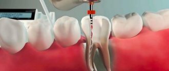
Stage No. 3: Obturation of the tooth canal.
After the canal has been treated, it must be hermetically obturated (sealed) to prevent re-infection. The channel cannot be left empty; it must be hermetically sealed.
Stage No. 4: Placement of a filling or crown.
After filling the dental canals, it is very important to effectively close the access of infection from the oral cavity to the dental cavity. Therefore, you cannot leave a tooth without a filling even for several days. Since the infection easily spreads through the dentinal tubules, which can cause re-infection of the tooth and the development of root resorption (root resorption).
Root canal disinfection
After removing the infected pulp, the doctor rinses and cleans the pulp chamber and root canals. If necessary, he expands the root canals so that they can later be filled more efficiently.
The dentist can then inject medication into the pulp chamber or root canal to permanently kill the infection. If it has spread to other areas of the mouth, you may be prescribed antibiotics. Also, on the recommendation of the dentist, other procedures may be prescribed.
If treatment needs to be continued at the next visit to the doctor, the dentist may install a temporary filling or a temporary crown. In the latter case, the restoration will help protect the tooth from food debris and saliva until a return visit to the clinic. Try not to bite or chew food with a tooth with a temporary crown until it has been fully treated and restored with materials designed to last.
Myths about root canal treatment.
MYTH No. 1. Removing the nerve of a tooth is painful as hell.
Modern anesthetics make it possible to achieve complete pain relief even if the tooth has been bothering you severely and for a long time and there are signs of the inflammatory process spreading to the surrounding tissues with the formation of pus. This is a matter of experience and qualifications of the doctor.
Myth No. 2. If the nerve is removed, the tooth will be “dead” and begin to quickly deteriorate.
This misconception originates from the times when toxic materials and substances were used to treat tooth canals, weakening tooth tissue and making them more fragile and less resistant to environmental influences. Modern treatment methods are aimed at preserving natural strength or even strengthening tooth tissue during the treatment process.
Myth No. 3. After treatment, cysts form under a dead tooth or the bone tissue of the jaw is destroyed.
High-quality cleaning and root canal treatment is the key to the health and longevity of the tooth. A well-treated tooth lasts exactly as long as a living tooth. The development of inflammation “under the tooth” is a consequence of poor-quality treatment and cleaning of the root canal, which previously often occurred due to the lack of high-quality enlargement and imperfection of dental materials.
Myth No. 4. It is easier to remove a tooth than to treat its root canals.
We often hear objections from patients, saying, “Why carry out such a long, unpleasant and costly treatment when it’s easier to just remove the tooth?” This approach also has a right to exist, but it is necessary to understand that the chewing load that was previously borne by the extracted tooth will be taken over by neighboring teeth, which will negatively affect their stability and durability.
Stages of periodontitis
Acute
It is characterized by the rapid development of the disease. The tooth may change color and become mobile; swelling of the gums and unbearable pain are often observed. Often the cause of the disease is tooth trauma.
Chronic
Chronic forms of periodontitis occur much more slowly and are partly asymptomatic. It often occurs with caries, under a filling or crown, so visually detecting the disease can be quite difficult. Sometimes a fistula is observed on the surface of the mucosa in chronic periodontitis. However, the disease can be easily diagnosed using x-rays. In addition, chronic periodontitis in the acute stage causes pain, as well as the feeling of an “overgrown tooth”.
Why, in general, is it necessary to treat root canals?
The main task of an endodontist is to preserve the tooth and return it to the ability to perform its function for the maximum period. The main task of endodontic treatment is to eliminate the source of chronic infection from the tooth canals, preparing it for further restoration, i.e. The endodontist returns the tooth to service. If you do not treat a tooth with an inflamed nerve, the tooth will eventually collapse and the infection from the root canals will spread to the bone tissue surrounding the tooth, which will lead to its loss and deformation. Severely damaged teeth must be removed.
Endodontic root canal treatment under a microscope.
Dentist Daria Dmitrievna Shevtsova performs endodontic treatment of tooth root canals under a microscope.
High-quality treatment is only possible when the doctor clearly sees what he is doing. It is impossible to carry out quality treatment by touch. The use of a dental microscope allows you to control the entire process of passing and cleaning the root canals. In the binocular microscope, the endodontist sees the smallest details of the canals: bends, branches, microcracks and the most secret “nooks” where infection can remain - which ensures high-quality cleaning of the root canals and eliminates the possibility of damage to the tooth walls (tooth perforation).
Using a microscope:
- increases the likelihood of treatment success;
- allows the use of biocompatible materials that strengthen the walls of the tooth;
- allows you to re-treat previously unsuccessfully treated canals;
- remove foreign bodies and broken instruments from root canals;
- close tooth perforations, etc.
The canals of the teeth have a complex anatomical structure with a mass of extensions and branches along their entire length, which significantly complicates the high-quality cleaning of the canals from infection. The infected pulp remaining during tooth treatment will sooner or later lead to the development of inflammation and the tooth may become sick. It is quite difficult to see the channels with the naked eye due to their small size. Previously, dentists had to treat tooth canals “by touch,” relying on their own experience and luck. In order to definitely kill the infection that remained after instrumentation of the canal, toxic materials were used that significantly weakened the walls of the tooth. The likelihood of an unfavorable outcome with this approach is extremely high: teeth treated in this way often simply had to be removed due to subsequent complications.