The dentofacial system is an important component of the human masticatory apparatus. It meets morphological and functional characteristics, such as number, size, shape, color, structure of dental units. There are a number of genetically determined or external factors that lead to disruption of these characteristics. Dental anomalies can have various consequences: some lead to periodontal pathologies, various forms of caries, others seriously limit chewing function, cause injury to the mucous membrane, and others cause aesthetic problems.
Improper growth of teeth results in speech and bite defects and breathing problems. As a result, the patient’s general health seriously suffers, and psychological problems arise.
Reasons causing anomalies
Anomalies of the dental system are very diverse and include both anomalies of dental units and incorrect formation of the dentition, as well as abnormalities in the development of the jaws. The most frequently diagnosed abnormalities of dental units. They can be caused by a wide variety of reasons, including both exogenous (caused by external factors) and endogenous (formed as a result of the physiological state of the patient).
Endogenous causes
Endogenous anomalies are formed as a result of genetic and endocrine disorders.
- Many structural features of the dental system, leading to improper development of teeth, are genetically determined. This can occur through direct inheritance (abnormal number and shape of teeth, edentia, diastema), through inheritance of a mismatch in the size of the jaw bones, through inheritance of a mismatch in the size of the jaw and teeth (crowding of teeth or sparse arrangement of teeth). Dental anomalies of this type include disorders associated with hereditary and congenital pathologies (clefts of the soft and hard palate, cleft lip, Down syndrome, Vaanderburg syndrome, Seckel syndrome, Shershevsky-Turner syndrome).
- Another part of the endogenous causes of dental anomalies are problems of the endocrine system, such as hypothyroidism (in later stages), hyperparathyroidism, hypocortisolism. Their frequent symptoms may be delays in the eruption and replacement of teeth, and disturbances in the formation of the enamel layer.
Exogenous causes
Exogenous (external) causes of dental anomalies are, for the most part, external unfavorable factors that affect the baby’s body during the formation of tooth germs. There are prenatal (prenatal), postnatal (postpartum) and intranatal (associated with childbirth) periods.
- An example of a prenatal factor is a pregnant woman living in poor environmental conditions. Various developmental disorders of the fetus can also occur as a result of the mother’s poor lifestyle.
- Factors in the intranatal period may include complicated labor, birth injuries, consequences of asphyxia and oligohydramnios.
- In the postnatal period, dental anomalies develop as a complication of pathologies in early childhood. These include childhood infections, rickets, hypovitaminosis, and micronutrient deficiency.
All these reasons are of a common nature. Local factors that affect dental development include everything related to improper oral care, feeding errors or bad habits. Among them are feeding too soft food, prolonged sucking of a pacifier or finger in early childhood.
Childhood injuries and caries with complications lead to dental anomalies.
The death of tooth germs or the development of supernumerary teeth can occur due to osteomyelitis.
Installation of crowns
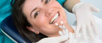
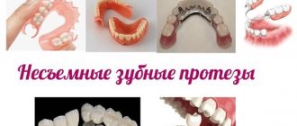
These are non-removable structures that can be used to eliminate dental defects. The installation of crowns is also indicated in case of severe destruction of dental units. The essence of the method is to insert an artificial tooth into the gum using a pin made of metal.
Installing crowns requires some preparation. The patient's natural teeth are prepared and, if necessary, pulp removal is performed. Crowns take several days to make. During this period, the patient is provided with temporary structures. The finished crowns are completely identical to the patient’s teeth. They differ only in width, which makes it possible to hide large gaps between chewing units.
The following conditions are contraindications: loose teeth, allergic reaction to the components used, periodontal pathologies. In addition, crowns are not placed on anyone under 16 years of age.
Anomalies in the number of teeth
All anomalies in the number of teeth are divided into three types:
- hyperodontia (excess of dental units above normal);
- edentia (complete absence of teeth);
- hypodontia (insufficient number of teeth).
Hyperodontia
Hyperodontia is a disorder of the formation of the dentition, which is expressed in the presence of supernumerary teeth (over 32 dental units in an adult). The cause of the disease may be a violation of the mechanism of formation of tooth germs in childhood or infancy. Hyperodontia mainly affects the incisors of the upper jaw. Other teeth are involved in the pathological process less frequently. Extra dental units grow small and have a cone shape.
The disease can be true or false.
- With true hyperodontia, the formation of extra dental units occurs in the human jaw.
- False hyperodontia occurs when the loss of primary teeth is delayed, which interferes with the normal growth of permanent teeth.
With polyodontia, teeth may erupt away from the dentition or next to a correctly positioned dental unit, which leads to its gradual displacement. The dentition can be seriously deformed, which leads to serious diction problems.
If supernumerary teeth interfere with the development of normal teeth, they must be removed. Broken teeth are corrected by installing braces.
In those rare cases when a row violation does not occur, the supernumerary tooth is preserved. Prosthetics are used to correct the shape.
Hypodentia
Hypodentia is a deficiency of dental units. This disorder occurs most often due to the death of tooth germs in the fetus. A small number of teeth causes a shift in the midline, the formation of wide gaps between teeth (diastema and three), and shortening of the dentition. All this adversely affects the bite. Possible ways of correction are prosthetics.
Edentia
Adentia is a complete or partial absence of teeth. With edentia, the continuity of the dentition is disrupted, which leads to difficulties in chewing and speaking. The reason is a violation of the development or death of tooth germs under the influence of genetic factors or under the influence of unfavorable external factors during the formation of the dental plate in the fetus (the formation of the germs of permanent teeth in the fetus occurs after the 17th week of intrauterine development). The disorder is corrected using prosthetics or dental implantation.
Supernumerary molar: review of a rare case
Supernumerary teeth or hyperdontia is a dental anomaly that is defined as the presence of a tooth or any dental tissue in excess of the set of 20 primary teeth and 32 permanent teeth. Supernumerary teeth can occur singly, in groups, unilaterally, bilaterally, they can erupt or be impacted on one or both jaws, both in the primary and permanent dentition. The frequency of occurrence in primary dentition varies from 0.1% to 3.8% and from 0.3% to 0.6%. In permanent dentition, the anomaly is more common in men than in women in a ratio of 2:1. However, this gender disproportion is not observed in the primary dentition. There is also evidence that the Asian population is more susceptible to the anomaly. Single supernumerary teeth occur in 76-86% of cases, double teeth in 12-23% and multiple teeth in less than 1%. Multiple hyperodontia rarely occurs in people without any other concomitant diseases and syndromes. Typically, this anomaly is part of systemic disorders such as cleft lip and palate, cleidocranial syndrome, Gardner syndrome, Fabry-Anderson syndrome, chondroectodermal dysplasia, Euler-Danlos syndrome, and tricho-rhinophalangeal syndrome.
Supernumerary teeth can be found in almost any area of the dental arch. Localization on the upper jaw is much more common than on the lower jaw, especially in the anterior region (80%). Somewhat less frequently, supernumerary teeth can be located in the distomolar zone, lower and upper premolars, in the area of the upper canines and lower incisors.
The crowns of abnormal teeth have a normal appearance or an atypical shape, and the roots are also fully or partially formed.
The position in the dental arch varies: mesiodens, paramolar, distomolar and parapremolar. Mesiodens is the most typical localization between the central incisors on the upper jaw, paramolar position is an additional molar, usually rudimentary, small in size and located on the buccal or palatal side in relation to one of the molars on the upper jaw. Most often found in the interdental space of the second and third molars on the buccal side; distomolar position is the fourth permanent molar; parapremolar localization is mainly found in the interdental space on the buccal side between the first and second premolars in the upper jaw. Variations in the morphological shape include different conical type, number of tubercles, and odontome. Supernumerary teeth may be small, conical with a normal root; teeth with multiple cusps are usually short, with a barrel-shaped crown and an invaginated rudimentary root. Another variant of a supernumerary tooth - an additional one - resembles one of the existing ones and is located behind it. Most supernumerary teeth in primary dentition are of the accessory type.
Odontomas are any tumors that develop from tooth tissue. Many authors are inclined to believe that odontomas are a hamartoma or malformation rather than a neoplasm. Compound and compound odontomas are two different types described. Complex odontomas are characterized by diffuse dentin tissue that is completely disorganized, while compound odontomas are malformations that have superficial anatomical similarities to a normal tooth.
According to their shape, supernumerary teeth are classified into additional (eumorphic) and vestigial (dysmorphic). If supernumerary teeth have normal morphology, they are classified as “additional”; if the morphology is abnormal, the teeth are classified as vestigial. The position of the supernumerary teeth can be between the central incisors, overlapping, and the orientation is described as vertical, inverted, or transversal.
This article presents a clinical case of the presence of an additional molar in a somatically healthy patient. A review of the literature regarding the incidence, classification, etiology, complications, diagnosis and treatment strategies of this pathology is also presented.
Description of a clinical case
A 22-year-old man came to the Department of Conservative Dentistry and Endodontics with complaints of pain in the posterior segment of the upper jaw on the left. Hereditary anamnesis and disease history are unremarkable; no signs of systemic diseases or syndromes have been identified.
Intraoral examination revealed Class I occlusion and no pathological tooth alignment. In addition to the full set of permanent teeth, one supernumerary tooth was found, located on the palatal side between the upper first and second molars on the left (Figure 1).
Figure 1: Intraoral photograph showing the paramolar position of the supernumerary tooth between the upper first and second molars on the left.
The supernumerary tooth is defined as a paramolar. The paramolar crown had two cusps and very much resembled the structure of a permanent premolar. The tooth is rotated axially, with the buccal surface distally and the mesial surface buccal. A carious lesion was found on the mesial side of the paramolar (Figure 2). Examination of the soft tissues revealed periodontal inflammation between the first and second molars and paramolars. X-rays were taken: panoramic, sighting and occlusal. Reading the panoramic image was difficult due to the palatal position of the tooth. On sighting and occlusal photographs, it was discovered that the supernumerary tooth was affected by caries and had one root (Photos 3 and 4).
Figure 3: Spot X-ray showing a paramolar with a fully formed tooth (indicated by arrow).
Figure 4: Occlusal radiograph of the maxilla showing the supernumerary tooth (arrow).
The patient was informed of the existing situation. It is recommended to remove the paramolar due to its inconvenient location for hygiene, possible food retention, recurrence of caries and damage to periodontal tissue. The patient was sent to the Department of Maxillofacial Surgery for paramolar removal.
The extracted tooth is cleaned, disinfected and analyzed. The morphology of the tooth is normal. The length of the root corresponds to the size of the crown. The root apex is fully developed. X-ray examination revealed type I canal configuration (Vertucci). Actual tooth dimensions: mesiodistal and bucco-palatal crown width 6 and 10 mm, respectively, crown length 6.5 mm, root length 12 mm. Morphometric measurements showed a high similarity of the supernumerary tooth with the premolar (Photo 2).
Photo 2: Photographs of the extracted tooth: (a) occlusal view, (b) mesial, (c) distal, (d) buccal, (e) palatal.
Discussion
The appearance of paramolars is a fairly rare occurrence. The etiology of this anomaly is not fully understood. Several theories have been proposed: phylogenetic, dichotomous, dental lamina hyperactivity theory, and a combination of genetic and environmental factors.
Phylogenetic theory refers to the process of atavism (evolutionary return). Atavism is a return to an earlier morphology or type. In past centuries, the third molar was almost always present in the permanent dentition; it was comparable in size to the second molar. Moreover, the fourth molar was also quite common. However, as a result of the evolution of phylogeny, the size of the dental arches gradually decreased, which led to a reduction in both the number and size of human teeth. This was one of the stages of preferential development of the cerebral skull over the facial skull. Thus, the appearance of additional paramolars can be considered an example of atavism, the genetic memory of the fourth molar in previous generations. It is worth saying that this theory was rejected by many authors.
The dichotomous theory explains the appearance of supernumerary teeth by splitting the tooth germ. The rudiment splits into two equal or unequal parts, from which morphologically normal independent teeth subsequently develop.
The lamina hyperactivity theory is the most accepted theory. She explains the appearance of paramolars as a result of local, independent, due to special stimulation, increased activity of the dental plate. According to the theory, lingual expansion of the accessory tooth bud leads to the development of a morphologically unchanged tooth, and the vestigial forms arise from the proliferation of epithelial lamina remnants, which is induced by the pressure of permanent teeth. Others are inclined to believe that hyperodontia is associated with multifactorial causes, which are still based on hyperactivity of the dental plate. Remnants of the dental lamina may remain in the jaws in the form of epithelial pearls or islands. When exposed to inducing factors, supernumerary teeth or odontomas can develop from additional rudiments. The best supported hypothesis is that the development of supernumerary teeth is associated with a complex of genetic and environmental factors. This is confirmed by the presence of similar anomalies in close relatives. However, despite the literature data, similar pathology was not found in the relatives of the described patient.
A careful analysis of the literature revealed very little information about the appearance of paramolars. Paramolars are somewhat less common in the upper jaw, very rarely bilaterally and almost never in the primary dentition. They are usually vestigial and located buccally between the second and third molars, although in some cases they can be located between the first and second molars. Fusion of paramolars with normal teeth is also incredibly rare. The literature describes the only case of endodontic treatment of a fused second left molar in the lower jaw and a paramolar with a split crown.
Diagnosis also requires differentiation of other structures that may appear in the molar area, such as an additional cusp or a fused supernumerary tooth. Bolk in 1916 first described an additional cusp on the buccal surface of the upper and lower permanent molars, which he called the paramolar cusp. Dahlberg in 1945 proposed the term paramolar cusp to refer to any abnormal cusp, supernumerary inclusion, or elevation on the buccal surface of both maxillary and mandibular premolars and molars. He presented a paleontological nomenclature in which he classified these structures as “protostylid” if they are on the lower jaw and “parastylid” if on the upper jaw. It is widely accepted today that such formations originate from the cervical region of the tooth and are variable in appearance. Often these structures appear on the buccal surface of the mesiobuccal tubercle and quite rarely on the distobuccal tubercle. It is believed that the paramolar tubercles may originate from the remains of their own epithelium or be a genetic remnant from mammals and lower primates.
Supernumerary teeth may erupt normally, remain impacted, or appear axially rotated or with other abnormalities. Supernumerary teeth with a normal position in the bone usually erupt. However, only 13-34% of supernumerary teeth in the permanent dentition erupt normally, compared to 73% in the primary dentition. The rest may remain impacted and cause complications.
The development of complications can cause a delay in the eruption of associated permanent teeth, retention, ectopic eruption, disposition, rotation of adjacent teeth, crowding due to insufficient space for eruption, malocclusion due to a decrease in space in the dental arch during the eruption of paramolars, tremors in the molar area, traumatic bite and ulceration of the buccal mucosa with buccal placement of paramolars, difficulties in orthodontic treatment, pathological development of the root of associated permanent teeth, formation of follicular cysts from the follicular sac of a supernumerary tooth, trigeminal neuralgia due to compression, pulp necrosis and root resorption due to excessive pressure of the paramolar , caries due to plaque accumulation, gum inflammation and localized periodontitis. As can be seen from the described case, due to plaque retention, carious lesions of the paramolar and inflammation of the surrounding periodontium occurred.
Most supernumerary teeth are impacted and are usually discovered incidentally during radiographic examination. However, if a patient presents with complications that are often associated with the presence of a supernumerary tooth, the dentist should consider this anomaly in the differential diagnosis and insist on appropriate x-ray examination.
The most valuable radiographic examination is the OPG with additional targeted photographs and photographs of the upper and lower jaw in the occlusal plane. To clearly localize an unerupted tooth, use the vertical or horizontal parallax technique. Parallax is a change in the view of an object against a specific background based on the movement of the viewer. This technique can be carried out by taking images of the same area, but from different angles, with two different devices. When using this method, as a rule, the reference point is the root of the adjacent tooth. In addition, cone beam CT may be used. This technology provides a three-dimensional image of the structures of the specified zone and is incredibly informative for the described anomaly.
Clinical management of patients with paramolars depends on the position of the tooth and its effect on surrounding tissues and important anatomical structures. Treatment offers two options: removal or observation. Observation does not include any manipulations other than clinical and radiological monitoring of the patient. This method is preferable if the presence of a paramolar is asymptomatic and does not cause any inconvenience. If any complications arise, tooth extraction is advisable. In the described case, we resorted to tooth extraction in order to maintain the proper level of hygiene, prevent the carious process and preserve the surrounding periodontium.
Conclusion
The dentist needs to know about the different types of supernumerary teeth for proper diagnosis and timely detection of this anomaly. Each such case requires careful diagnosis and subsequent appropriate treatment that causes minimal complications.
Authors: Gurudutt Nayak, Shashit Shetty, Inderpreet Singh, Deepti Pitalia Department of Conservative Dentistry and Endodontics, Kanti Devi Dental College and Hospital, Mathura, Uttar Pradesh, India
Anomalies of magnitude
There are two forms of this type of deviation, including macrodentia (increase in the size of dental units) and microdentia (small teeth).
Macrodentia
With macrodentia, the size of dental crowns is significantly increased. The cause of the disorder is, in most cases, dysfunction of the endocrine system, which is characterized by the fusion of several rudiments together. The disease is localized mainly in the area of the upper incisors. Large teeth interfere with the process of eruption and growth of neighboring dental units, which leads to their abnormal arrangement and crowding. Giant teeth are found on or outside the dentition line. This is a serious cosmetic defect that causes serious functional impairment and psychological disorders. Pathologically enlarged teeth are removed, and the growth of adjacent teeth is corrected using prosthetics or braces.
Microdentia
Microdentia – teeth that are too small. The disorder mainly affects the upper lateral incisors, but may affect the incisors of the lower or both jaws. The cause of the anomaly has not been studied, but it is reliably known that it develops in genetically predisposed patients. With the problem of small teeth, patients develop too large interdental spaces, which significantly disrupts the aesthetics of the dentition. To correct the pathology, incorrectly growing teeth are removed or covered with crowns.
Methods for correcting bite
The methods used in dentistry to correct bites are divided into 2 groups - surgical and non-surgical.
Surgical methods
Surgical techniques are necessary if the patient is diagnosed with complex anomalies of the dentofacial apparatus, high crowding of teeth, or if treatment needs to be carried out quickly. The intervention is carried out under sterile conditions. For pain relief, both general anesthesia (if necessary, incisions of bone tissue) and local anesthesia (if necessary, incisions on the gums) are used.
The rehabilitation period after surgery is up to 3 weeks. After surgery on the bone tissue, the patient will need to develop the jaw. Additional braces may also be required.
Surgical techniques are only suitable for treating adults - when the jaw is already fully formed. In severe situations, tooth extraction and implantation may also be required.
Non-surgical methods
If, during the diagnosis, the doctor identified mild to moderate jaw pathologies in the patient, then correction of malocclusion in adults can be done without surgical intervention. For this, orthodontists use:
- bracket systems,
- orthodontic devices,
- trainers,
- veneers,
- crowns,
- mouth guards
The first option is often chosen, as it is the most effective.
Shape anomalies
There are pathologies in the development of dental units, due to which they acquire an unnatural shape. These disorders are named after the scientists who first described them. The following types of dental shape anomalies are found: spiny teeth, Pflueger teeth, Fournier teeth, Hutchinson teeth.
- Spiked or awl-shaped teeth. With this pathology, the teeth acquire a cone-shaped shape. Wide at the base, they gradually narrow and, towards the cutting edge, become sharpened into a spike shape. This problem is combined with microdentia. There are irregularities and stains on the surface of the teeth. The disease affects the front and lateral incisors. The disease occurs in childhood and is caused by genetic factors in combination with external factors. Among them are vitamin D deficiency and endocrine system problems.
- Hutchinson's teeth are characterized by a modified crown shape of the incisors. Externally, they have the shape of a barrel, since the neck is significantly thickened. The cutting edge of the teeth acquires an arched notch. The enamel layer also suffers, which is present only on the sides and absent in the center.
- Fournier's teeth are a form of systemic hypoplasia of dental units associated with metabolic disorders at the stage of intrauterine development of the fetus. The barrel-shaped shape of the teeth is preserved, but the notch of the incisal edge is absent. The color of the enamel is not disturbed, but the enamel layer is underdeveloped, which can be seen during microscopic examination.
- Pflueger's teeth are a disorder that affects permanent dental units. Their crowns take on a conical shape. Thickening develops in the cervical region, and the chewing surface is significantly underdeveloped. The chewing function of the teeth is completely preserved.
- Turner anomaly. With this pathology, there is no enamel on the teeth. They have an abnormally lumpy surface, and replacement tissue, dentin, forms in the exposed areas.
Systemic hypoplasia with a violation of the shape of the teeth has three degrees of development. The third degree is the most dangerous, in which the crown is severely deformed and the enamel layer decreases. In this case, the defective teeth are removed and replaced with dentures. It is also possible to restore affected teeth using reflective components.
Anomalies of hard tissue structure
An altered shape, abnormal size or color of enamel is formed against the background of an abnormality in the structure of the hard tissues of the dental unit. Among such anomalies are:
- Hypoplasia (underdevelopment of tissue). The initial degree of the disorder is manifested by the presence of chalky spots on the enamel and areas where tissue deficiency is observed. Subsequently, all kinds of pits, grooves, and recesses appear on the surface of the enamel. The defect affects all teeth in the dentition.
- Hyperplasia (excessive tissue formation). Pathology also affects all teeth at the same time. It is characterized by areas where tissue growth is observed - tubercles, sagging enamel. The consequence of the pathology is a change in the shape and size of the teeth, a violation of the occlusion (the line of contact of the upper and lower jaws).
- Anomalies of amelogenesis (enamel formation) are expressed in the presence of brown or yellow spots on the surface of the teeth. Areas where the natural composition of the enamel is disrupted become especially sensitive. Microdentia develops against the background of pathology. The cause of the development of pathology is a deficiency of microelements that take part in the formation of dental tissue. Treatment consists of replenishing this deficiency. Additionally, local remineralizing therapy and physiotherapy are performed.
- Disturbance of dentinogenesis consists of dysfunction of the mechanism of dentin formation. The main symptoms of the disease are as follows: teeth become yellow-brown or grayish in color. The fragile dentin in these areas quickly wears off, causing tooth decay and then tooth loss. The disease occurs in genetically predisposed patients. The problem can only be solved by replacing the units destroyed by the disease with prosthetics.
Sealing
Currently, it is one of the most affordable ways to treat rare teeth. Its wide distribution is due to its low cost and ease of implementation.
The essence of the method is as follows: fillings of the same color as the natural chewing units are applied to the teeth. The procedure is considered a jewelry procedure, since the doctor must literally mold new bone structures. As a result, the teeth become wider and the gaps between them disappear. Only a specialist can visually distinguish fillings from natural dental units.
During the procedure, orthodontists use only high-quality materials. These are ceramics and porcelain.
After filling rare teeth, the functioning of the gastrointestinal tract is normalized and a correct bite begins to form.
Color anomalies
Healthy, young teeth look snow-white due to a thick dentin layer and high-quality enamel, which has sufficient characteristics of whiteness, shine and transparency. Lifestyle, age, genetic factors, and ecology can slightly change the color of teeth from bluish to yellow. Small deviations are not considered pathology, although they indicate a change in the quality of the enamel.
Various pathological processes occurring in the body have a more significant impact on the condition of the enamel. The cause may be carious lesions, the use of medications, or a lack of certain microelements in the body. Teeth can take on a gray, pinkish, brown, purple and even black tint.
When starting to treat a patient, the dentist excludes the development of chronic pathologies. After a course of therapy and stable remission is achieved, other causes of discoloration of the enamel are eliminated. The final stage of treatment is a course of teeth whitening.
Specialists at the Consilium Dent choose the enamel lightening technique together with the patient. The selection criteria are age, condition of the enamel, and the wishes of the patient.
Position anomalies
Incorrect position of teeth always develops as a consequence of another pathology and forms an incorrect bite. May affect one or both jaws. Incorrect position of teeth makes chewing food and oral hygiene difficult. This can lead to digestive problems and the development of cavities. There are several options for the pathological position of teeth within or outside the dentition:
- External or vestibular position means that the tooth is located outside the dentition, closer to the vestibule of the mouth. This position is typical for canines and incisors and develops as a serious cosmetic defect.
- Oral or internal position. The teeth are deviated inward closer to the tongue. The disorder is typical for canines, incisors and premolars. May cause tongue injuries and dysfunction of the temporomandibular joint.
- Mesial and distal position. Dental units are displaced forward or backward along the dentition, which leads to its shortening.
- Supra and infraocclusion are high or low positions of teeth relative to the occlusal curve. The cause of the disorder is underdevelopment of the alveolar process. It appears due to the presence of some obstacle that interferes with the normal formation of the tooth germ. Effective therapy is surgery.
- Tortoanomaly. The tooth is rotated around a vertical axis. The disorder is typical for incisors, canines, and premolars. The anomaly can cause injury to adjacent teeth. Dental units are rotated into the correct position using orthodontic structures.
- Transposition. The teeth change their location with each other. The disease affects canines that exchange places with premolars or lateral incisors. The disorder develops at the stage of tooth germ formation. The therapy is as follows: problematic teeth are removed followed by prosthetics.
- Crowding of teeth occurs due to lack of space when tooth germs are located too closely. Crowded teeth are closely adjacent to each other and rotate around their axis. Occurs in combination with macrodentia or with an undeveloped basal part. To eliminate the violation, some dental units are removed.
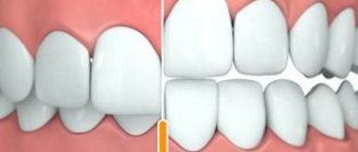
- Diastemas and tremas are wide spaces between teeth. The disorder develops as a consequence of an abnormal shape and size. The problem is treated with orthodontic methods or by installing veneers, which helps restore the aesthetics of the smile.
Misaligned teeth cannot always be cured using braces. Severe pathology, which is characterized by a skeletal disorder of the maxillofacial region, is treated with surgical reconstruction. The pathological fragment is transferred to the desired position and then secured. A fragment containing part of the dentition or the entire dentition can be used.
Correction of bite in adults: features and contraindications
Jaw defects cannot always be corrected during active tooth growth (up to 18 years of age). But treatment is possible even after 40 years. Bite correction in adults is carried out using the same methods as in children. But you will need to take into account that with age, body characteristics and contraindications appear.
Contraindications for bite correction:
- oncology,
- severe pathologies of the respiratory system,
- mental disorders,
- blood diseases,
- pathologies of the cardiovascular system,
- missing several teeth in a row,
- disruption or reduction in the process of bone tissue regeneration, the patient does not properly maintain oral hygiene.
Each case is unique, so the doctor’s task is to conduct a full diagnosis, select orthodontic plates, braces, aligners and trainers that will help solve the patient’s problem.
Diagnostics
An experienced dentist diagnoses dental abnormalities after an external examination and questioning of the patient. To clarify the diagnosis and identify the causes of the disease, a more detailed examination of the dental system is carried out:
- The construction of diagnostic plaster models and odontometric measurements make it possible to accurately identify changes in the size of dental units and their characteristics, which indicate the presence of pathologies.
- The color and transparency of the enamel are determined using a photographic method.
- Computed tomography or teleradiography makes it possible to obtain information about the condition of abnormal dental units in order to determine further treatment tactics.
- Panoramic radiography of the jaws is an important diagnostic method
- Electromyography of the jaws helps to assess the functional state of the facial muscular system.
To carry out differential diagnosis, the patient is referred for consultation to specialized specialists: endocrinologist, otolaryngologist, geneticist. Based on the diagnostic results, the attending physician determines treatment tactics. Depending on the type of disorder and the complexity of the disease, the patient may need the help of a dentist, surgeon, orthopedist, or implantologist.
Types of bracket systems
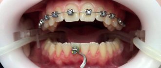
Braces are a system that consists of a power arch and brackets attached to the teeth. Since the arch presses on the teeth, they begin to gradually shift, occupying a physiologically correct position. This design is worn constantly. It can only be removed by an orthodontist for correction.
Braces systems differ in the method of fixation and location, and in the materials from which they are made.
According to the method of fixing the arc
To correct dental anomalies, the dentist can use 2 types of brace systems that differ in design:
- non-ligature (self-ligating) - in this case, the arc will fit into a special groove, and not be fastened with ligatures; this design looks more aesthetically pleasing;
- ligature - clasps on the arch are fixed using ligatures.
The type of braces also affects the length of time they are worn and the frequency of visits to the dentist. Thus, ligature ones require visiting a doctor for correction every month, and self-ligating ones - once every 1.5–2 months.
At the location of fixation
Bracket systems can be attached:
- on the outside of the dentition - such braces are called vestibular or external, they look less aesthetically pleasing, but are effective and have a low cost;
- on the inside of the teeth - these are lingual braces; their main advantage is high aesthetics. It is impossible to see braces from the outside, but wearing them affects diction; they are more expensive than vestibular ones.
The type of brace system is selected based on the patient’s situation, as well as taking into account his wishes. So, if a person works with people, and a lot depends on his appearance, he should choose lingual braces to correct his jaw bite.
By material
Depending on the material from which the braces are made, they can be:
- metal, their base is medical steel, titanium or gold; the design is hypoallergenic and does not react with substances that may enter the oral cavity;
- plastic - more aesthetic than the previous option, they are practically invisible on the teeth, but they can be stained and have low strength;
- ceramic - their advantage is a strong fixation, they are also not visible on the teeth, but can be stained if a person often drinks coffee, tea or wine;
- sapphire, based on aluminum oxide, they are aesthetic, invisible on the teeth;
- combined - combine the strength of metal systems and the high aesthetics of ceramic and sapphire.
To correct malocclusion, metal, combined or ceramic braces are often used.
Prognosis and prevention
Consilium Dent clinic uses advanced methods of therapeutic, surgical, and orthopedic treatment of dental anomalies in patients. By skillfully combining them, experienced specialists are able to restore the aesthetics of a smile and ensure the normal functioning of the dental system, even if the degree of deviation is significant.
Prevention of anomalies in the formation and development of dental units begins during the period of intrauterine development of the fetus and is carried out throughout a person’s life. The main stages are as follows: monitoring the successful course of the prenatal period, caring for harmonious postnatal development, organizing a correct lifestyle while the baby is growing up, caring for dental health and general health throughout life. By conducting an annual preventive examination, the dentist identifies dental anomalies at an early stage, when the pathological process is still reversible.
Expert of the article you are reading: