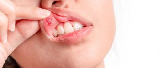
Periostitis of the jaw (flux) refers to dental diseases. The localization of the source of inflammation occurs in the periosteum. A subperiosteal abscess is formed, and the peri-maxillary soft tissues swell. A person experiences painful sensations radiating to the temporal region, ear, eye; his health worsens: weakness, sleep disturbances, fever, and headaches occur.
Periostitis, aka gumboil
The disease is diagnosed by inspection and palpation; an x-ray is required for confirmation. The doctor opens and drains the subperiosteal abscess and removes the tooth that is the source of infection. After this, the patient must undergo a course of physiotherapy and antibiotic treatment. It is also necessary to rinse your mouth regularly.
What kind of diagnosis is this? According to the international classification ICD-10, accepted throughout the world, periostitis of the lower jaw is assigned code K10.2.
Reasons for the development of periostitis
The causes of periostitis of the jaw are usually infection or injury. According to statistics, in practice this disease is observed in 5.4% of patients with inflammation in the face and jaw. Moreover, 95% of patients have an acute form, 5% have a chronic form. Localization of periostitis is observed one and a half to two times more often in the lower jaw. The clinical picture, both local and general, is peculiar. If the treatment process is started on time, it will end successfully, otherwise the disease progresses, which carries a high probability of severe purulent complications.
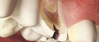
Why does this disease occur? There are several reasons. And the main one is complicated caries, in which pathogenic microflora penetrate through microchannels into the tooth cavity and then spread to the tissue surrounding the root.
Often periostitis is caused by chronic apical periodontitis. Diseased microorganisms move further and further through the channels over time. If the disease is not treated, the soft tissues are first affected, then the periosteum.
Staphylococcus always lives in the oral cavity. If the immune system is weakened, the population begins to grow and the periosteum becomes inflamed.
The hematogenous and lymphogenous form is usually caused by ENT diseases, ARVI, and infectious diseases such as measles and scarlet fever. Traumatic periostitis may occur after tooth extraction or trauma, surgical intervention, jaw fractures, infected wounds on the face, etc.
In many patients, periostitis is caused by previous hypothermia or overheating, as well as emotional or physical overload.
There are other reasons, less common: tuberculosis and other diseases that are fraught with complications spreading to the jaw periosteum.
The etiology and pathogenesis of periostitis in modern dentistry has been studied quite well. Treatment usually does not cause problems.
Clinical manifestations
Periostitis begins with pain and local swelling. These phenomena usually develop in the area of the tooth, which is the cause of the disease. The disease can begin with a serous form, but in many cases a purulent focus forms initially.
The general condition worsens. The patient experiences severe pain, which makes it difficult to eat, talk, and open the mouth. Swelling forms, which spreads to the soft tissues of the face, and flux forms.
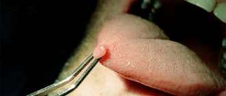
When examining the oral cavity, redness of the mucous membrane is noted. At the site of localization of the pathological process there is a clearly visible infiltrate. Over time, pus accumulates and exits into the oral cavity through the resulting hole - a fistula.
The chronic form proceeds smoothly. Pain occurs periodically, swelling is mild. There may be enlargement of regional lymph nodes and changes in facial contours.
For differential diagnosis, determining the condition of the jaw bone tissue and identifying affected teeth, an X-ray examination is performed. In acute periostitis, the bone tissue is not changed; in the chronic form, changes are observed in the form of areas of regeneration. X-rays provide diagnostically valuable information, so one cannot do without an image.
Periostitis can occur in isolation or be combined with other inflammatory diseases: periodontitis, osteomyelitis, cysts. It is also important to identify them, since a complicated process requires different treatment tactics.
Types and stages of disease development
The classification of odontogenic periostitis depends on the phase of the disease and the nature of its course. In addition to examination and palpation, the dentist prescribes an X-ray examination. The resulting image allows you to assess the condition of the roots and periapical areas. The image does not show thickening of the periosteum in the first three days from the onset of the disease.
- Various types of periostitis are classified according to the international classification ICD 10 depending on the route of infection into the periosteal tissue:
- acute odontogenic periostitis of the jaws (cause - diseased tooth);
- hematogenous (infection enters through the blood);
- lymphogenous (spread through the lymph system);
- traumatic (for injuries of the periosteum).
According to the course and pathomorphology, the disease is divided into two forms: acute and chronic.
In the acute form, the symptoms are pronounced. There is swelling of half the face, he is tormented by severe throbbing pain, and pus forms. The acute serous form is characterized by such clinical manifestations as infiltration in the periosteum, serous exudate is formed in the lesion. In acute purulent periostitis of the jaw (popularly called gumboil), a limited subperiosteal focus of inflammation is formed, fistulous tracts are formed, and pus comes out through them. The course of purulent periostitis is much more severe, with bursting pain that increases when drinking hot foods. If a fistula tract does not form, the dentist cuts the periosteum to allow the pus to escape.
The course of the chronic process is sluggish, stages of exacerbation occur periodically. Young bone tissue begins to grow on the surface of the jaw. With the development of the ossifying form, ossification and hyperostosis occur quite quickly. There are two degrees of spread of this process: limited (covering one to three teeth) and diffuse purulent (almost the entire jaw is covered).
If the wisdom tooth erupts with difficulty, retromolar periostitis may develop in the lower jaw. The pus does not come out on its own due to anatomical features, which requires dissection.
How to recognize periostitis
The most striking signs of gumboil are swelling and throbbing pain in the area of inflammation, which intensifies with pressure on the tooth. Over time, pain does not respond to painkillers, the cheek and jaw swell due to inflammation, the gums become red, pain can radiate to the eye, ear or throat (difficulty swallowing, turning the neck), body temperature rises, lymph nodes enlarge, weakness in the body is felt .
Causes of inflammation:
- neglected or untreated caries is the most common cause;
- dental intervention - flux after tooth extraction, poor quality treatment;
- sinusitis;
- angina;
- hypothermia;
- infections and injuries of teeth and gums;
- insufficient hygiene;
- Oral diseases - periodontitis, pulpitis, gingivitis and others.
Characteristic symptoms
Symptoms of acute periostitis of the jaw are determined by the form of the disease. The state of the patient’s immune system and the diseases he has are also influenced. But there are also characteristic signs that make it possible to make an accurate diagnosis.
The disease develops gradually. At first, the gums swell a little, and pressing on the tooth causes pain. Having discovered such signs, the patient should visit a doctor on the same day, otherwise the cheek may become swollen by the morning.
Signs of the form of the disease with serous infiltrate are:
- red color of the mucous membrane:
- the appearance of painful swelling in the fold between the gum and cheek;
- tolerable pain;
- temperature may rise to 37°C;
- facial asymmetry;
- submandibular and postauricular lymph nodes enlarge (lymphadenitis).
With a purulent infection, the condition becomes much worse:
- signs of intoxication and pain are observed;
- temperature (up to 38°C);
- the cheek becomes swollen;
- pain radiates along the trigeminal nerve;
- pulsation is felt in the swollen part;
- a fistula may occur;
- When you press in the area between the cheek and gum, you can feel the vibrations of the fluid.
Symptoms
At first, the symptoms of periostitis may not attract attention, and the disease may develop inconspicuously. If a tooth is affected by caries, then over time the inflammatory process affects the pulp. Then the pus gets under the periosteum and provokes the development of an inflammatory disease. At this stage, the symptoms of the disease become so acute that they can no longer be ignored.
Main symptoms:
- swelling of the gums in the area of the tooth affected by the disease, initially painless, over time the pain intensifies;
- formation of an abscess under the periosteum;
- swelling of the soft tissues of the lips and cheeks, which extends to the submandibular or infraorbital areas;
- acute pain radiating to the temples, ears, eyes;
- increase in body temperature.
How is periostitis diagnosed?
For an accurate diagnosis, the doctor collects anamnesis, conducts an external and internal examination, prescribes an x-ray and examines the x-ray. Since the symptoms of some dental problems are identical, the surgeon must have a good understanding of their symptoms.
When conducting diagnostics, doctors look for similarities and differences between chronic periostitis of the jaw or acute and other diseases of the oral cavity. An x-ray helps differentiate the disease from others (apical periodontitis, phlegmon, abscess, inflammation of the salivary gland, osteomyelitis).
Apical periodontitis is characterized by the presence of a purulent focus located at the top of the root.
Cellulitis and abscess are manifested by external changes on the skin. An infiltrate appears on the affected area, the skin over it turns red and becomes shiny.
With sialadenitis, it is necessary to palpate the salivary gland to determine its density. Osteomyelitis is diagnosed by X-ray - in the early stage of the disease, it shows decaying bones, in the late stage - formed sequesters.
Stages of the operation
When treating periostitis of the jaw, the surgeon must cleanse the cavity of pus so that the process does not spread deeper. To do this, he cuts the periosteum. The operation consists of several stages:
- Anesthesia . The affected area is numbed using modern drugs.
- Periostotomy . The surgeon makes a soft tissue incision along the fold between the gum and cheek, capturing the periosteum. This ensures the release of purulent exudate to improve the patient’s condition.
- Wound drainage . A rubber or gauze graduate is installed into the incision. This ensures the drainage of pus.
After opening the purulent lesion, the surgeon asks the patient to rinse his mouth with a disinfectant solution.
After this, chlorhexidine and similar solutions are used to wash the wound. Measures such as irrigation with dimexide with oxacillin and 15-minute applications of dimexide liniment are also effective.
After examining an x-ray of the tooth that caused periostitis, the doctor decides whether it can be preserved and treated in the future to eliminate the source of infection, or whether tooth extraction is necessary.
Treatment of periostitis of the jaw
Therapy includes the elimination of not only the source of inflammation itself, but also the reason why it arose. If periostitis is odontogenic in nature, treatment of the affected teeth is mandatory; in most cases, they have to be removed.
Serous periostitis is treated conservatively. The cause that caused the inflammation is eliminated, physiotherapy is carried out and mouth rinsing with antiseptics is prescribed. In most cases, these simple measures are enough for a quick recovery.
The purulent process requires surgical treatment. The lesion must be opened, cleaned of pus and dead tissue, and thoroughly sanitized. Drainage is installed in the wound, and regular rinsing with antiseptic solutions is carried out. To more effectively fight the infection and prevent its spread, a course of antibiotic therapy is prescribed. Anti-edema and painkillers are used as symptomatic therapy. This allows you to improve the general condition of the patient. After surgical treatment, a diet is indicated; solid foods, as well as spicy and salty foods, should be excluded.
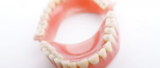
The issue of tooth preservation in acute periostitis is decided individually. Baby teeth in children, as well as permanent teeth that are severely damaged, are always removed. If it is possible to save a tooth and at the same time successfully stop the inflammatory process, a professional dentist will cope with this task. In case of chronic periostitis, the tooth must be removed in any case.
Treatment of periostitis of the jaw lasts for 5-10 days, depending on the stage and extent of the infectious process. With timely measures taken, the prognosis is favorable. If the disease is not treated, the purulent process spreads and can transform into phlegmon, osteomyelitis, and sepsis. An advanced disease causes irreversible consequences and can be life-threatening. At the first symptoms, you should contact your dentist. In this case, the cost of treatment will be lower, and negative consequences will be avoided.
Surgical intervention
With serous inflammation, as a rule, conservative therapy is limited. Pulpitis or periodontitis is treated, the patient is prescribed a course of physiotherapeutic sessions (UHF), rinsing with disinfectants. This leads to resorption of the infiltrate.
The acute purulent form requires surgical intervention to open the subperiosteal or submucosal abscess.
The milk tooth that caused periostitis must be removed. Severely damaged permanent teeth are also removed. Teeth that are capable of performing their functions need to be treated.
Drug treatment
The second stage of treatment after surgery is drug therapy. The patient is washed with modern antiseptic compounds (Chlorhexidine, Ethacridine, Dimexide with Oxacillin), and sterile bandages are applied. To impregnate them, use a disinfectant solution or ointment. Antibiotics (Oletetrin, Oxacillin, Lincomycin) must be prescribed to prevent complications and relapses. Nitrofurans (Furadonin), sulfonamides, painkillers, antihistamines (Suprastin) may be prescribed; vitamins. In the period after the operation, the patient must follow a gentle diet and rinse with antiseptics.
Physiotherapy
After surgery, the patient is recommended to undergo a course of physiotherapy to stop the inflammatory process, solux lamp, ultrasound. These are procedures such as: UHF, fluctuarization, microwave, laser, electrophoresis. Usually 5 to 7 sessions are prescribed.
Is it possible to do without it? Such a serious disease as periostitis of the jaw cannot be cured at home; qualified professional help is needed from a professional who will eliminate the source of infection. You can use folk remedies only in cases where the pain comes on suddenly, and you need to somehow improve the patient’s condition. But you need to visit a doctor as soon as possible. Before this, you can rinse your mouth with soda, salt, a decoction of herbs that have an anti-inflammatory effect, apply cold compresses to reduce swelling and relieve pain.
What to do for prevention
The main preventive measure is to constantly monitor the condition of the oral cavity. Preventive measures include:
- daily hygiene of teeth and gums;
- visiting the dentist twice a year;
- use of professional hygiene procedures, removal of tartar;
- timely treatment of caries and other pathologies;
- If you have problem gums, you need to be systematically treated by a periodontist.
To prevent periosteal disease, doctors recommend having an x-ray every year.
Periostitis of the jaw often develops even in children.
If you notice swelling on the face of a child complaining of pain, immediately take him to the dental clinic. Self-medication is unacceptable! Taking some medications may disrupt the picture of the disease, which will complicate the diagnosis and may result in serious complications. Moscow metro station Zvezdnaya, Danube Avenue, 23
Prevention
Periostitis occurs mainly due to untimely treatment of dental diseases. If caries and gum inflammation are not treated in time, they are likely to transform over time into this dangerous disease. If you brush your teeth regularly, remove any leftover food after each meal, and visit the dentist twice a year, this disease will most likely not affect you. During the examination, the specialist will conduct professional oral hygiene, identify existing problems and successfully solve them at the initial stage.
Diseases of other organs also need to be treated promptly. ENT pathology, furunculosis, as well as foci of inflammation in other organs are a source of infection, which can enter the oral cavity through the blood or lymph. Pay more attention to your health, eat right, strengthen your immune system and consult a doctor if there are pockets of infection in your body.
Injuries to the maxillofacial area can always provoke inflammation. If such a problem does occur, contact the dental clinic as soon as possible. The doctor will take measures to help prevent the development and progression of the infection.
The first symptoms of periostitis are a reason to immediately consult a doctor. Wasted time and useless self-medication can lead to serious complications. You should not hope that the symptoms will go away on their own, but that traditional treatment methods you read on the Internet will help you. This is a serious disease, so only a highly qualified dentist can cure it.