Recurrent aphthous stomatitis
Recurrent aphthous stomatitis is a chronic pathology characterized by inflammation of the oral mucosa. Its main symptom is a rash of aphthae or ulcers, which are noticeably painful. Aphthae usually appear periodically in the following areas:
- Language;
- Cheeks;
- Hard and soft palate;
- Mucous membrane of the lips.
If ulcers are periodically injured, they can develop into full-fledged, difficult-to-heal wounds, in place of which noticeable scars remain. The normal healing time for aphthae is no more than a week from the moment of its appearance.
The prerequisites for the occurrence of aphthous stomatitis are as follows:
- Chronic colitis;
- Diseases of the nervous system;
- Chronic stress;
- Injury to the mucous membrane;
- Hormonal imbalance during menstruation.
With a normally functioning immune system, ulcers heal on their own in one, or in extreme cases, two weeks. If healing does not occur, you must visit a doctor to prescribe the correct treatment. As a rule, the basis of therapy is taking vitamin C, which has a positive effect on the immune system.
Results and discussion
A study of the prevalence of TS showed that most often TS occurs in the first 1.5 months from the start of orthodontic treatment ( p
≤0.01), and subsequently the frequency decreases (Fig. 3).
Rice. 3. The incidence of traumatic stomatitis during orthodontic treatment in children and adults at various times during the active treatment period. Thus, according to the data we obtained, the incidence of TS was significantly less common than according to the literature [3–5], and amounted to 6.62–15.38% in children during different periods of the active period of orthodontic treatment with fixed appliances, and 4.69% in adults. -11.63% (see Fig. 2)
.
Before the start of treatment, TS of moderate and severe severity was detected in both children and young adults (Fig. 4, 5).
Rice. 4. The number of patients in the 1st and 2nd study groups depending on the severity of TS at different follow-up periods.
Rice. 5. The number of patients in the ٣-th and ٤-th study groups depending on the severity of traumatic stomatitis at different periods of follow-up. As a rule, the pain syndrome was mild, and the mucous membrane of the cheeks was most often affected, and less often the lips. Typically, in patients, regardless of age, with severe swelling and hyperemia, erosive and ulcerative lesions of varying severity were observed in the area of projection of orthodontic equipment (braces and arches).
The use of topical agents to treat TS has been effective in both children and adults. More pronounced positive dynamics of the reparative process of oral mucosa was noted when using gum gel with propolis in all age groups ( p
≤0.05) and observation period (
p
≤0.05).
At the same time, it should be noted that if in children on the 3rd day from the start of treatment TS of severe severity remained (see Fig. 4), then in young people (see Fig. 5) there was no such degree of TS ( p
≤0.05).
By this time, in adults, regardless of the drug used for the treatment of TS, there were no pathological changes in the mucous membrane in the area of the mucous membrane previously injured by the brace system ( p
≥0.05).
In children of groups 1 and 2, the effectiveness of treatment on the 3rd day was 34.66 and 48.96%, respectively ( p
≤0.01);
on the 5th day - 78.66 and 83.33%, respectively ( p
≤0.05). Similar data on the effectiveness of treatment of TS caused by injury to the mucous membrane of the cheeks and lips by braces and arches were obtained in young adults (Fig. 6).
Rice. 6. The effectiveness of treatment of traumatic stomatitis in patients of different groups at different periods of dynamic observation. Thus, in individuals included in the 3rd and 4th study groups, the effectiveness of treatment on the 3rd day was 60.29 and 68.03%, respectively ( p
≤0.05);
on the 5th day 91.18 and 95.08%, respectively ( p
≤0.05), which also indicated the greater therapeutic effectiveness of the gum gel with propolis for the treatment of TS caused by wearing fixed orthodontic appliances.
On the 10th day, no TS was detected in patients of all four study groups.
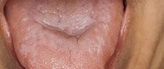
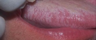
Herpetiform stomatitis
Herpertiform stomatitis is characterized by the appearance of numerous ulcers on the oral mucosa, which in appearance resemble ordinary herpes. Girls under 30 are at risk - they are the ones who suffer from pathology more than others. Locations of the disease:
- Underside of tongue;
- Floor of the oral cavity.
Ulcers heal in no more than 10 days from the moment they appear, leaving no scars. Treatment, as in the case of the previous type of stomatitis, is based on the use of vitamin C. The dosage and regimen of medications is prescribed by the attending physician.
Treatment
Like any serious disease, traumatic stomatitis requires immediate treatment. First of all, the doctor will have to eliminate the source of injury:
- In the case of a broken or chipped tooth, it is necessary to cure or remove this tooth, depending on the depth of the damage.
- Braces and dentures are improved or remade from higher quality material.
- Tartar is removed and the sharp edge of the tooth is ground down.
- If there are minor injuries, wounds and cracks are washed with antiseptics (peroxide, furacillin, infusions of calendula or chamomile).
- More serious injuries are numbed and treated with anti-inflammatory drugs.
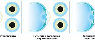
- For quick healing of wounds, ulcers and erosions, your doctor may recommend applications with rosehip or sea buckthorn oils, vitamin A oil solution and other keratoplasties.
- In cases of thermal burns or frostbite, applications of an anesthetic drug may be necessary, but in case of a chemical burn, you must first thoroughly rinse the damaged area with a neutralizer. For an acid burn, use an alkali solution, and for an alkaline burn, accordingly, use an acid solution. And only after eliminating the traumatic substance is further treatment with antiseptics and anesthetic rinses possible.
Recurrent necrotizing peryadenitis
Recurrent necrotizing peryadenitis is also called Setton's aphthae. Its symptoms are:
- Seals appear in the submucosa of the oral cavity;
- Instead of compactions, ulcers with raised edges develop over time;
- The ulcers become inflamed, causing blood and lymphatic discharge.
Places where such afts accumulate are the upper and lower lips, cheeks and sides of the tongue. The pathology is characterized by extremely severe pain. Patients find it so difficult to eat that many give up eating completely. Difficulties also arise during conversation. The healing process of aphthae lasts a long time - sometimes up to several months, and the pathology itself persists for up to several years.
Afty Bednar
Bednar's aphthae is the appearance of small wounds on the oral mucosa. The pathology manifests itself exclusively in children due to poor hygiene or accidental injury.
The location of the ulcers is the palate. They have a slightly yellowish coating and heal quite quickly - up to 5 days. For the pathology to go away, it is enough to normalize oral hygiene.
The gums become inflamed due to insufficient oral hygiene and lack of regular visits to the dentist. The result is an excessive accumulation of plaque on the teeth and pathogenic microorganisms. Pathogenic microorganisms trigger a process in which their own toxins and inflammatory mediators are generated. Treatment of diseases of the mucous membranes and gums with the Vector device
Types of traumatic stomatitis
Traumatic stomatitis can be conditionally classified according to the nature of damage to the mucous membrane and soft tissues of the mouth. All injuries are divided into three categories:
- Mechanical. They can be obtained as a result of falls, blows, or due to bad habits, such as biting the inner surfaces of the cheeks or lips, chewing hard objects, etc. In addition, the cause of traumatic stomatitis can be teeth with chipped parts, tartar, poorly fitting dentures or braces.
- Chemical. In children, chemical burns often occur when trying to taste any inedible substances. Among adult patients, traumatic stomatitis, provoked by chemical damage to the mucous membrane, can be caused by smoking and drinking alcohol.
- Physical. Such injuries most often occur as a result of a thermal burn. The risk of such damage is quite high at any age - all you have to do is sip too hot a drink or eat something scalding.
Traumatic mouth ulcers
The name of traumatic ulcers speaks for itself. This is the appearance of wounds in the oral cavity due to injury to the mucous membrane. The causes of injury may be:
- Bite of the mucous membrane;
- Accidental contact with a toothbrush;
- Inaccurate dental treatment.
One of the types of traumatic ulcers are prosthetic ulcers - wounds formed due to incorrect sizes of removable dentures. They are localized precisely under the dentures.
Healing of traumatic ulcers lasts 1-2 weeks. As a rule, this is a one-time pathology that goes away on its own without additional treatment. Such aphthae are painless and small in size.
If a complication of the disease nevertheless begins, it is necessary to visit a doctor so that he can prescribe anti-inflammatory and antibacterial medications to completely eliminate the pathology.
Tuberculosis of the oral mucosa
Tuberculosis of the oral mucosa is a complication of standard pulmonary tuberculosis. The cause of the pathology is the penetration of bacteria into the oral cavity through damaged soft tissues. Localization of ulcers occurs in the following places:
- Cheeks;
- Language;
- Floor of the oral cavity.
At the affected sites, standard tuberculous tubercles first form, and then ulcers appear in their place. They do not heal and gradually increase in size. Aphthae are shallow, but very loose with slight bleeding. Their edges are soft, but despite this, the ulcers are noticeably painful.
Usually, along with the appearance of tuberculous ulcers, the patient feels a general deterioration of his condition, which manifests itself in the following symptoms:
- Loss of appetite and weight;
- The appearance of a white coating on the tongue;
- Sweating;
- High body temperature.
Treatment of such a pathology involves staying in a special closed dispensary. As a rule, during the period of weakening of the symptoms of the disease, sanitation of the oral cavity is carried out, as well as treatment with anti-inflammatory and antibacterial drugs.
Syphilis
Syphilis is an infectious pathology caused by Treponema pallidum. Mouth ulcers are a typical symptom of syphilis that appears throughout the entire period of the disease. Moreover, the ulcers themselves undergo a certain development process:
- The ulcers are round in shape and have dense edges. They are painless and have a white coating.
- Over time, the ulcers begin to bleed slightly.
- After about 1-3 months, the ulcers heal, and in their place strong scars form, around which dense bluish edges are concentrated.
After defeating syphilis, dense scars still remain in the oral cavity. They are the most eloquent sign of a recent illness.
Treatment of syphilis is carried out in a closed venereology clinic. During remission, the oral cavity is treated with anti-inflammatory and antibacterial drugs.
Acute necrotizing gingivostomatitis
Acute necrotizing gingivostomatitis is a viral infectious pathology in which ulcers form on the surface of the entire oral mucosa, including the tonsils. The prerequisites for this disease are:
- Deterioration of the immune system;
- Injury to the oral mucosa;
- Lack of vitamins and minerals in the body;
- Chronic severe fatigue;
- Sudden hypothermia;
- Complication of ordinary stomatitis or viral pathologies.
The risk group is mainly men under 30 years of age. Ulcers are wounds with a loose bottom, uneven edges and a dirty green coating, which has an unpleasant odor and can be easily removed if desired. All the soft tissues around them are swollen and inflamed, and the wounds themselves bleed slightly. As a rule, the appearance of such ulcers is accompanied by the following symptoms:
- Acute pain while eating and talking;
- Very bad breath;
- Heavy secretion of saliva;
- A sharp increase in body temperature;
- Swelling and soreness of the gums;
- Bleeding gums;
- Yellowish plaque on the surface of the gums.
Treatment of the disease should be carried out under the strict supervision of the attending physician. The treatment regimen depends on how poorly the patient feels and at what stage the disease is. Typically treatment involves the use of:
- Broad-spectrum antibiotics;
- Antiallergic drugs;
- Vitamins C and R.
Additionally, the inclusion of high-calorie food and drink in the diet is prescribed. Sometimes, if necessary, heart medications are added to the treatment regimen.
Local treatment of pathology is also carried out by surgical removal of damaged tissue under anesthesia. After this, the oral cavity is treated with anti-inflammatory, antiseptic drugs, as well as a solution of white clay. When a positive result is achieved, professional oral hygiene is performed.
Treatment of traumatic stomatitis
The choice of treatment tactics for traumatic stomatitis is largely determined by the nature of the damage that caused the inflammation. Therefore, first of all, the specialists of the Doka-Dent clinic eliminate the traumatic factor:
- Irritating dentures and braces are adjusted or remade;
- if the cause of traumatic stomatitis is a broken tooth, it is treated and restored;
- Tartar deposits are removed with professional cleaning.
Then the dentist proceeds directly to the treatment of traumatic stomatitis. If there are minor injuries, the wounds are washed with antiseptics. More serious lesions are anesthetized and then treated with anti-inflammatory drugs. If the wound is penetrating and deep enough, stitches may be required.
Further treatment of traumatic stomatitis is carried out mainly at home. The doctor prescribes various products for rinsing, applications and other procedures that should ensure the speedy healing of ulcers, erosions and wounds. In addition, the clinic specialist gives the patient the necessary recommendations that will help avoid relapses. Then a date for a follow-up appointment is set, at which the doctor will assess the success of the treatment.
HIV infection
Oral ulcers are a common symptom of HIV infection and occur in approximately 30% of patients. In this case, their treatment is very specific. An infectious disease doctor selects a treatment regimen and medications for each patient individually. Sometimes ulcers are considered normal and do not require treatment, but this happens extremely rarely and only in advanced stages of the disease.
The patient can also go to any public dental clinic for surgical removal of ulcers. But before that, you need to make sure that there are no negative consequences after such surgery. If removal is justified, it is carried out in accordance with all precautions.
Prevention of oral ulcers
Prevention of any type of ulcers is always the elimination of the root cause of their appearance. If ulcers are a symptom of infectious diseases, it is necessary to prevent the pathogen from entering the body. If this is the result of poor oral hygiene, you need to pay more attention to it. It is also worth visiting your dentist for advice on proper hygiene.
At least once a year, experts recommend visiting a dental clinic for professional oral hygiene. So, mouth ulcers can be a sign of many diseases. To determine the reason for their appearance in more detail, it is necessary to pay attention to the location of the ulcers and their appearance. To treat diseases, it is better to consult a doctor. Self-medication may not only not lead to the desired results, but also aggravate the situation due to the lack of correct therapy.
Traditional management of trophic ulcers
- Cleansing the wound should be done daily.
- The first step is to remove any dirt or dead tissue from the ulcer, the second is to apply an appropriate dressing. This provides the best conditions for healing.
- Use mild soap and water as a cleanser. The method of cleansing with saline solution has also proven itself well.
- Antiseptic solutions are also used to wash the ulcer, for example, chlorhexidine, a weak solution of furatsilin, a decoction of chamomile or string.
- Avoid using antiseptic cleansers such as iodine and hydrogen peroxide, which often damage sensitive skin and can interfere with healing.
- Some ulcers improve with the use of moist gauze dressings that dry after being applied to the wound. Dead tissue adheres to the gauze and is removed when you change the dressing.
- Daily hot tub or hydrotherapy may also be helpful as a method of cleaning the ulcer and reducing tissue that is dead or dirty.
- To better remove dead tissue, chymotrypsin is added to the wound after washing and covered with a napkin. The drug has anti-inflammatory, antiviral and healing effects. This dressing is done twice a day.
- To relieve inflammation, hormonal ointments are used (they are used for no more than 5 days and are not rubbed into the wound, but applied in a thin layer under a dry cloth).