Reasons for appearance
In medicine, a hole in the gum is called a fistula. A fistula appears as a consequence of the activity of harmful microorganisms that are located in the area of the alveolar process. As a rule, the causative agents of pathology are coccus bacteria.
That is, the main reason for the formation of a fistula is a large amount of pathogenic microflora in the mouth. Lack of hygiene, the accumulation of soft plaque and the formation of tartar provoke the appearance of a fistula in the gums. Over time, tartar grows and begins to put pressure on the gums. In addition, soft dental deposits spoil the aesthetics of a smile; pigment spots may appear on the enamel.
Another reason for the appearance of a fistula near a tooth may be infectious inflammation. Diseases such as tonsillitis, ARVI, and whooping cough reduce immunity, creating all the conditions for the proliferation of bacteria.
How is tooth extraction performed?
It all starts with a consultation. The doctor will take a three-dimensional photograph, assess the location of the tooth in the jaw, the number and direction of the roots - wisdom teeth are famous for their unusual anatomy. All other human teeth are classified and have their own standard structure options, while the shape of wisdom teeth is random.
By the way, why wisdom? The point is the timing of the formation of tooth germs. All other teeth are formed in utero and after birth only continue their development and eruption. The formation of figure eights in the jaws occurs at the age of 4-5 years, and the beginning of teething occurs at an older age, hence the connection with wisdom.
After assessing the situation based on the image, you will be scheduled for surgery. At the appointment, the doctor administers anesthesia and removes the tooth. Most often, you need to make a small incision in the gum and cut the tooth into several parts; you can read more in detail here.
After removal, the additional incision is sutured, but the hole itself remains. The doctor inserts a membrane made from the patient’s venous blood into it - before the operation begins, the blood is taken and the tube is sent to a centrifuge, where the membrane is formed. Experience has shown that this will heal better and faster. A blood clot forms in the hole with the membrane, through which it will heal.
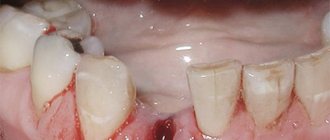
Fistula in the soft periodontal tissues after tooth extraction
The formation of a hole after tooth extraction is normal. As a rule, the hole heals within one to two weeks. If the operation was performed on wisdom teeth, the tightening process may take several weeks. Impaired socket tightening can occur due to infection or in case of injury to the area where the tooth was removed.
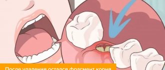
In a situation where the hole has tightened and a gap has formed in the periodontium, it may indicate that the tooth was not completely removed. Small fragments of the tooth gradually begin to decompose, forming a fistula.
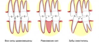
Forms of alveolitis
Depending on the course of the complication, three stages are distinguished:
- Serous.
It makes itself felt
2-3 days
after tooth extraction. At this stage, pain occurs when eating, headache. Lymph nodes increase in size. - Purulent.
This is the next form that occurs after the serous one, if timely treatment is not carried out. Diagnosed a week after the tooth was removed. The pain becomes unbearable and is also felt in the head or ear. The hole becomes covered with a purulent, dirty yellow coating. There is an unpleasant odor from the mouth. Swelling and lymph nodes enlarge and become painful. Opening your mouth and eating food is extremely difficult due to pain. - Hypertrophic.
At this stage, it seems that the symptoms are subside: the condition is normalized, the temperature decreases. However, atrophied tissue grows, and when pressure is applied, pus is released from the inflamed wound.
If you notice any of the above symptoms, you should not self-medicate, but rather consult a dentist.
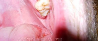
Fistula between tooth and gum
A hole between a tooth and periodontal tissue indicates the development of cervical caries or the complications it caused. Cervical caries can be located between the teeth and not be visible to the naked eye. Due to its inaccessible location, such caries often turns into pulpitis.
Pulpitis is an inflammation of the blood vessels and nerve endings located in the pulp chamber of the tooth. The chronic stage of the pathology may be accompanied by the formation of a purulent focus in the soft tissues. If pulpitis progresses to the next stage of periodontitis, several fistulas may appear at once.
Stages of healing
The process of tissue restoration consists of the following stages:
- During the first 24 hours after surgery, a blood clot forms in the socket. The formation closes the wound and protects against infection and food debris. Subsequently, the clot takes part in healing, so specialists urge not to touch or rinse the wound for the first day;
- After 3-4 days, the wound becomes covered with a white film, which indicates the formation of epithelial cells. The wound may have a yellow or grayish tint due to food pigments. If the wound is green, dark, or there is a putrid taste in the mouth, then you should visit a doctor;
- After 7-10 days, the gums become covered with a white coating, under which young granulation tissue is formed;
- On days 18-25, the wound heals and heals;
- Next, bone formation occurs, which can last 4-6 months.
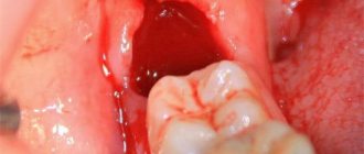
The appearance of a hole in the periodontium in a child
The formation of a hole in the gum near a tooth in a child can be due to reasons such as: periodontitis caused by complicated caries, or injury to the oral mucosa. With periodontitis, the lesion reaches the jaw bone. A cavity appears inside the periosteum, which is filled with pus. When there is an excess of pus, it breaks out through the fistulous tract, thereby bringing severe pain.
Frequent mechanical damage to the mucous membrane and gums causes infection to enter the oral cavity. Bacteria entering the periodontium cause inflammation and fistula formation.
What can you do to speed up the healing process?
Only “sterile conditions” in the oral cavity will allow the wound to heal quickly, although this is a rather relative concept. There are both harmful and beneficial bacteria in the mouth. In addition, internal diseases can sometimes negatively affect the gum healing process:
- intestinal dysbiosis;
- pathological phenomena in the gastrointestinal tract, esophagus.
The main thing is to prevent the development of secondary infection of the hole, adhering to certain rules so that the wound heals and heals. So, you need to do the following:
- hold the gauze swab placed in the hole by the doctor more tightly in the first few hours after tooth extraction, squeezing with the jaws;
- stop smoking, drinking alcohol and hot solid foods for the first 2-3 days until the healing process and formation of new bone takes place;
- do not wash out blood clots, which create a kind of plug (protection) from the penetration of harmful bacteria and microorganisms;
- avoid accidental injury to the socket, i.e. it is better to chew food on the other, healthy side;
- stop playing sports, visiting saunas and overheating in the sun for 3-4 days;
- It is advisable not to sleep on the sore side of the jaw in the first days;
- rinse your mouth with herbal decoctions, although doctors do not recommend doing this to avoid washing out blood clots.
Often, out of ignorance and mistake, patients begin to vigorously rinse their mouths with various traumatic means at hand (soda, salt, hydrogen peroxide) so that the hole space heals faster, thereby aggravating the process and significantly delaying the healing time of the hole. All that happens is irritation, destruction and washing out of blood clots, and increased pain. All this may be a reason to return to the dentist and perform a secondary operation.
Of course, some adherents of traditional medicine try to minimize unpleasant symptoms using herbal infusions, decoctions and lotions. Dentists approve ointments and gels with anti-inflammatory and wound-healing effects so that the wound begins to heal faster.
The main thing is not to neglect the frequency and duration of home treatments. You can make gentle infusions and lightly rinse your mouth to remove food particles after each meal. Although in the first days it is extremely undesirable to do this. Such leaching can lead to the liquefaction of blood clots, a process of “gurgling” in the mouth.
You can use weak solutions of herbal decoctions (chamomile, sage) or Furacilin, Chlorhexidine, as pharmaceuticals. It is better to first consult a doctor and apply it carefully, without overdoing it, so that the healing of the hole occurs comfortably and gradually.
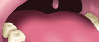
Symptoms
Signs of fistula formation include:
- aching pain that increases in the evening;
- burning and itching of gums;
- swelling in the affected area;
- gum hyperemia;
- increased body temperature;
- general weakness, chills;
- increased irritability;
- apathy.
The appearance of symptoms of pathology is individual for everyone. Some may experience very severe pain and discomfort in the mouth, while for others the symptoms may be almost invisible.
Why perforation occurs and how to avoid it
Such consequences can be avoided by properly planning removal using a CT scanner and following gentle removal protocols .
There is a group of patients with the anatomy of roots whose apices penetrate into the sinus, and if such patients are treated as a standard removal protocol, perforation and all associated complications will certainly occur. Only this is visible in the image created by a computer tomograph in a special ENT mode, and this is not possible in any clinic. In our Center this can be done with the latest SIRONA-SIEMENS equipment and the GALILEOS diagnostic software package.
Having studied the CT image, the surgeon must be prepared for an emergency situation. If perforation does occur, it is important to perform immediate microsurgical closure in a sterile operating room to avoid the development of an inflammatory process.
Even an experienced doctor without a good image on a modern expensive tomograph may not notice the details and skip the patient to a regular dental surgeon, not pay attention to the anatomical features and not carry out the necessary actions. The patient will find out about this later by the characteristic symptoms:
- passage of air, whistling and squelching in the socket of the extracted tooth;
- foamy, bloody or yellowish nasal discharge;
- strange, unreasonable organic odors in the nose and mouth,
- change in voice timbre (nasality).
If you do not carry out timely emergency closure of the anastomosis after tooth extraction, the hole itself may never heal . The gum tissues heal, epithelialize, and shrink, but the bones in the area of such a fistula between the nose and mouth never grow together due to the difference in the growth periods of the bone and gum, the gum will instantly take up all the free space, the slowly growing bone simply will not have time to fill the defect.
Within 2-3 weeks, a thin fistulous tract forms from the oral cavity to the sinus. In this case, the diameter of the hole is reduced naturally due to the formation of scar tissue; the symptoms of perforation may temporarily disappear or not appear. But this does not prevent infection from entering the sinus due to food entering the sinus.
As a result, the sinus becomes inflamed, signs of unilateral sinusitis appear, which should confuse a thinking patient (sometimes there are coincidences, but sinusitis-sinusitis, which comes as an accompaniment of the flu or a cold, is always bilateral).
Other causes of perforation
The remaining 5% of clinical cases of sinus perforation injuries occur for the following reasons::
- Not ideal endodontic treatment of tooth canals - the therapist treated the canals, was in a hurry, used excessive force when filling the canal, and under the influence of a rotating canal filler with pressure, the filling material fell beyond the tooth root. We often encounter cases where even fragments of endodontic instruments are sometimes found in the thickness of the filling material, which jam in the canal and tear into pieces, leaving metal fragments in different places of the tooth root;
- Punitive sinus lifting - an inexperienced or rude doctor does not feel the density of the tissues, breaks through the Schneiderian membrane, the bone material is forced into the gap and enters the sinus;
- Author's implantation - it happens that due to the lack of skills in performing a sinus lift and the desire to restore the missing bone, a decision is made - the implant is placed in the residual bone that exists, without bone grafting. Result: the implant completely fails or partially comes out into the sinus.
A fragment of an endodontic instrument in a tooth canal
Treatment
If you find a fistula on the gum, you should immediately go to see a dentist. Based on the diagnosis and identification of the causes of the disease, appropriate treatment is prescribed.
Treatment can be carried out in two ways:
- drug treatment;
- surgical treatment.
If the cause of the formation of a fistula is a carious process or pulpitis, then the following treatment is carried out:
- destroyed dentin is removed;
- The cavity is processed and a filling is applied.
Then, to eliminate inflammation and remove pus, the following is prescribed:
- rinsing with antiseptics;
- antiseptic and analgesic gels and ointments;
- Antibiotics may be prescribed if necessary.
If the cause of the hole is periodontitis or an infection that has penetrated into the root of the tooth, surgical intervention is performed.
In the first case, the fistula is opened, the accumulated pus is removed through drainage, and the cleaned hole is treated with antiseptics and covered with a medicinal bandage.
When an infection occurs, not only the gums are opened, but also the alveolar process. After treatment of the tooth root, bone augmentation is performed. If the root is severely damaged and cannot be treated, the tooth must be removed.
Diagnosis and treatment methods
Diagnostics at a dentist's appointment will help confirm the symptoms of a dry socket. After an examination, the doctor will prescribe treatment. As a rule, it depends on the stage of inflammation. In the case of a mild form, drug treatment with antiseptics and anti-inflammatory drugs is possible. At the middle stage, you will need antibacterial therapy, as well as cleaning the hole from pus and filling it with an anti-inflammatory drug. All actions are performed under anesthesia. If necessary, antibiotics are prescribed.
At the third stage, the most advanced stage, the patient may need hospitalization and even surgical intervention. With proper care and no complications, the hole heals within seven days. And a month later there is no trace left of her.
Prognosis and possible complications
If the fistula is detected and treated in a timely manner, the prognosis is favorable. If you follow all the specialist’s recommendations, within a week the resulting wound will completely heal and heal.
If proper treatment is not carried out, the following consequences are possible:
- abscess formation;
- phlegmon;
- osteomyelitis;
- tooth loss.
Preventive measures
- regular teeth cleaning;
- carrying out professional hygiene in the dental office;
- preventive examinations by a dentist every six months;
- daily consumption of fruits and vegetables.
What can the patient do after removal?
To help the wound heal, the patient must take steps to prevent secondary infection. To do this, immediately after the intervention you need to accurately follow the general recommendations of the dentist.
- Press the cotton swab placed in the hole firmly with your teeth. This is important to stop bleeding and blood clot formation.
- For the same reasons, you should not eat or drink for 3 hours.
- Once a clot has formed, it is necessary to exclude the possibility of injury and removal. Do not rinse your mouth vigorously, inspect the wound with your fingers or a toothpick, smoke, drink alcohol, hot foods and drinks, or solid foods.
- Visits to the fitness center, sauna, and beach should be excluded for several days. Physical activity and overheating can lead to complications.