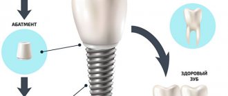
Inferiority of the row leads to impaired chewing function, bite pathologies, and bone loss. These processes can be stopped using implantation. The procedure involves implanting a titanium root into the jaw followed by fixation of an artificial crown. As a result, the functions of the masticatory apparatus are completely restored. However, after tooth extraction, the installation of a prosthesis must be carried out on time. Delay is fraught with atrophy of the alveolar ridge. To fix the titanium rod, additional interventions will be required to build up the bone and eliminate mucosal defects. When planning the removal of one or more units, the patient should discuss the timing of reconstructive measures with the implantologist in advance.
Implantation methods after tooth extraction
Dental implantation after extraction is carried out according to two protocols:
- One-stage - involves installing an implant immediately into the socket of the extracted tooth. There are techniques that allow not only the installation of an implant, but also the fixation of a crown on it.
- Delayed - requires complete healing of the hole and restoration of bone tissue after removal. The bone is restored in 2-5 months depending on the health of the patient. Only then is implantation carried out.
Implantation methods
How long after surgery can a permanent implant be placed? The answer to this question depends on the type of implantation. In addition, the type of implant affects the method of its installation: transgingival, patchwork, or in the socket immediately after tooth extraction.
There are different types of surgery:
- one-stage;
- one-step;
- two-stage.
Regardless of the method, it will take time for the pin to heal. This usually takes from 2 to 6 months. At this time, the patient wears a removable denture, which is fixed on adjacent teeth or a provisional crown on the implant. The temporary structure is then replaced with a permanent zirconia crown. Let's look at each method in more detail.
One-step method Single or multiple dental defects are eliminated in one visit to the dentist. The method of implant installation is transgingival. The doctor makes a puncture through which he screws a pin into the deep tissues of the jaw. Due to the fact that the procedure is minimally invasive, a temporary crown can be placed in just a few days and replaced with a permanent one after 3-5 months.
There are 4 technologies that are used within this method:
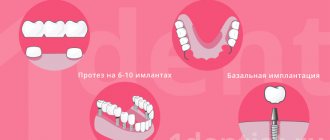
- Basal. Used when there is insufficient bone tissue. Implants are fixed not in the spongy layer, but in the basal layer of the jaw, which is practically not subject to atrophy. This procedure is performed only if three teeth are missing in a row, otherwise there is not enough space for implants.
- All-on-4. Four implants are installed - two frontal and two lateral. Next, removable or fixed dentures are attached to them. The technology is not suitable for jaw bone atrophy.
- All-on-6. Six implants are fixed. Two are in the frontal zone, and four are in the lateral region at the site of the chewing teeth. Then dentures are installed on them.
- Complex. In this case, different types of implants and implantation technologies are used. Suitable for non-standard situations, for example, when the patient needs to have his front and molars installed.
Two-stage method Used when there is a risk of complications. The installation method is patchwork. This means that the gums are peeled away to gain access to the bone. A bed for the implant is formed in the jaw, a pin is inserted and the gums are sutured. The artificial root remains without load for a period of 3 months. At this time, the patient wears a removable denture, which holds onto the adjacent teeth and does not load or loosen the pin.
There are 2 options for implementing this method - classic and beam.
- Classical. A bone bed is formed, a pin is installed in it, which is closed with a plug, and the gum is sutured for 3-6 months. Then the gum is opened, the plug is removed and a gum former is installed for a month. Afterwards the implant is placed.
- Beam. 2 or 4 implants are installed in the most favorable areas of the jaw. 3-6 months after the operation, the implants are combined with a metal beam, onto which the prostheses are subsequently fixed.
Regardless of the chosen method, it is necessary to take into account the characteristics of bone tissue - density, height, width. The pin survives if the bone is of sufficient size - more than 10 mm. If there is not enough bone tissue, then an implant with the selected size and shape that can cope with the chewing load will not be installed.
When it is possible to perform implantation - the doctor chooses after a complete diagnosis
It is necessary to study the condition of the bone tissue, its size and density. Based on the results, the doctor makes a decision - delayed or immediate implantation. Our Center is equipped with diagnostic equipment, so results can be obtained within 15 minutes . But this is only a preliminary assessment; the final decision is made after tooth extraction.
Levin Dmitry Valerievich
Chief physician, Ph.D.
Indications and contraindications for immediate implant placement
Indications
- Mechanical damage to the root - fracture, dislocation
- Restoration of frontal units
- Therapeutic treatment did not bring results
- Sufficient bone volume
- No contraindications
Contraindications
- Deficiency, looseness of bone
- Inflammation at the root apex
- Enlarged socket after removal
- Inflammatory processes of the oral cavity, gum pathologies
- Reduced immunity, systemic diseases
- Pregnancy, breastfeeding period
How to do dental implantation immediately after extraction
After a tooth is removed and an implant is immediately placed, the process of osseointegration occurs faster. The hole with the artificial root begins to heal, the blood clot turns into bone tissue that surrounds the implant in the early stages of healing. This promotes accelerated osseointegration of the implant with the jawbone, ensuring reliable fixation and stability.
Stages:
Diagnostics
Full diagnostics, choice of design in the absence of contraindications.
Tooth extraction, artificial root installation
Local anesthesia or sedation is used. A bed is formed in the hole, an implant is installed, and the space around is filled with osteoplastic material. After the procedure is completed, the gums are sutured. A temporary removable prosthesis is installed. Engraftment lasts 4-6 months.
Prosthetics
After osseointegration of the implant with the jaw bone, the gum is opened, and a gum former is fixed to the implant for 10-14 days to create a gum contour. Then the former is replaced with an abutment, and a crown made from the impression is installed.
In rare cases, it is possible to fix the crown to the implant immediately after its installation. At the same time, the crown is removed from the bite so that the implant does not move during chewing. This method is recommended for the anterior parts of the jaw in order to quickly restore the defect. Not suitable for lateral use due to active chewing loads.
Stages of treatment
The duration of one-stage implantation is about 4-6 months. Restoration of bone tissue after tooth extraction is not required; bone tissue augmentation as a separate procedure is also not performed. Therefore, after installing the implants, a temporary prosthesis is immediately fixed, and after a few months, when the implants and bone tissue have completely fused, a permanent dental crown is installed.
Galina Vladimirovna, 57 years old
“I had my natural teeth removed and implants were immediately placed. Within a week my smile was back. I really like the way I look and the way I talk. My diction hasn’t changed, I’m comfortable. I am very pleased and grateful to the doctors!”
- we give SMILES, HEALTH and new LIFE
- smile and enjoy every day
- start CHEWING immediately
- forget about the unpleasant smell and pain
- complete the entire treatment in a WEEK
watch a video with the patient
Preparation for implantation (from 2 days to 2-3 weeks)
When a tooth is removed and an implant is installed at once, sometimes there is very little time left to prepare for treatment, since most often teeth that are in very poor condition have to be removed. However, implantologists pay great attention to this stage, since it is necessary to conduct a thorough examination of the patient’s oral cavity and body, to exclude possible contraindications that could cause rejection of the structure.
Preparation for immediate implantation is absolutely standard and includes taking a series of blood tests, taking an X-ray or panoramic photo of the jaw, as well as dental treatment and removal of deposits. If there are acute problems with the body, we can refer the patient to an appointment with a highly specialized doctor to clarify the diagnosis, assess the state of health and carry out appropriate treatment
Installation of implants and bone grafting (1-2 hours)
Installation of an implant with simultaneous tooth extraction is carried out in a fairly simplified form: additional tissue incisions and bone drilling are not required to form a bed for the implant. The tooth is removed from the socket as carefully as possible, with minimal trauma to the bone tissue. Next, the cavity is thoroughly sanitized. If necessary, an x-ray is prescribed so that you can make sure that the cavity is empty and there are no pieces of instruments or parts of the tooth left inside it. Next, a dental implant is fixed in the hole, and the space around it is sprinkled with artificial bone tissue. The gums around the implant are sutured.
Temporary prosthetics (3-6 months)
Depending on the initial situation, both one-piece and two-piece implants can be used for simultaneous implantation. Both of them can be instantly loaded with a temporary prosthesis.
Three types of structures can be used as temporary dentures (they are selected based on the quality of bone tissue, the degree of primary fixation of the dental implant and the number of missing teeth):
- removable immediate denture (for front and side teeth),
- lightweight plastic crown (only for front teeth with single restorations),
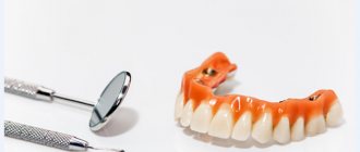
- acrylic prosthesis with plastic crowns (with complete edentia).
A resin crown is created that is slightly shorter in length than the adjacent teeth in the row. This allows you to significantly reduce the load on the prosthesis, and hence the implant itself. At the same time, the appearance of the smile is maintained at the highest level. From the point of view of further aesthetics, a single crown is a more preferable option for single restorations, since it allows you to shape the position of the gums.
It is important! Installation of a single crown is possible only on single-rooted teeth, which do not bear much pressure, and they mostly perform aesthetic functions - these are incisors and canines. If you chew food with the restored tooth, the implant simply will not take root, since it will shift due to the load.
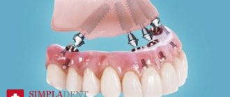
In case of complete edentia, when it is necessary to restore the entire dentition, several implants are installed at once. If there are teeth that need to be removed, some of the implants can be fixed into their sockets. In this case, a fixed prosthesis with a metal base, acrylic base and plastic crowns is fixed on top. It firmly connects all installed implants, which prevents them from moving. Read more about immediate loading methods >>>
Permanent prosthetics (1-2 weeks)
Permanent prosthetics are the final stage of treatment. It can be started only after the doctor is convinced that the implants have completely fused with the bone tissue - measurements are carried out with special devices and x-rays.
The temporary crown is removed, impressions are taken - the data is transferred to a dental laboratory, where an individual prosthesis is created. The dental technician must take into account the position of neighboring and antagonist teeth located on the opposite jaw. The manufacturing process takes about 1-2 weeks.
Metal-ceramics or zirconium can be chosen as materials for permanent dentures - these crowns are more aesthetic, stronger and more durable.
Advantages of the method
- shortened treatment times,
- no bone grafting required
- instant restoration of aesthetics,
- no bone tissue augmentation is carried out,
- lower cost of treatment,
- The service life of installed implants is more than 20 years.
Disadvantages of the method
The only disadvantage of one-step implantation is that it is not always possible to carry out it. You need to prepare for treatment: assess the condition of the body, exclude contraindications, select an implant model depending on the volume and quality of bone tissue. But the removal of your own tooth quite often has to be done urgently, which does not make it possible to conduct a full examination and understand whether it is possible to install an implant at the same time.
Advantages and disadvantages of one-stage dental implantation
pros
- One operation is performed, waiting time is reduced
- Simplified installation process
- Minimal loss of mucous and bone tissue after surgery
- If the technology is followed, the percentage of successful operations is 99.6%
Minuses
- Contraindicated in chronic inflammation, diseases
- Not performed if there is insufficient bone
- Strict rehabilitation period, frequent visits to the implantologist
For a favorable outcome, entrust the operation to a surgeon with extensive experience in this protocol.
Is it possible to place an implant if a tooth has been lost for a long time?
Whether it is possible to place an implant if the tooth was removed a long time ago is decided only by the surgeon after a complete diagnosis. Already 3 months after removal, the bone is so absorbed that it may not be enough to install an implant and augmentation will be required. The recommended implantation period is no later than 2-3 months.
In case of bone atrophy, osteoplasty is performed:
- Guided bone regeneration . Most often performed on the lower jaw, bone materials, biomembranes, and bone growth stimulators are used. It is used if the width and height of the patient’s bone is not enough for implantation. If bone tissue atrophy is insignificant, it is possible to perform NCR simultaneously with the installation of implants. Otherwise, it is carried out in a separate preliminary stage. It takes 3-4 months for the grafted material to fuse with the bone.
- Sinus lift. Intervention in the bone structure of the upper jaw in the area of the maxillary sinuses. Using instruments, tissue is punctured to raise the bottom of the sinus. The resulting space is filled with osteoplastic material. The operation can be closed or open. A closed sinus lift is performed for minor bone atrophy simultaneously with the installation of implants. Open - for moderate and critical atrophy in a separate stage.
Alternatives
Single or segmental row defects can be eliminated using alternative methods - removable or bridge dentures. However, their use is possible only after complete tissue healing. Removable orthopedic devices do not stop bone atrophy; fixed bridges slow down the process, but do not completely eliminate it. 3-4 months after the operation, the alveolar ridge decreases, gum recession occurs, which is especially noticeable in the incisor area.
The use of removable structures is accompanied by unpleasant sensations as a result of rubbing soft tissues, and also reduces the patient’s quality of life due to restrictions on the use of solid foods.
Price
The Center for Private Dentistry Dr. Levin provides a case payment system.
- When implanting Nobel Biocare PMC Select or Groovy, the cost includes the surgical stages, including the design model, surgery, anesthesia, and all necessary procedures. The price is indicated taking into account the preparatory stage.
- Removal is paid separately, the price includes surgery, inflammation relief, socket rehabilitation, consumables, anesthesia.
- The case for installing a metal-free zirconium dioxide crown includes all stages - impressions, manufacturing, installation.
- Sinus lifting and any bone tissue augmentation operations are paid separately.
Sinus lift
Bone atrophy occurs when teeth are lost due to reduced weight bearing. When the roots of the teeth are missing, the bone cells do not experience pressure and are no longer saturated with nutrients. Gradually, the soft layers of bone dissolve, the bone becomes thin, and the gums collapse.
It is impossible to fix the pin in a short and narrow bone. Therefore, before implantation, the dentist may prescribe a sinus lift to the patient - an operation to restore the volume of bone tissue in the upper jaw in the place of the missing tooth.
Types of surgery
There are two types of sinus lift: closed and open.
- In the first case, the operation is performed under local anesthesia. First, the doctor makes a small hole in the bone of the upper jaw, then lifts the bottom of the maxillary sinus, fills the resulting space with bone material and sutures the wound. This type of operation is suitable for bone volumes of 7 to 8 mm.
- In the second case, the operation is performed through the bed created for the implant without additional incisions. Using a special surgical instrument, the dentist lifts the bottom of the maxillary sinus and introduces osteoplastic material that fills the missing volume of bone tissue. This type of surgery is suitable for bone volume less than 7 mm.
Sometimes pin implantation is carried out simultaneously with bone tissue augmentation. The procedures are combined if the bone is of sufficient thickness and its height is at least 7 mm, that is, with a closed sinus lift.
The open type of operation is more difficult and the risk of complications is higher. In this case, implantation is carried out after the rehabilitation period. As a rule, after six months, when the recovery period of soft and bone tissues is completed.
Contraindications
Like any surgical operation, sinus lift has contraindications. The operation cannot be performed in the presence of diseases of the ENT organs during the period of exacerbation, treatment of oncology in the head and neck area, blood clotting disorders, diseases of the immune system and after a heart attack.
Before performing a sinus lift, it is necessary to undergo an examination for the presence of contraindications. If the risk of complications is high, then it is better to refuse the operation and choose another type of dental prosthetics.
Alternative options for tooth restoration after extraction
After removal, doctors recommend restoring the tooth in a short time. This is caused by bone tissue atrophy, which begins in the absence of chewing load, which complicates recovery in the future.
If for some reason the patient is not ready for implantation, alternatives can be offered:
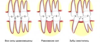
- Bridge prosthesis. It is a non-removable structure of interconnected crowns. For installation, two supporting teeth are depulped (nerve removal, canal filling) and ground down for crowns. At the same time, the load on the support increases, mobility develops, which is why the bridge will need to be replaced. The bridge structure is not able to completely prevent bone tissue atrophy, but it prevents rarefaction, displacement, and curvature of the dentition. The rate of bone atrophy and the development of mobility depend on the patient’s health.
- Removable dentures . They represent a structure made of artificial gum with soldered crowns, attached to adjacent teeth with hooks. The supporting teeth are ground down if the fastenings are metal. The prosthesis has a range of mobility and requires careful care, since food particles can get under it. Removable dentures do not stop atrophy, but they do prevent displacement of adjacent units.
Implantation prevents all problems, including bone atrophy. An implant is an artificial root on which a crown is placed. Neighboring teeth are not ground down or damaged. The chewing load is maintained. The survival rate of dental implants is 98%. Implantation is considered the most reliable way to prevent bone resorption.
Re-prosthetics for severely damaged teeth
Every dentist in his practice very often encounters the task of repeated dental prosthetics. And as a rule, after removing old structures, a thin stump of a tooth that has been aggressively treated for a crown is discovered. This is due to outdated principles of prosthetics, when there was no talk of adhesive fixation.
Repeated prosthetics are caused by a number of problems awaiting the doctor, such as: root caries and violation of biological width, lack of ferrule effect, as well as complete destruction of the stump.
The question arises, how can one predict recovery and accurately outline a treatment plan for the patient before removing the old structure?
The answer is simple - this cannot be done for sure! Therefore, in my practice I apply the principle of “diagnostic autopsy”. Patients with such a clinical situation, after being informed of possible treatment options, are offered a diagnostic visit, after which a final decision will be made whether to preserve or remove the tooth.
What does a “diagnostic autopsy” consist of: old structures are removed, the condition of hard tissues, effect ferrules and periodontal tissues is assessed, as well as the possibility of their restoration. Temporary structures are made for the patient using the key made before the old structures were removed and further actions are discussed.
Let's look at the stages of treatment using a clinical example.
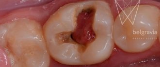
Photo 1. Metal-ceramic crowns connected together were previously installed on teeth 1.1, 2.1. Probing revealed overhanging edges of the crowns, as well as a deep subgingival location of the crown border on tooth 1.1.
Photo 2. Before removing the crowns, a silicone key was made.
Photo 3. After removing the old structures, the condition of the root and stump of the teeth was assessed. A violation of the biological width is also visible.
Photo 4. The remains of the composite and areas of carious lesions were removed with a bur. An ultrasonic nozzle was used to clean the cervical area of the teeth. CBCT shows normal endodontic status. Therefore, retreatment of the canals would be required only in the case of depressurization on the endoaccess side. An attempt to extract such a volume of pins is fraught with excessive thinning of the root and, as a consequence, the impossibility of preserving the teeth.
Photo 5. To restore the biological width, as well as enhance the ferrule effect, surgical lengthening of the crown part of the teeth was performed. Gingivoplasty in area 2.1 and gingivosteoplasty in area 1.1. After suturing, temporary restorations were made using a silicone key for 2 weeks.
Photos 6, 7. After 2 weeks, the stumps were restored with Core composite after sandblasting with aluminum oxide. Impressions were taken for laboratory temporary restorations.
Photo 8. Condition of soft tissues at the time of fixation of temporary single structures made of CAD/CAM composite.
Photos 9, 10. Condition of soft tissues after 1 year of wearing temporary structures.
Photo 11. The CAD/CAM system allows you to repeat the formed perigingival contour of temporary restorations in permanent structures, which is undoubtedly a big advantage and ease of use.
Photo 12. Immediately after fixation of ceramic crowns made of solid milled press ceramics, tooth 1.2 was temporarily restored with packable composite in a layer-by-layer technique, for financial reasons of the patient.
Subsequently, it is possible to make an exact copy of a ceramic veneer for tooth 1.2 by comparing digital scans before and after preparation.
Photos 13, 14. Follow-up examination after 1 week. The structural, functional and aesthetic parameters of the teeth are restored, and a healthy gum condition is achieved.