A targeted photograph of the tooth is taken using a radiovisiograph. It is a compact device that operates locally and provides a clear image of an individual tooth for immediate, on-site diagnosis. The image appears on the computer screen immediately after the procedure. This diagnostic technique is used in modern clinics and is considered a mandatory step in dental treatment.
When is it necessary to take a targeted photograph of a tooth?
Radiovisiography is used in the following situations:
- diagnostic search - the image visualizes the carious process and its depth, you can also detect pulpitis or periodontitis;
- the image allows you to assess the condition of the periodontal tissues, investigate the spread of the problem, its clear localization;
- During endodontic treatment (root canal treatment), several targeted images are required to monitor each stage of treatment.
Indications for orthopantomogram
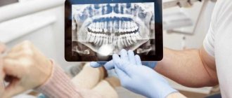
An orthopantomogram (or “OPTG”, “panoramic image of the dental system”) is one of the types of diagnostic radiography. In dentistry, OPTG is of key importance - many types of treatment cannot be started without this diagnostic method.
Method of performing an orthopantomogram:
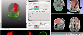
Technically, it is carried out as follows: the beam source (X-ray tube) and its receiver (film or digital sensor" move around the object under study in opposite directions. As a result, a very limited part of the object of study is in focus, everything else is blurred. Panoramic views are made photographs using orthopantomographs.The volume of radiation is such that panoramic photographs can be taken every day for a month without significant harm to health.
Before the procedure, the patient is put on a special lead apron to protect him from unwanted exposure to X-rays. An orthopantogram is performed from a standing position. A disposable cover is put on a special tube. The tube is clamped by the patient himself with his front teeth. The X-ray tube will rotate around the patient's head for a few seconds. Information from the sensor will be sent to a computer, corrected using special programs, and then this image can be printed on paper or film, and also saved in digital format. No other preparation is required.
Contraindications: first and second trimester of pregnancy, lactation.
Why do modern dentists use radiovisiography?
Compared to classic X-rays, which previously provided images on film, computer radiovisiography has a number of advantages that have forced massive X-ray machines out of dental practice:
- lower radiation exposure to the patient (in order to obtain an image on a computer sensor, less X-ray radiation is needed than to develop an image on film);
- saving time for the doctor and the patient (no need to develop the film);
- a high-quality image that the doctor can enlarge on a computer, examine it more closely, perform color correction, etc.;
- the image file can be stored on the computer for a long time, transferred and reused;
- The image is transferred to the doctor at the dental unit directly during treatment - this significantly shortens the treatment process.
A competent doctor will never begin treatment without confirming his diagnosis and treatment plan with an objective diagnostic method. Sighting image data is an indication for many treatment tactics and explains the need for dental procedures.
General description of the sighting shot
A targeted X-ray is a familiar type of x-ray, in which one tooth or a row of three adjacent units is illuminated. The format of the captured image is a 2D image.
Possibilities for reviewing problem areas include:
- dentin with its cavity;
- root canal system;
- vessels close to the diseased tooth.
Instructions for prescribing a targeted photo
The patient is sent for an x-ray if necessary:
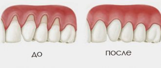
- examine the condition of the gums for the presence of periodontal disease;
- establish the causes of acute toothache;
- confirm caries or pulpitis discovered during sanitation;
- examine the integrity of bone tissue after injury;
- check the filling of the canals after depulpation;
- monitor the development of wisdom teeth.
Types of targeted shots
List of existing X-ray images:
- Standard shot. It is produced using analog X-ray equipment to ultimately obtain a planar image. The main disadvantage of the outdated methodology is the short period of preservation of the clarity of the basic result.
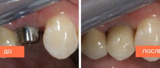
- Digital photo. A more modern method of performing x-rays involves the use of a radiovisiograph. The device digitally records the area of the problem area, which can then be saved on your personal PC resources.
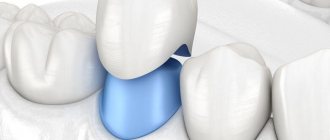
- Interproximal image. Such an image is prescribed, if necessary, to examine the adjacent surfaces of teeth for the possibility of hidden carious cavities between them.
- Periapical image. It is done to obtain data on the condition of the tooth root and the adjacent soft tissues.
- Intraoral image. This type of x-ray is the best way to check the integrity of dentin and diagnose the initial stages of caries.
The voiced types of targeted photographs are not suitable if we are talking about significant problems with teeth. In this case, a panoramic x-ray is prescribed, which has much greater diagnostic capabilities.
Advantages of X-ray
- safety of diagnostics at permissible radiation dosage;
- the ability to take several pictures (up to 3-5 pieces) at one time;
- affordable cost of this service (350-500 rubles);
- the reality of taking a targeted photo in almost all clinics.
Cons of X-ray
- limited viewing range of problem areas;
- presentation of the image in a single-plane perspective;
- ineffectiveness when wanting to plan corrective actions for malocclusion;
- the inability to detect literally all cracks in the tooth root and diagnose nerve inflammation;
- the minimum term for the preservation of a photograph when it fades.
Sight Shot Limitations
The prohibitions do not apply to pregnant women in the 2nd - 3rd trimester, whose dental condition may deteriorate during pregnancy. For breastfeeding mothers, problems arise not from the procedure itself, but from increased sensitivity to myths about the dangers of radiation for milk. As a result, they lose teeth and then have to be removed.
If unwanted spots on the enamel were discovered during sanitation in a child, then he will definitely be prescribed a targeted scan. If the clinic client’s age is too young, the procedure is prescribed only for serious damage to the baby’s jaw (fall, difficult birth of the mother).
However, there is a list of patients who are prohibited from diagnosing using X-rays:
- children under 2 years of age, if it is possible to avoid the procedure;
- pregnant women in the first trimester;
- people during a serious illness and in the postoperative period;
- clinic clients with reduced immunity and severe anemia;
- patients with bleeding of any etiology and intensity.
Execution steps
Interproximal radiography is one of the types of intraoral examination that allows you to obtain a picture of 1 section of the oral cavity with an image of the upper and lower dentition. To obtain the image, a special holder is secured between the closed teeth. It allows you to identify interdental caries and various changes in bone tissue due to gum disease. And also check the correct installation of crowns, dentures or fillings.
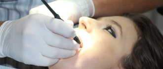
An X-ray of the tooth is taken by a doctor in an office specially equipped for this purpose. Before taking an X-ray of a tooth, the doctor gets acquainted with the problem and studies its location.
To obtain a clear image of the problem area, the patient's head is fixed in the required position. The process of visiography takes a matter of seconds, but during this time the patient is required to remain completely still. The video in this article shows how to take targeted photographs of teeth so that the image is as informative as possible.
Using a digital sensor, the radiologist directs the beam of rays to the desired area. The photograph is taken either from the inside of the mouth or from the face. During the manipulation, the patient does not feel any pain or discomfort.
Features of the procedure in children
Contact intraoral radiography can be prescribed for children in cases where tooth damage cannot be examined in any other way. The technique allows early detection of disturbances in the process of teething, bone diseases, and prescribing effective treatment.
In addition, this method allows you to control the implementation of orthodontic manipulations if the child has problems with the formation of the jaw.
The study is carried out in the same way as in adult patients. Children under 2 years of age are recommended to undergo x-rays only in case of urgent need. For example, in case of injury during childbirth, to monitor the development of the jaw, or after a fall from a height, to assess the integrity of the teeth.
Features of the technique and differences from standard photography
The difference between this technique is the replacement of conventional X-ray film with an electronic matrix of the visiograph sensor. The sensor connects to a computer and records x-ray radiation. The operating principle is similar to using X-ray film, but has its own differences:
- The radiovisiograph has a sensitive sensor, this reduces the radiation dose to the body, since the device requires less X-ray radiation to obtain an image;
- in one visit, the dentist can take 9–10 photographs without causing harm to the patient’s health;
- control of each step during the procedure, which makes it consistent;
- maximum speed of image acquisition, which is ahead of any other technique;
- the procedure does not cause discomfort to the patient, is easily tolerated and does not cause pain;
- the ability to quickly adjust the parameters of a targeted tooth image on the screen to obtain the most accurate data;
- the doctor can take measurements of the necessary structures automatically;
- storing images in a computer database and quickly searching them;
- Pictures can be transferred via e-mail, within the system and recorded on removable media.
How is a targeted tooth image taken?
This technique does not cause pain to the patient. It is carried out in a short time and consists of the following stages:
- protecting the patient with a lead apron;
- installation of the sensor inside the oral cavity;
- the patient should not move for several seconds;
- The healthcare professional takes the photo and it automatically appears on the computer screen.
Some patients may experience a gag reflex if the sensor touches sensitive areas. The employee who performs the procedure will give recommendations on how to avoid unpleasant sensations. If you breathe deeply through your nose during the procedure, you can avoid the reflex. Moreover, you need to be patient for a few seconds. Sometimes, difficulties arise under the influence of anesthesia - this can interfere with holding the sensor. In this case, a clinic employee will also help by fixing the device in the patient’s mouth.
The described technique is a necessary diagnostic step in dentistry, which will make treatment safe and comfortable. Entrust your dental treatment to professionals and forget about toothache.
Thanks to the use of a radiovisiograph installed at the Le Dent clinic, the radiation dose is significantly lower and amounts to about 0.2 mSv. That is, even if the patient needs to take 10–15 targeted photographs of the tooth in one visit, he will receive the same radiation dose as if he had taken one picture on a regular X-ray machine.
To take targeted images of individual teeth, our clinic uses a Kodak 2100 with a visiograph. The radiation dose of this device is 30% lower than that of other similar models of radiovisiographs. Today, this is one of the safest and most convenient installations.
Contraindications for the study
Sight radiography has such a low radiation dose that there are practically no contraindications to its implementation. It is not recommended to conduct radiographic examinations in the first trimester of pregnancy, therefore preparation for bearing a child should begin with sanitation of the oral cavity. Also, the procedure may not be comfortable for patients with an increased gag reflex in cases where examination of large molars is necessary. However, specialists carry out it very carefully, which most often does not cause any discomfort.