A 60-year-old patient came to the Research Center clinic with a complaint that she had mobility of the front tooth of the central incisor. Purulent discharge appeared from the gums in the area of the front teeth from the fistulous tract. From her story it became clear that the patient had previously had metal-free structures installed, crowns on her front teeth, the so-called. central incisors. She had them done in another clinic, where before installing crowns she had intra-canal teeth whitening
, which may have triggered a number of pathological processes.
The decision was made based on the CT scan:
- remove 2 central incisors, remove two upper front teeth
- and install implants with immediate loading.
Immediate loading
means that prosthetics of the upper anterior teeth will be performed on our patient also on the day of surgery.
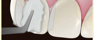
It was also decided for the patient to leave and use as temporary crowns the veneers of those crowns that she had on her front teeth at the time of her visit to our clinic.
Therefore, the teeth, two central incisors, were removed very carefully. The photograph clearly shows that both incisors have a lesion
: the root part of the teeth was destroyed, eaten away, and these teeth could not be saved.
The anterior teeth were removed atraumatically,
with preservation of holes. And - with the preservation of the old crowns as temporary prostheses for the upper front teeth, which were useful to us for securing them to the installed implants in the future.
In this photo you can see the sockets of the teeth
. I can note that the removal of the upper front teeth was atraumatic, but in the area of tooth 11 (the first incisor of the first segment), due to a fistulous tract on the gum, severe destruction of the root occurred, as a result of which the cortical plate was lost. When such a fistulous tract occurs in the area of the front teeth, it means that the process has already started, a lot of unpleasant things have already happened. In this case, the cortical plate was destroyed. Before the removal of the front teeth, at the planning stage, individual templates were made for the patient, with the help of which the implants were subsequently installed.
Lower and upper teeth
Teeth consist of 3 parts: crown, neck and root. Their tissues are enamel, dentin, pulp. The teeth of the upper and lower jaws are mirrored. This limits the similarity between the organs of the upper and lower jaw.
The characteristic features of the lower teeth are as follows:
- The lower jaw bone around the teeth being removed is denser
- Diseases develop more often. The reason is the settling of more food debris.
- The frontal incisors are smaller and weaker than the upper ones.
The small and large molars (premolars and molars) have smaller roots than the upper ones. All of the above explains why the removal of lower teeth occurs more often and more difficult than the upper ones.
Indications and contraindications
The factors that determine the removal of the upper front teeth include:
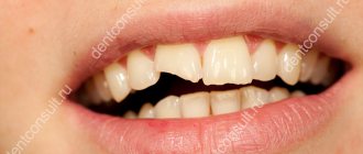
- Destruction of the unit, allowing access to pathogenic bacteria and allowing the possibility of infection of root canals and bone tissue;
- Fracture of the crown structure, accompanied by exposure of pulp tissue;
- Ineffectiveness of endodontic treatment of chronic periodontitis;
- Complete destruction of the tooth structure, in which the preserved root cannot be used as a basis for installing a prosthesis;
- Impacted position of the unit, leading to systematic tissue inflammation;
- Development of severe periodontal disease, causing tooth mobility;
- Mechanical injuries, including jaw fractures;
- Abnormal development of the dentition, including crowding of units.
The procedure may be temporarily or completely limited due to the following contraindications:
- Pathologies of the cardiovascular system;
- Acute forms of chronic diseases;
- Oncological diseases;
- Functional abnormalities of the central nervous system;
- Allergic reaction to anesthetic drugs;
- Diagnosed mental disorders.
The final decision on how exactly the removal of one or more teeth will be carried out is made by the dentist based on the results of a comprehensive examination.
Contraindications for removal
Extraction of teeth is undesirable, so dentists recommend saving every tooth. If removal is inevitable, then you need to know that this procedure is not carried out in the following cases:
- viral infections and fever;
- exacerbation of cardiovascular diseases;
- stomatitis;
- pregnancy in the last month.
Critical days in women are a relative contraindication. If the removal of the lower teeth is supposed to be done under anesthesia, the following is added to the above: pathologies of the endocrine system, epilepsy, heart failure, old age, pregnancy.
Contraindications
- absence of sixes and/or sevens in the dentition;
- correct placement in the jaw, not harming neighboring teeth;
- the possibility of conservative treatment of figure eight;
- loosening of neighboring units, which require a figure eight to splint.
In Russia, it is considered advisable to preserve wisdom teeth, if possible. Their removal is postponed for as long as possible, although most often they have to come back to it again.
Even if a wisdom tooth is treated and the canals are filled, it does not “live” for long. The reason is the inconvenient location, which complicates hygienic care. Food plaque accumulates on the enamel, and the tooth quickly deteriorates.
What teeth are removed in the lower jaw?
Depending on the type of teeth, extraction occurs with varying degrees of complexity. Single-rooted front incisors are easier to remove (2 pieces on each side). The canines (on either side of the incisors) have a chisel-shaped root hidden deep in the gum. Sometimes it becomes bent or bifurcated. Together with the incisors, the canines are involved in biting.
The lateral teeth that perform the chewing function include 2 small molars with one root, and 2 large ones with two even roots.
In the eighth position are wisdom teeth. Extraction is complicated by irregular growth and long, twisted roots, the number of which sometimes reaches up to five. These organs do not bear any functional load. Extraction of figure eights is difficult, and sometimes anesthesia is used.
Tooth extraction is practiced if they have an unaesthetic appearance, are a source of infection in the periodontium and cannot be treated, or the crown of the organ is completely destroyed. Extraction is used for growth pathologies, the formation of cysts and abscesses, and to correct bites.
Stages of the procedure
Teeth extraction in St. Petersburg is performed using the latest technologies, using modern instruments. The extraction takes place without pain or complications - effective medications are used for this.
Extraction begins 5-7 minutes after the administration of the anesthetic. The surgeon peels off the gum and uses forceps with wide cheeks to grab the tooth and then remove it. In simple cases, this takes no more than 1 minute. But sometimes it is pulled out in parts.
Complications
Complications sometimes occur during tooth extraction. The first is bleeding. Normally, it stops as soon as a blood clot forms in the socket (this takes up to 30 minutes). But sometimes gum bleeding begins hours later and even the next day. Provoking factors are the following:
- vasodilation after the anesthetic wears off;
- damage to blood vessels, soft or bone tissue, large arteries (rare);
- infection in the socket;
- other reasons caused by the characteristics of the patient’s body.
The next complication is the already mentioned infection (alveolitis), which causes pain and destruction of the walls of the socket, an abscess. If swelling or pain occurs, the patient should consult a doctor. Sometimes there is damage to the nerve endings, which causes loss of sensitivity. After extraction of lower molars, swelling often occurs, making it difficult to speak and eat.
Price
Prices for tooth extractions in St. Petersburg depend on the complexity of the procedure and range from 3000-7500 rubles. The minimum cost of anesthesia is 8 thousand rubles. in 1 hour. Due to the dense bone tissue in the lower jaw, more anesthesia is required, which also affects the price.
The MY ORT clinic employs highly qualified medical personnel who can solve almost any of your dental problems. Call us at our number and make an appointment. The first visit to the dentist is absolutely free!
Indications for care after removal of the upper tooth
- After removing a front tooth or root tooth, the doctor places a gauze swab into the hole, the purpose of which is to create compression and block small capillaries. This is necessary to stop bleeding. You can keep the tampon in your mouth for no more than half an hour. If it remains in the hole longer, the wound may become infected.
- The blood clot (thrombus) that forms in the hole should not be touched with the tongue, toothpick, hands, or try to rinse it out of the mouth. The blood clot protects the wound from penetration of microbes and promotes its healing.
- For two to three hours after the removal of the upper teeth, you should not eat or drink, so as not to infect the socket. Within three hours, the blood clot will be completely formed, then the likelihood of food getting into the hole will be negligible.
- For several days after surgery, you should refrain from spicy foods and hot drinks.
- On the day of removal of the upper tooth, it is not recommended to perform any hygienic manipulations in the oral cavity. You can brush your teeth the next morning, but avoid the surgical area. In the future, you can return to normal dental care.
We also advise you to read the article about the removal of upper wisdom teeth
Differences from the simple method
Simple tooth extraction is a procedure implemented in the presence of only one root, in case of looseness of the problematic elements, as well as their belonging to a replaceable set of mammary units. Such operations do not take much time and do not require special preliminary preparation. In difficult cases, a prerequisite is the use of anesthetic drugs that eliminate pain. The invasive procedure takes longer - one to two hours, and in some cases is divided into two stages. In addition, for complex operations, a special instrument is used, the choice of which depends on the indications of the clinical picture. The rehabilitation period and recovery treatment last longer and require strict adherence to medical recommendations.
Preparation and stages of the procedure
One of the main factors determining the final success of treatment is the quality of preliminary preparation. As part of the preliminary examination, a visual examination is carried out, radiography or CT is prescribed, and antibacterial therapy techniques are applied to prevent the spread of infection in the patient’s body. The choice of removal technology depends on the indications of the clinical picture:
- Extraction using forceps is a fairly gentle method recommended in cases where the structure of the crown has not been seriously damaged, and allows for fixation and subsequent loosening in a circular motion until the root part is separated from the alveolus;
- Elevator extraction – used for complex extraction of elements located outside the dentition, which eliminates the use of forceps. The dental instrument is inserted into the periodontal fissure and then twisted, which leads to rupture of the ligaments, after which the tooth is squeezed out of the socket;
- Extraction using a drill is effective in the presence of multiple roots that are defragmented and removed separately, as well as in cases where a filling was previously installed on the crown.
Quite often, the removal of a problematic tooth is accompanied by the presence of purulent foci, gum inflammation or periodontitis. In such cases, the doctor prescribes intensive therapy with the use of antibacterial drugs, and only after the inflammatory process has stopped, he sets a date for surgery. The recovery period lasts 10-12 days and is characterized by complex symptoms. After the effect of the anesthetic drugs wears off, the patient begins to experience severe pain, to eliminate which analgesics are prescribed.
About our clinic
It is best to entrust the removal of a complex tooth in St. Petersburg to professionals working at Magenta Dental Clinic. Our specialists are always ready to help and preserve the natural attractiveness of your smile. Carrying out express operations to restore a weakened bone structure with subsequent restoration of the dentition is carried out quickly and painlessly. The experience of doctors and a high level of service guarantee a positive treatment experience. You can make an initial appointment at a convenient time by phone or by leaving a request on the clinic’s website. Contact us - we will help!
Changes in bone
Bone tissue, in the area where there are teeth, constantly experiences a certain load, which is transmitted to it through the teeth during the process of chewing food. Thanks to this load, the proper functioning of the bone occurs, that is, the balance of osteoblasts (cells that provoke bone growth) and osteoclasts (cells that provoke bone breakdown) is maintained. If a tooth is missing, the balance of the above-mentioned cells is disrupted in favor of osteoclasts, and therefore, bone tissue atrophy. That is, the bone ridge, where a tooth has been missing for a long time, becomes narrow at the top, and upon lateral examination there is a significant dip on the buccal surface.
Rice. 1 – Bone atrophy
It should be noted that the longer a tooth is missing, the more pronounced is the atrophy of bone tissue. Everything would be fine, but further restoration of the missing tooth is not possible without restoring the volume of the bone. Bone grafting and gum grafting are required. Of course, modern dentistry has reached great heights in maxillofacial surgery and various types of plastic surgery, which are performed quite quickly and successfully. But in any case, this is a complex surgical intervention that could have been avoided by simply replacing the lost tooth in time. In addition, the final result, when interacting with the bone, is delayed, and six months, this is exactly the period required, for the bone to return to normal and become suitable for implantation.
Rice. 2 – The process of replacing a lost tooth