Many parents are concerned not only with the psycho-emotional development of the child and issues of general health, but also with such particular issues as, for example, the development of the child’s dental system. In many countries around the world, such attention is a common part of the dental culture, combining prevention, hygienic care and regular follow-up with appropriate specialists. Russia, unfortunately, is not in first place in terms of development of dental culture. In this article we will tell you more about the growth of a child’s jaws, possible developmental anomalies, what and at what age you need to pay attention, and which specialists you should contact if you notice any abnormalities.
The human maxillofacial apparatus develops along with the growth of the child and goes through various stages of development. Thus, the growth of the alveolar processes in width ends around 3 years of age. At this time, you can show the child to an orthodontist so that he can determine the presence or absence of any dental anomalies. By the age of 3, children usually finish erupting their baby teeth. From this moment on, the width of the alveolar processes no longer changes. Further, the alveolar processes will only lengthen. This process usually occurs between the ages of 6 and 12 years, when children's molars erupt.
The bite is formed in stages: temporary – replaceable – permanent.
Temporary milk bite
A primary dentition is a dentition made up of baby teeth. These teeth received this name because of their milky white color. The eruption of baby teeth begins around 6-8 months (earlier and later eruption is possible).
The appearance of the last baby tooth occurs at about 2 and a half years of age. There are a total of 20 teeth in the primary occlusion - 10 each on the upper and lower jaws. In the primary dentition, space is prepared for future permanent teeth: thus, at the age of about 5 years, the spaces between the incisors widen.
A panoramic image (orthopantomogram) shows not only grown teeth, but also the presence of the rudiments of permanent teeth.
Predicting growth using x-rays of the hand and vertebrae
The development of the dentofacial system correlates with the patient’s age. Skeletal age is determined by the degree of ossification (ossification) of bone structures. During the process of maturation, each bone undergoes changes that are determined radiographically. The sequence of changes is constant for all people.
The degree of growth of the lower jaw can be assessed by the condition of the cervical spine. The assessment is made visually and consists of two parameters: the shape of the vertebra and the presence of concavity at its lower border. The connection between the increase in the length of the lower jaw (body and ramus) and the stage of development of the cervical vertebrae has been scientifically proven.
As a vertebra grows, its shape changes, which makes it possible to assess the degree of maturity. An important point in the analysis is to determine the degree of concavity of its lower surface.
At the very beginning, young immature vertebrae have a rectangular shape. The lower surface of the vertebra is flat or slightly concave. (Fig. 1) Peak growth is expected no earlier than a year after this stage.
As they mature, the vertebrae become increasingly concave on the lower surface, remaining rectangular and horizontal in shape. (Fig. 2) The most active period of growth has already occurred a year or two before this stage.
Upon final maturation, the vertebra acquires a concavity of the lower surface, its shape becomes rectangular, vertical or square. (Fig. 3) This indicates that the peak of growth occurred no later than two years ago before this stage.
The use of this method is relevant when predicting the growth of the lower jaw, for example, when treating with functional devices.
Another method for assessing the degree of growth is an x-ray of the hand. To analyze skeletal maturity up to approximately 9 years of age, the degree of mineralization of the carpal bones, the development of the metacarpal bone and phalanges are used.
The assessment consists of determining the ratio of the sizes of the epiphysis (the widened end of the tubular bone) (Fig. 4) to the diaphysis (the central part of the tubular bone). (Fig.5)
The equal width of the diaphysis and epiphysis is designated as “=” (Fig. 6), the coverage of the diaphysis by the epiphysis in the form of a cap is “cap” (Fig. 7), with complete ossification – “unit(u)” or also “closed (c)” . (Fig.8)
In newborns, signs of carpal bones appear from the 3rd month of life.
An X-ray of a child’s hand is characterized by the “=” and “cap” stages. (Fig.9)
Rice. 9
An x-ray of an adult’s hand is characterized by the “closed” stage. (Fig.10)
Rice. 10
In modern orthodontic practice, determining height using an x-ray of the hand is performed, for example, before orthognathic surgery.
Changeable bite
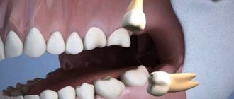
Between the ages of 6 and 12 years, a gradual change of teeth occurs when baby teeth fall out and are replaced by permanent ones. Doctors call this condition a mixed dentition. If your child is already being seen by an orthodontist, then changing the bite is an important stage, which should also take place under the supervision of a specialist. The process of changing the bite looks like this: the first molar erupts behind the last milk tooth. It is important that baby teeth are healthy - only in this case the change in bite occurs normally.
It is not recommended to remove baby teeth prematurely without special medical indications. It is also not recommended to ignore the treatment of baby teeth, believing that “they will fall out anyway.” The first large permanent molar—the “six”—erupts behind the baby teeth when baby teeth have not yet begun to fall out.
Using a panoramic photograph of the jaws, the doctor monitors the change of teeth, the presence of the rudiments of permanent teeth and the depth of their location.
The Dial-Dent clinic places great importance on dental treatment for young patients. Healthy baby teeth are the key to healthy permanent teeth. In addition, timely and regular observation of the child by a pediatric dentist will allow his dental and orthodontic problems to be resolved in a timely manner. Unfortunately, it is still not uncommon now for a desperate mother to come to the clinic and ask to have her baby’s bad teeth treated under anesthesia. It is difficult for a dentist to understand why the parents did not pay attention to the child’s condition earlier, when the treatment would not have been so difficult and traumatic for the little patient...
Typical problems.
The formation of the dentofacial system in this age range usually proceeds without any problems. At the age of 12, not a single baby tooth remains in the oral cavity, which makes this period the most favorable for orthodontic treatment with braces. By the age of 15, jaw growth begins to slow down, which is why it is advisable to carry out any orthodontic treatment before this age, as it allows you to do without tooth extraction.
PHOTO: Teeth of a 14-year-old teenager. There are 28 teeth in the mouth.
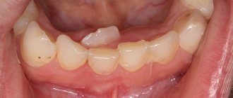
If there was an early removal of primary chewing teeth and no appropriate correction was carried out, there is a lack of space in the dentition for the eruption of permanent premolars and canines. Since the canine is the last one to erupt, it is the one that will not have enough space. Crowding of teeth is formed in the anterior part of the jaws: in this situation, fangs can erupt outside the dental arch closer to the lip.
If the wisdom tooth germ is incorrectly positioned in the lower jaw, existing crowding of teeth may form or worsen. By exerting pressure on a nearby tooth (second molar), it leads to a displacement of the entire dentition in the anterior direction. Complete eruption of an incorrectly positioned “Wisdom tooth” is not possible (there is not enough space for it in the dentition). Incomplete eruption (retention) of the “Wisdom tooth,” in turn, leads to deterioration of hygiene in this area and the rapid development of the carious process both on the 8th tooth and on the adjacent second molar.
Permanent bite
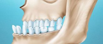
After all primary teeth have been replaced and the second molars (“sevens”) have erupted, the formation of a permanent dentition is considered complete. In an ideal permanent dentition, after changing teeth, the upper incisors should overlap the lower incisors by 2 mm. To assess the correct bite, it is also necessary to pay attention to the relationship and contact of the upper and lower teeth. At this age, your child is most likely already visiting a dentist, who, if necessary, will provide a referral for a consultation with an orthodontist. The last stage of teething will occur at the age of 18-20 years, when wisdom teeth appear. Sometimes there is not enough space for them to grow, or they may be in an incorrect position. In these cases, you may need to consult a dental surgeon.
A person needs a correct bite not only for beauty and beautiful speech, but also for the normal functioning of the maxillofacial apparatus. Poorly chopped food can lead to diseases of the gastrointestinal tract and the entire body as a whole. Over time, malocclusion can cause headaches and poor health. An uncomfortable psychological state also plays an important role for a person with malocclusion. That is why we recommend that you pay attention to the condition of your child’s teeth and promptly listen to the advice of doctors. The sooner you start orthodontic treatment, the better the results will be.
The Dial-Dent clinic has a newly created orthodontic department, headed by orthodontist O.A. Baranova. She specializes in correcting bites in adult patients and solves any, even the most complex cases. If you are concerned about your child’s malocclusion, then it is best to contact pediatric specialist E.V. Rada, the owner of numerous diplomas, certificates and certificates.
Dial-Dent specialists, whom you can contact for advice on the condition of your child’s oral cavity:
- Borisova Yulia Aleksandrovna, pediatric dentist.
- Nazarenko Evgenia Yurievna, pediatric dentist.
- Rada Elizaveta Valerievna, dentist-orthodontist, correction of bite in children.
- Selector Olga Nikolaevna, dentist-orthodontist, bite correction in children.
- Sleptsova Maria Pavlovna, dentist-orthodontist, bite correction in children.
- Tsukor Tatyana Borisovna, speech therapist - child psychologist.
- Smirnova Elena, dental hygienist.
Features of the period
The physiological replacement of temporary teeth with permanent ones is completed by the age of 12 years of a child’s life. At this moment, the teenager should have 24 permanent teeth in the oral cavity: canines, incisors, first molar, first and second premolars.
The eruption of the seventh tooth (second molar) begins at age 13. And by the age of 18, the “wisdom” tooth, that is, the third molar, may begin to emerge.
Causes of hyperplasia
(MG) is caused by overdeveloped, aggressive growth of the condyle. This condition causes abnormal jaw growth, which means one side of the jaw stops growing earlier. Juvenile idiopathic arthritis (JIA) and condylar fractures can cause growth and mandibular problems. Inflammatory and/or mechanical damage to the condylar cartilage often causes jaw hyperplasia.
Symptoms of inferior hyperplasia
Symptoms include slowly progressive unilateral growth of the condyle of the head and neck. This results in malocclusion, facial asymmetry, and a shift to the middle of the chin on the unaffected side. Characteristics of condylar hyperplasia may also include: a posterior open bite or tilted occlusal plane, depending on the time at which the hyperplasia develops, and asymmetry of the lower half of the face. Characteristic symptoms of lower hemisphere hyperplasia are asymmetrical facial bones (enlarged lower facial bones on one side).
Until what age can a child’s teeth be straightened?
Parents often ask the question: “At what age should a child’s bite be corrected? / At what age is it better to straighten teeth?”
It is best to have your first consultation with an orthodontist at age 6. Most often, it is at this age that the change of teeth begins: the sixth teeth erupt, the lower first incisors fall out. There are individual differences in the timing of eruption (early/late eruption). This is not a pathology! It is necessary to focus more on the situation in the oral cavity, rather than age.
6-7 years is the peak of a child’s growth, which contributes to the maximum result of orthodontic treatment and psychologically the child will be ready to wear any equipment . BUT! If at an earlier age parents see asymmetries or disproportions of the face, the lower or upper jaw is strongly pushed forward, it is worth going for a consultation with an orthodontist without waiting for 6 years.
Methods for straightening children's teeth vary greatly. It all depends on the specific clinical picture.
Most often, permanent teeth erupt crookedly and crowded due to lack of space for them and narrowing. The aggravation of the situation can be prevented by removable plate devices that stimulate the growth and expansion of the jaws, i.e. create space for teeth and they are aligned.
But straight teeth do not equal correct bite! Bite is the relationship of the jaws relative to each other, their proportional relationship and position in the skull. These parameters can be assessed by an orthodontist in person. Correction of bite in children is carried out using intraoral removable and non-removable plate devices, orthodontic trainers (guards) and extraoral devices. The choice of design is individual.
Remember that preventing pathology is much easier than correcting it in adulthood.
It should be said that orthodontic treatment is often necessary both in the mixed dentition (6-12 years old) and in the permanent dentition (from 13-14 years old). In mixed dentition, devices work to normalize the correct position of the jaws relative to each other, their growth and expansion. The effect on the position of the teeth is not so significant.
Braces/aligners are installed for a child after the eruption of all permanent teeth to completely straighten the teeth and create optimal interdental contacts. Such treatment, after the correct 1st stage at an earlier age, is easier, faster, without complex mechanics of movement.
You can correct an incorrect bite at any age (from 6 years and above), but the earlier you start, the easier it is to achieve maximum and stable results.
What is mandibular hyperplasia?
Hyperplasia is characterized by a constant or rapidly growing condyle when growth should be slowed or stopped. This condition causes development of the head, neck and jaw, causing significant functional and aesthetic deformities of the face. Disorders of the dental craniofacial structure affect the normal growth of the jaw, which leads to gross changes in the normal morphology and structure of the hard and soft overlying tissues. The result is asymmetry and significant functional deformities, which pose a serious problem for both orthodontists and maxillofacial surgeons.
Orthodontics Clinic
Patterns of development and growth of the dental system
Orthodontic dentistry is not only about correcting the position of teeth, but also about controlling jaw growth, correcting the shape of the facial skeleton, normalizing the functions of the dental system and restoring optimal facial aesthetics.
Prenatal period
The growth and development of the jaws is closely related to the formation of teeth, the alveolar process and is subject to the general development of the entire organism under the influence of endo and exogenous factors. At the 7th week of pregnancy, the baby begins to separate the oral cavity from the nasal cavity. It is during this period that the development of the fetal dental system can be adversely affected by endogenous factors of the mother - toxicosis, disturbances of hormonal regulation, water-salt and vitamin metabolism, exogenous influences, which can lead to nonunion of the upper lip, alveolar process and palate. During the period of intrauterine development, the alveolar process of a child grows by 55% of its future size, and over the next 24 years after birth by only 45%. This growth process occurs in waves, in impulses, with alternating periods of acceleration and deceleration of bone tissue formation. Therefore, when consulting a patient in the clinic, we realize that these nine months of intrauterine development are a little-known period for finding out the exact cause of the formation of an abnormal bite.
Newborn period
During the newborn period, the child experiences so-called infantile retrogenia (the lower jaw is displaced back in relation to the upper jaw), the sagittal gap between the jaws ranges from 5 to 10 mm. This is a physiological norm. Functional load during feeding (6 times a day for 30 minutes) promotes muscle training and growth of the lower jaw, which leads to normalization of the jaw ratio. Artificial feeding, in which the child receives milk quickly and in large quantities, does not contribute to the necessary load, leading to retarded growth of the lower jaw and the formation of a distal bite. If the growth of the lower jaw does not occur in the first year of the child’s life, then this lag will be observed from year to year, which will subsequently require long-term orthodontic treatment from 2 to 5 years.
Period of temporary occlusion
During the period of temporary occlusion from 7 months to 3 years, the most active growth of the child and dentoalveolar arches is observed. During the period of formed temporary occlusion from 3 to 6 years, sagittal growth stops and transversal growth of the jaws is noted, which leads to the appearance of three (gaps) between the teeth. In the absence of three, the crowded position of the anterior teeth occurs on the upper jaw in 62.5% of cases, on the lower jaw in 79%.
Period of early mixed dentition
During the period of mixed dentition, from the age of 6 years, a powerful impulse of jaw growth is associated with the eruption of incisors and first molars. The rudiments of these teeth move vestibularly and toward occlusion, subjected to pressure from the lips, cheeks, and tongue, leading to automatic restructuring of bone tissue and an increase in the distance between the primary canines. Violation of the myo-functional balance during teething due to improper breathing, chewing, swallowing, and speech leads to underdevelopment of the alveolar process and the appearance of crowding of the anterior teeth. It is known that a force of 1.7 grams is sufficient to displace the lower frontal teeth lingually with the lower lip. With myofunctional disorders, the lower lip can exert a pressure of 100-300 g on the front teeth of the lower jaw, and a pressure of 500 g on the tongue, which leads to the formation of malocclusion. Therefore, to eliminate functional disorders during this period, functional equipment, for example, a Frenkel apparatus or a lip bumper, is most effective. Considering that the frontal area of the lower jaw in an adult and a child after the eruption of the incisors has a constant size, great importance must be attached to the development of this area from 6 to 8 years, where all dental aesthetics are concentrated. An attempt to increase the lower dentition in the anterior section after 9-11 years leads to relapse. The correct position of the lower incisors at the age of 7-9 years contributes to the increase in the upper dentition in the frontal region, which prevents the appearance of a crowded position of the incisors in the upper jaw.
Period of late mixed and permanent dentition
In the final period of the mixed dentition, from the age of 9, there is no active growth of the jaws, so very important importance must be attached to the sequential change of teeth:
- Lower jaw - normally, the canines, first and second premolars change sequentially, the first molars can move mesially. Under the pressure of the lower canines, the frontal area moves forward and the bite increases.
- Upper jaw - first premolars, canines, second premolars, canines move distally.
The period of permanent dentition is characterized by active growth of the dentoalveolar arches, associated with the eruption of second and third molars. The beginning of mineralization of the cusps of the third molars is observed at 7-8 years; by the age of 9-13 years, the rudiments of these teeth are already revealed on the orthopantomogram.
Peaks of growth
During the formation of the dental system, the following growth peaks are observed:
- At 3-6 years old.
- At 6-7 years old in girls and 7-9 years old in boys and is associated with the eruption of incisors and molars
- At 9 years old in girls and 11 years old in boys and is associated with the change of 3,4,5 teeth.
- At 11-13 years of age in girls and 13-15 years of age in boys, it is associated with puberty and the eruption of second molars.
According to scientific research, at 11 years of age, the lower jaw can normally grow in the sagittal direction to approximately 3 mm per year, and at 14 years, up to 5.5 mm. The use of functional equipment at this age stimulates the growth of the lower jaw by 1.5 mm more than normal. From this we can conclude that the effect of functional equipment should not be overestimated during these periods of formation of the dental system; it is necessary to understand that in order to correct the pathology of occlusion in the sagittal direction, it is necessary, first of all, to create conditions for the physiological growth of the lower jaw, eliminating all adverse effects. A very good treatment result can be obtained by correcting the bite at the age of 6-8 years, during the period of eruption of the frontal teeth, when using functional equipment the pressure of the lower lip is eliminated, the teeth and lower jaw physiologically move in the anterior direction.
What can cause hypoplasia of the lower jaw?
Mandibular hypoplasia tends to be a congenital condition, however, it can definitely occur due to trauma or. Mandibular hypoplasia indicates an incomplete jaw. The radiographic appearance of maxillary and mandibular lesions often includes radiolucency with poorly defined or even irregular diffuse borders. Expansion of the orbital border usually creates hypoplasia and poor pneumatization of the ipsilateral maxilla. Possible hypoplasia of the body of the lower jaw, an abnormal coronoid process, as well as a zygomatic arch.