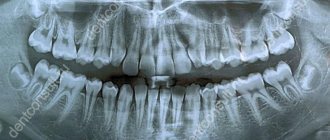
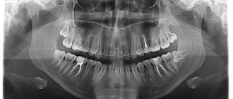
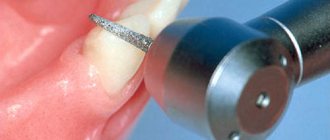
An x-ray is the dentist’s main tool in making the correct diagnosis. However, a conventional orthopantomogram or targeted photograph has limited diagnostic potential and does not provide complete data on the condition of the teeth and maxillofacial area. But technologies are constantly being modernized and today, more informative technology has come to the aid of conventional radiography - dental computed tomography (CT).
What does a 3D dental x-ray show?
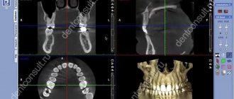
3D dental tomography is a highly accurate diagnostic method that makes it possible to obtain a three-dimensional image of the dental system in different projections. Volumetric images obtained with CT allow the specialist to enlarge, rotate and examine the area of interest from all sides and at different depths:
- The entire maxillofacial apparatus.
- A dentition or an individual tooth.
- Paranasal sinuses.
- Bone and periodontal tissues.
Dental CT allows you to detect inflammation, assess the homogeneity of the filling material and check the quality of installation of a filling, crown or implant, see the number of dental roots and their fragments, identify neoplasms, assess the degree of curvature of teeth, determine the exact parameters of bone tissue (height, width, density, etc.). d.). The information obtained allows the doctor to optimize treatment measures and predict the result.
Prescribing a CT scan of the upper and lower jaw before installing implants
Diagnosis of teeth and jaw structures
3D X-rays make it possible to examine areas of the maxillofacial system - from one tooth to a complete set of cross-sectional sections of the jaw and maxillary joint. Tomography before implantation allows:
- identify hidden carious cavities of neighboring teeth, curvature, length of canals, number of broken tooth roots to draw up a treatment plan before implantation;
- detect impacted, supernumerary teeth that may interfere with the installation of implants;
- evaluate bone septa, areas of pathological reorganization of bone tissue;
- visualize inflammation, cysts, granulomas, dental abscesses near the planned implantation area, which must be treated before surgery;
- identify inflammation in the maxillary sinuses and lacrimal ducts, which can become a temporary obstacle to implantation;
- evaluate bone tissue parameters - volume, density, degree of resorption, alveolar process inclination, thickness of cortical plates in order to correctly select the size and shape of the implant;
- clarify the anatomical structure of the maxillary sinuses, mandibular canal and other bone structures in order to plan the angle of inclination when installing an artificial rod;
- identify anomalies of the dentofacial system, pathologies of the temporomandibular joint for the correct modeling of the orthopedic structure for the implant;
- control the quality of implant and crown installation;
- assess the bone density around the installed implant;
- clarify the severity of the injuries received if the dentition is being restored after an injury
The information received in digital format is recorded on a medium (disk, flash drive) and sent by e-mail.
3D modeling of the operation CT results allow you to simulate the installation of implants of the required size, determine the angles of implementation bypassing the anatomical structures and simulate the final result of implantation.
The tomograph data is loaded into a computer program, and a 3D model of the jaw is created. The implantologist creates a virtual operation plan - selects the shape and size of artificial roots, their number, determines the installation location and angle of inclination. This allows you to take into account the nuances in advance.
A customized surgical template is printed using a 3D printer. This is an overlay with guides for future implants, which accurately determine the location and angle of installation of artificial roots. During the operation, the template is tightly applied to the gums, and the implants are installed with maximum precision.
Why are dental x-rays prescribed?
A 3D photograph of teeth is performed if the following indications exist:
- Injuries of the maxillofacial area.
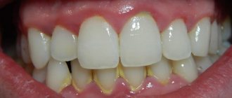
- Preparation for endodontic treatment (structure of root canals, pathological processes in the periodontium, degree of pulp damage, etc.).
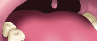
- Diagnosis of neoplasms (cysts, abscesses, granulomas, tumors).
- Anomalies of development and deformation of the maxillofacial apparatus.
- Quality control of filling and implant installation.
- Planning of orthodontic treatment (identification of impacted and dystopic teeth, analysis of the condition of the tissues around each tooth, etc.).
- Detection of hidden periodontal cavities and pockets.
- Implantation planning (assessment of jaw bone parameters, indications for sinus lift or osteoplasty, modeling the result of implantation).
- Endogenous pathologies of the maxillary sinuses.
Three-dimensional x-ray examination is the gold standard when planning any complex dental procedure or surgery. CT allows you to quickly make an accurate diagnosis, competently plan treatment or dental prosthetics, and monitor the results.
Indications for computed tomography
- Upcoming orthodontic or orthopedic treatment
- Planning for dental implantation (in this case, photographs are taken with markings of missing teeth)
- Diagnosis of hidden dental pathologies
- Diagnosis of cysts, tumors of the dental system, diseases of the temporomandibular joint
Contraindications
- Pregnancy
- Acute respiratory infections
How is the preparation carried out?
Computed tomography of teeth does not require special preparation. Before the examination, the radiologist will ask you to remove items that may interfere with the operation of the tomograph: earrings, hair clips, piercings, glasses, hearing aids. If you have removable dentures, you will need to remove them from your mouth. Before the procedure, the patient must wear a protective lead apron.
What equipment is used
To carry out 3D diagnostics, a three-dimensional computed tomograph SOREDEX Scanora 3D with advanced functionality is used. This is the latest generation equipment, which allows you to obtain three-dimensional images of the anatomical structures of the maxillofacial region in a few seconds, with the least radiation exposure for the patient.
The program analyzes the obtained multiplanar sections and builds them into a 3D model, thanks to which the specialist is able to accurately assess the condition of the dental system, detect all pathological processes occurring in this area and competently plan a treatment regimen.
A virtual 3-dimensional model of the scanned area can be recorded on any digital media (CD, flash drive), which allows the attending physician, if necessary, to view diagnostic data or involve related specialists in the analysis of the received information.
Manufacturers of intraoral scanners
Today, there are 7-8 companies widely represented in the world that produce intraoral scanners. Firstly, the ones I talked about. This is CEREC from the German company Sirona, this is the iTero scanner, which is currently produced and owned by Align Technology. This is a company that also makes Invisalign aligners. This is the American company Carestream. This is the European company 3Shape with a TRIOS scanner.
Recently a very worthy scanner has appeared on the market - in terms of scanning quality and convenience, and most importantly, with the most reasonable price. The scanner is called i500. From the Korean company Medit.
Star Smile is a partner of major scanner manufacturers
We, the Star Smile company, have established contact and cooperation with all of the above manufacturers, which allows our clients, when using these scanners, not to have to deal with sending stl files to our company, but simply to create an ID scan number in their personal account, and we, using our agreement, online connection with the servers of scanner manufacturers, we will independently gain access to the intraoral scan, to stl files and will be able to begin production of aligners.
One of our last contracts that we were able to conclude was a contract with Sirona, thanks to which we now have access to CEREC scanners. Any client who owns a CEREC scanner (we are primarily talking about the latest Omnicam models because the previous models are not very suitable for orthodontics, they are mainly intended for orthopedics) can take an intraoral scan of the patient and immediately send this intraoral scan to us at laboratory for the production of aligners.
This is a very important contract for us, because in both Europe and America CEREC occupies a leading position in the intraoral scanning market. I think that up to 50% of our new customers who register on our website in Europe and North America own CEREC scanners.
What other advantages of intraoral scanning for Star Smile clients?
Another advantage is the speed of data exchange between the clinic and us, as manufacturers of aligners.
If the doctor takes traditional impressions of teeth, then in order for us to begin production, these impressions must either be scanned in-house at the clinic, or sent to our office for digital 3D scanning.
Sending dental impressions and the process of scanning them into 3D dental models takes quite a lot of time. On average, it's probably two to three business days.
If the doctor works with an intraoral scanner and has scanned the patient’s oral cavity, then we can begin production of the virtual SetUp as part of the aligner production almost immediately after the doctor has completed the scan and registered the order in our system.
We see that on average this reduces the production time for a virtual SetUp by 2.5 days, which is very good, considering that the average production time for a virtual SetUp is just over 4 days. This means that those clients who start working with intraoral scanning receive a virtual SetUp in two business days, even a little faster.
Another key advantage of intraoral scanning and intraoral scanners over traditional impressions is online control over the quality of the scan, which can be carried out by the doctor.
Let’s imagine such a situation that the doctor took impressions of the teeth, looked at them, and they seemed to him without errors, without flaws. I sent these casts to the laboratory of any manufacturer. And then, during 3D scanning, it turned out that the cast was not of sufficient quality. Somewhere there are braces, for example, somewhere, perhaps, the gingival margin is not “awakened” well enough, somewhere there are air bubbles or saliva. All this affects quality.
When working with traditional casts, this is only visible when casting a plaster model and scanning this plaster model. This is a very painful situation when it turns out that the impression is not of sufficient quality and cannot be used in the production of aligners. This is unpleasant because you have to call the patient to have impressions of his teeth taken, and a lot of time is wasted. We spend at least two or three days taking new impressions and sending them back to 3D scanning and production.
If the clinic works with an intraoral scanner,
then the doctor or his assistant who is engaged in scanning, during the scanning process can visually, on the computer screen, control the quality of the resulting scan, the quality of the digital model. If he sees that there is a mistake somewhere, then this place can be immediately rescanned. This virtually eliminates errors in scans, meaning there is no need to return the patient for rescanning.
Undoubtedly, the intraoral scanner is the most promising and useful technique for 3D dental modeling in the present and near future. But it’s also very expensive. We have diligently avoided the cost of scanners in the article, because now you will be offered a service that will solve your scanning problems without purchasing a scanner. Why buy a scanner, since the cost of intraoral scanners starts from 15 thousand euros? Taking into account all the above advantages, our company Star Smile launched a new service in January 2022, which we call “On-site scanning”.
Possible harm
Cone beam dental computed tomography is the safest and fastest diagnostic method. Thanks to the use of a conical X-ray beam, the radiation dose received during the study is 10 times less than when using spiral CT. And the pulsating mode of the X-ray beam further reduces the radiation dose. The three-dimensional computed tomograph SOREDEX Scanora 3D is one of the safest devices in terms of X-ray radiation dose - only 0.035 m3v.
However, despite the safety of the study, CT also has contraindications. If we just talk about dental tomography, it is not performed during pregnancy (in the 1st trimester). 3D dental x-rays with contrast are prohibited for pregnant and lactating women, patients with endocrine disorders (diabetes mellitus, thyroid pathologies), renal failure and intolerance to iodine-containing drugs.
On-site 3D scanning from Star Smile company
While on-site scanning
operates in Moscow and St. Petersburg. Thanks to this service, all our clients - clinics or doctors - can call a Star Smile employee with an intraoral scanner, who, by agreement, will come to your clinic at the appointed time and scan the patient.
How much does on-site 3D scanning cost?
By the way, the on-site scanning service is free when ordering Star Smile aligners. It increases the accuracy of aligners, speeds up the process of their production, and eliminates the possibility of errors when taking impressions. And as an added bonus: your clinic will look more advanced, high-tech to your patients.
For now, this service is available in Moscow and St. Petersburg. Since we Star Smile operates in more than 70 cities of Russia, we are already considering other cities in which on-site scanning will be in demand by orthodontists and dentists.
Online consultation with a doctor
If you are concerned about the condition of your teeth. Painful sensations arise, gums bleed for a long time, and seals have appeared on the jaw. You are concerned about previously installed dental implants. And there was a need to take a 3D photo of the teeth. Then, after receiving three-dimensional visualization, it is better to go for an examination or consultation with a dentist to interpret the images and compare the results with your current complaints. It is impossible to independently understand the nuances of a 3D image, much less make a diagnosis. The specialist will explain the situation and give recommendations before the in-person appointment.
Advantages of the method
- The ability to rotate, enlarge, and examine images in any projection and section, which is impossible with conventional 2-dimensional scanning.
- The examination lasts only a few seconds (8-20 seconds).
- Complete diagnostic information.
- Maximum security.
- Digital information format.
- Detection of any pathological processes at an early stage.
- No prior preparation required.
- 3D reconstruction without distortion or artifacts.
- A wide range of purposes - from endodontic dental treatment and implantation to maxillofacial operations.
Is there an alternative to CT
There are many other diagnostic imaging methods (x-ray, orthopantomogram, ultrasound, etc.), but only CT provides the possibility of highly accurate, separate images of all types of tissue at different angles and to different depths. Although a panoramic dental photograph remains an equally important diagnostic tool for a dentist today, it can only provide a general overview. In turn, a 3D tomogram allows you to obtain not a single flat image of the jaw, but a whole series of sequential multiplanar images in different projections and without the distortions inherent in a panoramic image.
Example:
due to the different density of bone structures exposed to X-ray radiation, it is impossible to see less dense bone in a 2-dimensional image; accurate information is provided by a 3- D image of the teeth.
How does the procedure and decoding work?
To take a 3D photograph of teeth, a standing or sitting patient needs to bite a special plate and fix his position in the device using a fixing stand. During the entire scanning time, you must remain absolutely still.
The tomograph sensor makes a series of revolutions around the patient’s head for 8-20 seconds, producing about 200 images in different projections. Processing digital data takes 5-15 minutes, after which the information is written to a disk or flash drive. No preparation is required, you just need to remove all metal jewelry from your neck, ears, and hair before the procedure.