Fused tooth roots are a fairly rare occurrence. According to statistics, they are detected in approximately 4% of patients who seek help from a dentist. Experts attribute the causes of the problem to:
- tooth dislocation,
- severe damage to the jaw,
- infection of the rudiment during eruption,
- violation of root resorption when changing teeth,
- long-term inflammation of soft tissues,
- genetic predisposition.
Find out if it's dangerous, what symptoms you shouldn't ignore, and how to treat it.
What is ankylosis
Tooth ankylosis is a pathology characterized by fusion of the cement of the tooth with the jaw bone. The tooth root is deprived of part of the ligamentous apparatus, which often leads to adhesion or germination of bone tissue. The aesthetics of the teeth are disrupted, they look shorter and change their angle. Ankylosis usually occurs during tooth eruption. Pathology can also form at a late stage in the formation of the dental arch.
It is important to know. The process is accompanied by periodontal necrosis and the incorporation of root cement into the alveolar bone.
Ankylosis of a primary tooth is caused by loss of the periodontal ligament. It is observed in cases where a primary molar does not fall out due to the absence of a permanent dental unit. Ankylosed teeth are different in size from their neighbors, so bite correction will be required.
Correction methods
Several decades ago, a person with such an anomaly had two options: remove the merged specimens, or walk with them all his life, building up an inferiority complex.
Today, dentists, using modern techniques and materials, can successfully correct this problem while the main tooth remains intact.
What problems need to be solved?
When choosing a treatment method for this anomaly, the doctor is primarily guided by the tasks that must be solved through therapy.
The main tasks include:
- Correction of pathology while preserving the more significant half.
- Freeing up the necessary space to correct its position.
- Creating favorable conditions for the formation of normal occlusion and dentition.
- Preservation of the pulp chamber and the pulp of the main half to extend the life of the tooth.
What determines the choice of method?
Unlike the treatment of other dental anomalies, in which the fundamental factor in choosing a method is the patient’s age, in this case they rely only on the degree of pathology :
- To eliminate the problem in the first degree of anomaly, they resort to gentle treatment, which involves grinding off the adjacent part.
This method allows you to completely preserve the main one, without the use of additional equipment and special filling materials, since the affected units do not have a common pulp chamber.If necessary, the procedure ends with restoration.
- For the second type hemiresection , in which the supernumerary part is disconnected and removed. In this case, periodontal tissues are not injured.
- With the third type of anomaly, hemisection , which involves exfoliation of the gum tissue and dissection of the root part.
- Treatment for the fourth degree is prescribed exactly the same as for the third, but the dissection is carried out along the entire length .
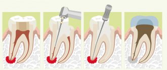
Sequence of procedure
The treatment takes place in several stages, the first of which involves the immediate removal of the secondary part.
If it is necessary to carry out removal with dissection of only the upper part, then conduction anesthesia is used. In case of complete dissection, general anesthesia may be administered.
Depending on the technique, the main points of the procedure may differ.
Hemiresection involves several steps:
- The dentist uses a diamond disc to cut off a smaller portion of the tooth.
- Performs extraction by rupturing the periodontal ligaments and dislocation.
- Grinding the cut areas with constant irrigation with an aseptic preparation.
- Washing the wound and applying a medicinal application with biologically active components that restore damaged tissue. The bandage is fixed for 2 weeks, after which it is removed.
How to recognize macrodentia, characteristic signs and treatment methods.
In this material we will look at why wisdom teeth are characterized by manifestations of dystopia.
Here https://orto-info.ru/zubocheliustnye-anomalii/zubov/polozheniya/krivyie.html you will see in the photo how effective the fixation of veneers is on crooked teeth.
Hemisection , which is a more traumatic operation, proceeds as follows :
- To gain access to the root part of the tooth, the dentist performs an excision of soft tissue by making an angular incision on the vestibular side of the jaw, plunging the scalpel all the way to the alveolar bone.
In order for the excised tissue to move to the side without any problems, it is cut on both sides of the problem tooth and at its cervical part.The result is a trapezoidal flap that is easily separated from the jawbone.
- It is then peeled off, exposing the cortical bone, from which a limited area is removed. This allows access to the roots.
- Using a special fissure-type bur and diamond discs, the root and then the crown part of the tooth are cut off. The cut off parts are removed.
- The disconnection site is polished, while the wound is constantly treated with saline solution, which ensures the safety of the main part of the pulp left in the alveolar arch.
- The cleaned area is covered with a special composite that restores the tooth wall.
- The mucosal and periosteal flaps are placed in place and sutured with simple surgical sutures, which are removed after 7 days.
- Next, the tooth is splinted for 2 weeks.
2 weeks after removal of the complementary half, the second stage of treatment begins - orthodontic. This type of treatment allows you to completely restore the normal position of your teeth and correct your bite.
Most often, braces with arches of varying degrees of rigidity are used for this, regular replacement of which leads to complete restoration of the row after 8–15 months .
To prevent the tooth from returning to its previous position after correction, retainers are additionally prescribed. The retention period, as a rule, lasts almost as long as the main treatment.
How to remove a tooth with root separation, watch the video:
Symptoms
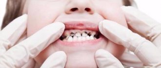
Parents should be wary of the fact that their child’s baby teeth are not falling out. It is fused to the bone and cannot easily separate from the alveoli, as is normal. If the pathology is not detected and treated in a timely manner, the child’s jaw begins to develop incorrectly. The diseased tooth is located lower than the others, and the neighboring teeth are placed at an angle in relation to it. Problems with bite occur.
Another scenario is also possible. The baby tooth does not erupt completely and is delayed in its development. Because of this, the permanent tooth also cannot develop, and after the milk tooth falls out, it becomes fused with the jaw. This is how ankylosis of permanent teeth occurs. The final diagnosis is made after analyzing the results of a computed tomography scan.
Good to know. The service life of ankylosed teeth is completely different. They can last for decades or fall out, thinning out the dentition.
Reasons causing anomalies
Anomalies of the dental system are very diverse and include both anomalies of dental units and incorrect formation of the dentition, as well as abnormalities in the development of the jaws. The most frequently diagnosed abnormalities of dental units. They can be caused by a wide variety of reasons, including both exogenous (caused by external factors) and endogenous (formed as a result of the physiological state of the patient).
Endogenous causes
Endogenous anomalies are formed as a result of genetic and endocrine disorders.
- Many structural features of the dental system, leading to improper development of teeth, are genetically determined. This can occur through direct inheritance (abnormal number and shape of teeth, edentia, diastema), through inheritance of a mismatch in the size of the jaw bones, through inheritance of a mismatch in the size of the jaw and teeth (crowding of teeth or sparse arrangement of teeth). Dental anomalies of this type include disorders associated with hereditary and congenital pathologies (clefts of the soft and hard palate, cleft lip, Down syndrome, Vaanderburg syndrome, Seckel syndrome, Shershevsky-Turner syndrome).
- Another part of the endogenous causes of dental anomalies are problems of the endocrine system, such as hypothyroidism (in later stages), hyperparathyroidism, hypocortisolism. Their frequent symptoms may be delays in the eruption and replacement of teeth, and disturbances in the formation of the enamel layer.
Exogenous causes
Exogenous (external) causes of dental anomalies are, for the most part, external unfavorable factors that affect the baby’s body during the formation of tooth germs. There are prenatal (prenatal), postnatal (postpartum) and intranatal (associated with childbirth) periods.
- An example of a prenatal factor is a pregnant woman living in poor environmental conditions. Various developmental disorders of the fetus can also occur as a result of the mother’s poor lifestyle.
- Factors in the intranatal period may include complicated labor, birth injuries, consequences of asphyxia and oligohydramnios.
- In the postnatal period, dental anomalies develop as a complication of pathologies in early childhood. These include childhood infections, rickets, hypovitaminosis, and micronutrient deficiency.
All these reasons are of a common nature. Local factors that affect dental development include everything related to improper oral care, feeding errors or bad habits. Among them are feeding too soft food, prolonged sucking of a pacifier or finger in early childhood.
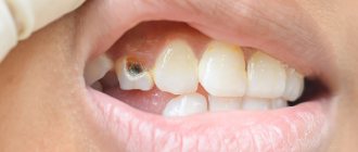
Childhood injuries and caries with complications lead to dental anomalies.
The death of tooth germs or the development of supernumerary teeth can occur due to osteomyelitis.
Treatment of ankylosis
Treatment of ankylosis with traditional orthodontic methods (braces) is impossible. The tooth grows into the bone and it is impossible to return it to its correct position. But restoring the functionality of the dentition is quite possible. In dental clinics, the method of treatment is selected individually in each individual case.
First of all, the patient is referred for a dental computed tomography scan. The dental surgeon studies panoramic images and draws up a treatment plan. Most often, surgical treatment methods are used: excess fibrous tissue is removed and joint mobility is restored. Crowns made of metal-ceramics, ceramics and zirconium dioxide are also used to treat dental ankylosis. With their help, the bite is corrected.
Anomalies of hard tissue structure
An altered shape, abnormal size or color of enamel is formed against the background of an abnormality in the structure of the hard tissues of the dental unit. Among such anomalies are:
- Hypoplasia (underdevelopment of tissue). The initial degree of the disorder is manifested by the presence of chalky spots on the enamel and areas where tissue deficiency is observed. Subsequently, all kinds of pits, grooves, and recesses appear on the surface of the enamel. The defect affects all teeth in the dentition.
- Hyperplasia (excessive tissue formation). Pathology also affects all teeth at the same time. It is characterized by areas where tissue growth is observed - tubercles, sagging enamel. The consequence of the pathology is a change in the shape and size of the teeth, a violation of the occlusion (the line of contact of the upper and lower jaws).
- Anomalies of amelogenesis (enamel formation) are expressed in the presence of brown or yellow spots on the surface of the teeth. Areas where the natural composition of the enamel is disrupted become especially sensitive. Microdentia develops against the background of pathology. The cause of the development of pathology is a deficiency of microelements that take part in the formation of dental tissue. Treatment consists of replenishing this deficiency. Additionally, local remineralizing therapy and physiotherapy are performed.
- Disturbance of dentinogenesis consists of dysfunction of the mechanism of dentin formation. The main symptoms of the disease are as follows: teeth become yellow-brown or grayish in color. The fragile dentin in these areas quickly wears off, causing tooth decay and then tooth loss. The disease occurs in genetically predisposed patients. The problem can only be solved by replacing the units destroyed by the disease with prosthetics.
Color anomalies
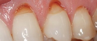
Healthy, young teeth look snow-white due to a thick dentin layer and high-quality enamel, which has sufficient characteristics of whiteness, shine and transparency. Lifestyle, age, genetic factors, and ecology can slightly change the color of teeth from bluish to yellow. Small deviations are not considered pathology, although they indicate a change in the quality of the enamel.
Various pathological processes occurring in the body have a more significant impact on the condition of the enamel. The cause may be carious lesions, the use of medications, or a lack of certain microelements in the body. Teeth can take on a gray, pinkish, brown, purple and even black tint.
When starting to treat a patient, the dentist excludes the development of chronic pathologies. After a course of therapy and stable remission is achieved, other causes of discoloration of the enamel are eliminated. The final stage of treatment is a course of teeth whitening.
Specialists at the Consilium Dent choose the enamel lightening technique together with the patient. The selection criteria are age, condition of the enamel, and the wishes of the patient.
A milk tooth in an adult: to remove or not?
If a baby tooth has not fallen out, is susceptible to caries or interferes with the growth of a molar, this does not mean that it is doomed to be removed. Dentists still recommend taking care of it and extending its service life for you, curing it if necessary. If only because there is no guarantee that the root one will then grow in place of the removed milk one, even if its rudiments are discovered.
To make a final decision about baby teeth in adults, it is necessary to take an x-ray. The image will allow the doctor to understand whether at least some rudiments of the molar tooth have formed, as well as in what condition the roots of the milk tooth are - whether they are resolving or not. If there are no buds and the roots are not going to resolve, removal is contraindicated. If the roots have resolved and the tooth is wobbly, then most likely it will have to be removed and you will have to wonder about the optimal prosthetic option.
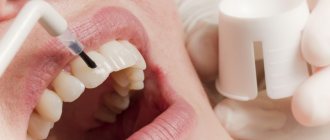
If the baby tooth is held firmly, but you are not very happy with its appearance and you would like to make it more aesthetic, the dentist will offer options for solving this problem. In this case, much also depends on the position of the “experimental” in the dentition. Restoration, veneers. Lumineers - modern dentistry has a sufficient arsenal of solutions specifically for your problem.
One way or another, having discovered that there is still a “piece of childhood” in your smile, go to the clinic, where specialists will take an x-ray, determine the presence and condition of the rudiments of a molar tooth, the condition of the roots of a milk tooth - and provide high-quality treatment, preserving your health and guaranteeing comfort.