For a long time, dental granuloma develops asymptomatically! There is a feeling of an overgrown tooth, slight pain when pressed. At the initial stage, there are no visual changes; inflammation in the apex is discovered by chance during an X-ray or computed tomography scan prescribed when visiting a doctor for other dental diseases. The question begins to worry, what is a granuloma?
Dental granuloma can be detected when a dense nodule of 5-8 mm appears at the base of the tooth, nearby tissues become red and swollen, acute pain begins to bother you, or in a photograph of the tooth before prosthetics. Because of the fear of opening the gums, many people put off visiting a doctor and are treated with rinsing decoctions.
| A nodular root formation is a source of infection; without treatment, inflammation spreads to the tooth socket, periosteum, subcutaneous tissue of the face, and lymph nodes. The periodontal tissue dissolves, in severe cases the tooth becomes loose and falls out. |
What is granuloma, its causes
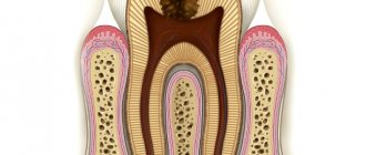
Dental granuloma is a focal proliferation of cells of the connective tissue that fixes the tooth. Externally, it appears in the form of a rounded nodule (up to 10 mm), usually localized at the root apex. A nodular neoplasm is formed as a result of prolonged inflammation in the pulp or periodontal tissues.
Granulation in the tooth, as a protective reaction, is triggered in response to infection and suppuration. A capsule is formed around the source of inflammation, isolating bacteria and dying cells. An infectious and inflammatory process occurs inside the nodule. The tissue that replaces the dead cells grows, and granulosa formation increases. The formation of a granuloma on the root of a tooth is provoked by an infection that has penetrated into the periodontium (periodontal tissue), which is preceded by:
- untreated caries and pulpitis, periodontitis;
- cracks, fractures;
- non-compliance with canal filling techniques;
- tooth extraction, which leaves an infected root tip.
The nodular neoplasm goes undetected for months and proceeds without pain! Exacerbation of dental granuloma is provoked by reasons not related to dental diseases - hypothermia, viral infections, physical overexertion. Granuloma of the tooth under the crown occurs due to secondary caries, insufficient oral hygiene after prosthetics. The infection progresses and enters the periodontium when the root bottom is perforated, carious tissue and pulp are incompletely removed, or the filling is not properly adhered to. When the edge of the crown moves away from the gum, bacterial plaque accumulates in the gap, the stump is destroyed, and inflammation develops in the soft tissues. Granuloma of the tooth after treatment of pulpitis develops when the canals are poorly cleaned and not tightly sealed.
Treatment methods
Granuloma can be cured in two ways, each of which has its own characteristics:
- In a therapeutic method that is possible at an early stage of the disease, the doctor uses antibiotics to prevent the development of inflammation. This allows you to preserve healthy dental tissues and subsequently carry out tooth reconstruction.
- With the surgical method, the dentist cuts the gum, releasing the pus that has accumulated in it. A drainage is placed in the wound cavity, and the patient is prescribed antibacterial, anti-inflammatory and antiseptic agents.
- In the most difficult cases, the tooth is removed, after which pus flows out through the wound and the granuloma disappears.
Treatment at the Family Dentist clinic
We have a good reputation in Minsk, consultations are conducted by dental therapists with 16 years of experience , specializing in the treatment of dental canals. They trust us; more than 80% of patients brought family members and friends to the clinic. Every second patient has been with us for more than 6 years. We do radiovisiography on site in the office, working with four hands together with an assistant to reduce the time of unpleasant manipulations. The effectiveness of dental granuloma treatment is ensured by premium class equipment. Even with serious damage to the crown and root, we strive to preserve the dental unit.
What are the possible complications?
The neoplasm itself does not cause any consequences. If an infection gets into the root of the tooth or if inflammation occurs again, it may fester. In this case, a person experiences gumboil, severe pain, swelling and distension of the gums. If dental granuloma is not removed in time, this can lead to troubles:
- The tooth will have to be completely removed.
- The neoplasm can develop into a malignant tumor.
- This pathology often causes osteomyelitis of the jaw.
Conservative therapy
Tooth granuloma grows for a long time without pain or visual signs. Patients turn to the dentist when the tumor increases in size, swelling and redness of the gums are clearly visible, or is discovered by chance in an image taken before prosthetics. But not all doctors undertake the treatment of dental granuloma and offer resection (removal) of the root apex.
In our clinic, a tooth can be removed from the source of inflammation without surgical intervention ; the cost of treating one root canal starts from 100 rubles. The doctor treats or re-treats the canals, disinfects them, and fills them with calcium hydroxide. The alkaline preparation kills bacterial flora and stimulates the formation of bone tissue. In difficult cases, we change the medicine several times.
The success of treatment can be assessed after 6-9 months using a control x-ray. If the granuloma in the tooth is eliminated, a permanent filling is installed and the coronal part is restored with composite material. If more than 50% of the dental tissue is destroyed, prosthetics with an inlay or crown are performed. We prescribe an additional antibiotic for dental granuloma to reduce the intensity of inflammation and prevent complications. In case of purulent inflammation, we install drainage and excise the nodular formation with the apex of the root.
We always warn that the result depends not only on our qualifications and experience, but also on the patience of the patient. After going through the canals, you need to prepare for several visits to the dentist to apply the paste, and do not miss appointments.
What is the difference between a dental cyst and a granuloma?
Many patients in dental clinics confuse cysts and dental granulomas, while these two pathological processes are significantly different. Seeing the difference is important because the diagnosis affects how the problem is addressed. Thus, treatment of tooth root granuloma is often conservative, but getting rid of the cyst can only be done through surgery - it is excised or removed along with the affected unit. So, how is a granuloma different from a cyst?
- Firstly, by its structure and structure. The first does not have a cavity and is a continuous formation of granulations, while the second is a capsule that is filled with pathological contents;
- Secondly, its size. Cysts are always larger than granulomas: they often reach one centimeter in diameter;
- Thirdly, symptoms. Granuloma is more prone to exacerbation, the development of pain symptoms and exacerbations than a cyst. But if the latter reaches a large size, the unit becomes mobile, which is not observed with granulomas;
- Fourthly, in the condition of the surrounding tissues. With cysts, the mucous membrane does not look inflamed and does not change its color, but the bone tissue, as the process progresses, becomes more pliable and decreases in volume. With granuloma, the mucous membrane reddens and swells, the formation of a fistula is possible, but the bone tissue is almost not affected.
On an x-ray, the cyst has clear contours and boundaries, which cannot be said about the granuloma, which looks like a darkened round shape.
Treatment of granuloma - the story of our patient
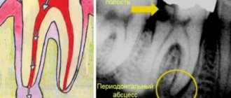
| The patient came to the appointment with complaints of the destruction of the 47th tooth and pain in the lower jaw on the right, which intensified when biting. A diagnostic radiograph taken after the examination revealed a granuloma in the area of the distal root. |
| On the first visit, after mechanical and medicinal treatment, the canals were temporarily filled with calcium hydroxide paste. The medicine has a bactericidal effect and stimulates the restoration of bone tissue. We took a control x-ray to make sure that there were no voids left in the root canals. |
Due to the presence of purulent discharge, anti-inflammatory therapy and antibiotics were prescribed. After the filling, the patient was bothered by pain when biting, which went away within a day.
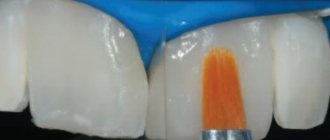
| At the second appointment two weeks later, the patient did not complain of pain. The paste was removed, the canals were filled with gutta-percha pins (permanent filling), and a third control photograph was taken. To make sure that the inflammatory process has stopped, it was recommended to return for the next examination with a control x-ray in 6 months. |
Symptoms of dental granuloma
In the very initial stage, the symptoms of granuloma are mild or absent altogether. When a nodule with a focus of inflammation increases, it can already be felt in the mouth with the tongue in the form of a foreign body. This is accompanied primarily by:
- redness of the gums and severe swelling,
- body temperature rises,
- at an advanced stage, pus appears between the tooth and gum,
- acute pain and general malaise occurs.
Our doctors are always ready to help and cure granuloma.
| Pankratieva Alesya Georgievna. Practicing dentist of the first qualification category. More than 15 years in the profession. Founder of the Family Dentist clinic, Member of the Belarusian Dental Association. View profile | Bobkova Irina Leonidovna. Practicing dentist of the highest qualification category. More than 20 years in the profession. Assistant professor. Co-author of a patent for a method of treating periodontitis. View profile |
Feedback from our patient:
Thank you very much for your attentive attitude towards patients. Quality services. I have been discussing at the Family Dentist for a long time and the doctors always have a good mood and a smile on their face. I will recommend your dentistry to my friends and acquaintances.
With respect, Gameza N.A.
| Do not leave the source of infection untreated - sign up for the Family Dentist clinic. The sooner you start treatment, the cheaper and easier it will be. | Call to make an appointment +375 29 604-61-61 |
Laser tooth removal
Laser therapy is a popular trend in modern dentistry and is used in many dental procedures. The most common methods of using a laser are: cleaning enamel, removing various neoplasms such as cysts, excision of gums, deepening the vestibules of the oral cavity, stopping bleeding of soft tissues and removing teeth.
The increasing popularity of laser equipment in dentistry is evidenced not only by its distribution in most Moscow clinics, but also by the constant increase in positive reviews of procedures where the laser was used.
Benefits of the procedure
Laser technology is rapidly gaining popularity in dentistry due to several unique advantages. Laser tooth removal has the following advantages:
- Sterile intervention, antibacterial action of the laser - thanks to the use of this technology, the likelihood of infection of the future hole is practically absent.
- Quick stop of bleeding, ensured by the temperature effect of the equipment.
- High precision of manipulations of modern laser systems.
The popularity of laser technologies is also ensured by their versatility and ability to solve various types of clinical problems. With their help, performing various therapeutic and surgical procedures becomes much easier, and there are no additional risks when using these techniques.
When should you remove teeth?
In order to remove a molar, sufficiently serious reasons are required. It’s not just the presence of various diseases or defects – the doctor also needs to establish the impossibility of saving the tooth using therapeutic methods.
- Damage to most of the tooth, when not only the crown is destroyed, but also the root canals, which can no longer become the basis for dentures. In most cases, under such circumstances, restoring a tooth by filling or installing a crown is not so effective - installing a full-fledged implant will be much more effective.
- A serious inflammatory process inside the tooth, which can lead to more serious diseases such as periodontitis. Such pathologies contribute to damage to the soft and bone tissues surrounding the tooth, and also create a risk of blood poisoning. Since after a certain stage of the inflammatory process, its treatment involves the removal of a significant part of the crown and root system, a common practice is complete extraction and subsequent installation of an implant.
- A severe degree of retention or dystopia that cannot be corrected by orthodontic correction methods, that is, through the use of braces or aligners. A fairly common procedure is laser removal of wisdom teeth, as they are often susceptible to similar defects. Thanks to the effects of laser equipment, it is possible, without any risks, to open the gum tissue, which often covers the dystopic or impacted figure eight, and then carry out its extraction.
Contraindications to the procedure
Despite the fact that certain prerequisites may be present for deletion, deletion is not always possible. Since extraction is a surgical operation, not every body is able to endure it. When a specialist works to analyze a clinical situation, he needs to exclude the presence of the following pathologies:
— autoimmune diseases or other factors that seriously suppress the immune system;
- severe diseases of the cardiovascular system;
- diabetes;
- serious damage to the endocrine system;
— oncology;
— various diseases of the neuropsychiatric spectrum.
Preparing for removal
The dentist is required to carefully analyze the situation and only then make a decision about extraction. For a complete understanding, a preliminary diagnosis is necessary - in addition to a standard visual examination, X-ray imaging is often used.
Modern X-rays allow you to obtain detailed visualization of any part of the jaw system. Thanks to them, doctors accurately determine the pathology, as well as the complexity of the future operation.
The specialist needs to exclude some contraindications that could negatively affect the possibility of future extraction. It is important for patients to mention possible pathologies that negatively affect their body.
Order of operation
If there are no contraindications, dentists can begin the procedure immediately. Removal begins with sanitation and antiseptic treatment of the oral cavity - such measures reduce the likelihood of pathogenic flora entering the formed hole.
After sanitation, doctors administer anesthesia - depending on the volume and duration of work, as well as the location of the tooth, the dosage may vary. Once key areas are completely numbed, doctors begin removal.
The extraction procedure itself is divided into several large stages. In accordance with clear regulations, the dentist performs the following actions:
- Dissection of soft tissues - this step is most often used when removing a dystopic or impacted wisdom tooth, since it is covered by a dense layer of gum. During the dissection, the gum tissue is moved apart, allowing the dentist to gain access to the tooth.
- In order to conveniently remove a complex tooth, the dentist crushes it using a drill and other special tools. He removes the remaining fragments in the hole using forceps.
- After removing the fragments, the doctor re-examines the resulting cavity to make sure there are no tooth fragments.
- If no dental tissue is found, the doctor treats the wound with antiseptic and anti-inflammatory agents.
- To prevent pathogens from entering the new wound, a special protective membrane is placed.
- The final stage of the operation is to return the soft tissue to its previous position and its subsequent fastening with sutures.
In total, the operation takes no more than two hours, but the time of work always depends on a number of circumstances. So, quite often the dentist needs to not only carry out extraction, but also stop the inflammatory process inside the tissues of the oral cavity.
Recommendations after removal
To minimize any post-operative health risks, you should follow the following rules:
- limit food and liquid intake for the first few hours after extraction;
- for the first few days, carefully clean the oral cavity, without touching the empty socket;
- carefully monitor your health and if there is an increase in temperature, pain, weakness or other sensations that cause discomfort, immediately consult a doctor;
- do not delay replacing a lost tooth to prevent possible atrophy of bone tissue, unless we are talking about a wisdom tooth, which usually does not bear any load.
Price
Laser dentistry will require certain costs from the patient. Every year the price for such services becomes more and more affordable for most people, but you will still have to spend money. Before determining the final price for laser surgery, it is worth considering the following factors:
- Various components of the clinical picture that influence the course of treatment: the root cause of removal, concomitant diseases of the oral cavity, tooth structure. If the root cause is mechanical destruction, the doctor does not need to fight the inflammation, which means the operation will be easier. Also, if the tooth has many roots, a complex extraction will occur with a corresponding increase in cost.
- Pricing policy of the clinic. Depending on the location of the medical center and the level of services provided, the final price of the work may differ by tens of thousands of rubles.
If you need laser tooth extraction in Moscow as soon as possible, make an appointment at our medical center. One of the methods of tooth extraction in our center is laser surgery - our specialists have a proven set of techniques for the safe and painless removal of problematic parts of the dentition. At the first consultation, the doctor will analyze the clinical situation and, together with you, prescribe the optimal course of treatment. It is possible to make an appointment through the website or by phone.
Author:
Head, one of the founders of the clinic. Chief physician. Maxillofacial surgeon, implantologist