Osteosynthesis of the jaw is a method of surgical treatment of bone fractures using special, most often metal, structures. Osteosynthesis surgery is performed if the fracture site cannot be secured with splints, and also when it is difficult to fix bone fragments. There are different methods of performing the operation, they can be used depending on the type of injury and its severity.
Osteosynthesis allows you to maintain the required level of blood circulation in the damaged bone, which means that such a fracture will heal faster. Not only the integrity of the bone is restored, but also its structure, and this will take only a few weeks.
During the rehabilitation period, it is important to follow certain rules and monitor your well-being and the condition of your bone tissue. Violations of doctor's instructions can lead to pain and even loss of chewing functions.
Indications for osteosynthesis
Osteosynthesis may be prescribed in the following cases:
- if there are not enough stable molars at the fracture sites;
- upon impact, the fragments moved significantly;
- broken jaw bone behind teeth. With such an injury, individual parts of the bone tissue are displaced;
- the injury occurred as a result of the development of inflammatory diseases that thin the bone tissue;
- in case of a fracture of the lower jaw, if too small or massive fragments have formed;
- if the branches and body of the jaw are incorrectly positioned relative to each other;
- it is necessary to perform reconstructive surgery or osteoplasty.
Discussion
Despite the development of medicine in general and maxillofacial surgery in particular, the problem of providing emergency medical care for fractures of the facial skull remains not fully resolved. Due to the increase in the number of road traffic accidents and domestic conflicts, and the passion of young people for traumatic sports, there has been an increase in the number of victims with injuries to the facial skull, among which fractures of the lower jaw occupy the first place [12].
The difficulty of early diagnosis of fractures of the angle of the mandible is associated with the insufficient information content of routine methods of radiation examination (radiography of the skull in frontal or lateral projection), late referral of victims to specialized maxillofacial hospitals and, as a consequence, the choice of irrational methods of treatment, which in turn leads to development of various kinds of complications, reduction in the quality of treatment and life of patients.
Types of osteosynthesis
There are several methods of osteosynthesis; the doctor decides what type of operation the patient needs. Most often, surgeons combine several methods with each other to achieve better results.
Osteosynthesis of the jaw can be:
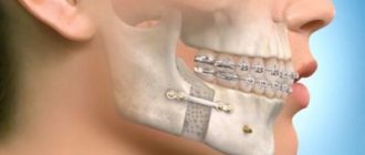
- Open. It is usually used for severe fractures. During the operation, soft tissue is cut and bone fragments are exposed. They are connected to each other and non-functional small fragments are removed, compressed soft tissues and fascia are released. However, with such an operation, there is still the possibility of tissue peeling off from the bone, then the callus at the fracture site will not be formed correctly. And this can affect the patient’s quality of life. In addition, stitches remain on the skin and paresis (decreased activity) of facial muscles is even possible. Depending on the type of fastening device, it is possible that the incision on the face will have to be made again to remove the fastener.
- Closed. The doctor combines bone fragments without cutting facial tissue;
- Focal. The fixing fastener is applied directly to the fracture site;
- Extrafocal. Fasteners are placed on top of the skin, above the fracture site.
The essence of osteosynthesis and what kind of procedure it is
With osteosynthesis, the doctor not only connects the fragments of the damaged jaw, but also securely fastens them with metal structures or glue.
Open focal osteosynthesis is carried out using:
- Bone suture. If the lower jaw or cheekbone is broken and the injury is recent, it can be fastened with a bone suture. For this, stainless steel, titanium wire or nylon thread is used.
During the operation, the doctor dissects the soft tissues of the face and fixes the fragments with wire. If the fragments are connected using a bone suture, the patient retains chewing function and can continue to care for the oral cavity. This method is contraindicated if the fracture site is inflamed or the patient has an infectious or purulent bone lesion.
- Installation of periosteal metal mini-plates. This method has proven itself in the treatment of all types of jaw bone fractures, except those in which a large number of fragments have formed. In this case, it is enough to make an incision only on one side. The doctor aligns the fracture sites, applies mini-plates to them and screws them on. Nowadays, mini-plates are most often fixed inside the oral cavity.
- Fixation with quick-hardening plastics. It is practiced only for fractures of the lower jaw. After exposing the bone fragments, the doctor makes a bone groove and places a special fixing compound in it. Excess plastic is removed with a cutter, after which the wound can be sutured.
- Osteoplast glue. This composition for fixing bone fragments is made of purified epoxy resin with an organic antiseptic - resorcinol. After application, the glue hardens in 8-12 minutes and reliably fixes the combined bone fragments. Nowadays this method is rarely used.
- Metal staples. For osteosynthesis, staples made of nickel-titanium alloy are used. At low temperatures, the alloy becomes plastic and is easy to shape into the desired shape. Then, at room temperature, the staple regains its previous shape. During the operation, it is first cooled, then inserted into the prepared holes on the bone fragments. As soon as the staples warm up, they straighten and securely fix the fracture site. Metal staples are especially convenient for fixing a fracture of the angle of the lower jaw.
Closed focal osteosynthesis is practiced for healing fracture sites where bone displacement has not occurred. Methods:
- Installation of Kirschner spokes. The doctor inserts metal needles into the bone fragments using a surgical drill. For reliable fixation, the needles are fixed into each fragment to a depth of up to 3 cm. There is no need to dissect soft tissues; the operation is performed through the mouth. This is a low-traumatic practice, but wearing needles creates a lot of inconvenience for the patient.
- Applying a surrounding suture. Applicable when the fracture gap is displaced forward or backward along the jaw. In this case, the suture passes through the central part of each bone fragment. If there are a lot of fragments, the operation takes a long time, but this is one of the most reliable methods of osteosynthesis. It allows the patient’s jaws to be restored even after very complex injuries.
Introduction
Injuries to the maxillofacial region continue to remain one of the pressing surgical problems, which is associated with an increase in the number of patients with fractures of the facial skull bones as a result of road accidents and domestic conflicts, the aggravation of this type of pathology, and the increase in multiple and combined injuries [1, 2]. According to specialized literature, the share of maxillofacial trauma in the structure of various injuries among the urban population is 3.2–8.0% [3]. Fractures of the lower jaw account for up to 85% of the total number of fractures of the facial skull [4–7]. The development and implementation of new methods for fixing bone fragments have significantly increased the effectiveness of surgical treatment in the category of patients under discussion; however, according to a number of authors, complications range from 5.2 to 38.4% of cases [8–11].
Many of the proposed methods are successfully used in everyday practice for performing osteosynthesis for facial skull fractures - titanium plates on the bone, bone suture with stainless steel or tantalum wire, Kirschner wires, a possible combination of bone suture and wires, fixing structures made of shape memory materials. The purpose of this article is to describe the clinical use of titanium nickelide (nitinol) shape memory brackets for mandibular angular fractures.
Clinical case
Patient T.
, 41 years old, was admitted to the clinic of maxillofacial surgery of the University Clinical Hospital (UCH) No. 2 of the First Moscow State Medical University named after. THEM. Sechenov with complaints of pain in the lower jaw on the right, aggravated by chewing and opening the mouth, swelling of the face on the right, and malocclusion.
From the anamnesis it was established that the injury was received as a result of a fight with an unknown person 8 hours before going to the hospital. The patient did not report any compression phenomena. He went to the trauma center, where an X-ray of the skull and intermaxillary fixation were performed using Tigerstedt dental splints. The patient was taken by an ambulance team to the clinic of Clinical Hospital No. 2 of the First Moscow State Medical University named after. THEM. Sechenov, was hospitalized on an emergency basis.
On admission: general condition is relatively satisfactory. Somatic status without features. Upon examination, pronounced swelling of the soft tissues in the parotid-masticatory, buccal and submandibular areas on the right was determined. It was difficult to gather the skin into a fold; local pain was noted on palpation. Regional lymph nodes are not enlarged. The symptom of direct and indirect load is positive in the area of the angle of the lower jaw on the right. Vincent's sign was positive on the right side. From the side of the oral cavity: opening is limited to 2 cm, there was a violation of the closure of teeth such as an open bite on the right. Ruptures in the mucous membrane of the alveolar part of the lower jaw in the area of 4.7–4.8 teeth, as well as the presence of hemorrhagic clots in the oral cavity, were visualized. Swallowing is free, moderately painful (Fig. 1, 2,
Rice. 1. Patient T.’s appearance during hospitalization.
Rice. 2. Orthopantomogram of patient T. during hospitalization. There is a violation of the integrity of the bone tissue in the area of the angle of the lower jaw on the right with the presence of tooth 4.8 in the fracture line. 3).
Rice. 3. X-ray of the skull of patient T. in a direct projection.
Based on complaints, anamnesis, clinical examination and x-ray examination, the diagnosis was made: “Fracture of the lower jaw in the area of the angle on the right with displacement of fragments.”
After additional examination and preoperative preparation, on the day of admission to the hospital, the patient underwent surgical intervention: osteosynthesis of the lower jaw in the area of the right angle using external access using titanium nickelide brackets.
The operation was performed as follows: with premedication under local anesthesia with Sol. Lidocaini 1% 20 ml, on the side of the fracture parallel to the edge of the lower jaw, retreating 2 cm in the area of the angle, a skin incision 4 cm long was made. The skin, subcutaneous fat, superficial fascia of the neck and m. Platisma. The masticatory muscle itself was cut off at the point of attachment to the angle of the lower jaw. The lower jaw in the area of the angle is skeletonized. After visualization of the fracture line, small bone fragments were removed, soft tissue interposition was eliminated, and blood clots were removed. Before performing osteosynthesis of the lower jaw, a loose bone fragment and tooth 4.8 were removed from the fracture line (Fig. 4),
Rice. 4. A removed bone fragment not connected to the periosteum, and tooth 4.8 from the fracture line. Reposition of bone fragments was performed. The bite was fixed in the patient’s usual position using rubber rods and osteosynthesis was performed using two Ω- and S-shaped titanium nickelide brackets under bite control (Fig. 5).
Rice. 5. Operation stage. Osteosynthesis of the lower jaw using Ω- and S-shaped titanium nickelide brackets.
The brackets were installed as follows: holes were formed bicortically on each fragment using a drill, then the legs of the bracket, pre-cooled to +1-3 °C, were bent to the sides (the bracket was activated) and inserted into the corresponding milling holes. When the bracket was heated to 35-36 °C, the original shape was restored - the legs of the bracket were brought closer together, which led to dosed compression of the bone fragments. The postoperative wound was treated with antiseptic solutions, and a latex graduate was installed. The wound was sutured in layers. Hemostasis was performed during the operation.
The postoperative period was uneventful. The patient was discharged for outpatient observation on the 7th day after surgery, after removal of the sutures. Mouth opening at the time of discharge was 3.0 cm.
On the 2nd day after surgery, control radiographs of the skull in a direct projection and orthopantomography were performed. The position of the bone fragments and fixation structures was correct, no secondary displacements were detected (Fig. 6,
Rice. 6. Orthopantomogram of patient T. after surgical treatment. 7).
Rice. 7. X-ray of the skull of patient T. in a direct projection after surgical treatment.
During a follow-up examination after 1 month, an increase in mouth opening to 4.2 cm was noted; the patient’s chewing function and appearance were completely restored (Fig. 8).
Rice. 8. a — appearance of patient T. 1 month after surgery; b — amplitude of mouth opening of patient T. 1 month after surgery. As a result of dynamic observation and interdisciplinary treatment together with a physiotherapist, the prescription of physiotherapy and vitamin therapy on the affected side, the sensitivity of the skin and teeth in the zone of innervation of the inferior alveolar nerve was restored after 6 months.
Over a three-year observation period with examinations once every 6 months, a stable state of occlusion, absence of pain in the lower jaw and neuropathy of the inferior alveolar nerve were noted.
Osteosynthesis using ultrasound
With the help of metal fasteners, the jaw bones can be fixed quite firmly. But during surgery, it is most often necessary to cut through facial tissue and the salivary glands or branches of the facial nerve can be damaged.
It is less traumatic to perform osteosynthesis using ultrasound. In this case, the bone-fixing devices can be inserted shallowly into the bone and a small amount of scars remains on the patient’s face.
The doctor uses low-frequency ultrasound on a titanium plate with spikes. A plate with holes for the dental bur is placed at the fracture site and adapted to the shape of the jaw. An instrument is then used to make shallow holes in the bone through the plate. After which low-frequency ultrasound vibrations are directed to the base of the spines. In this way, the spikes gradually sink into the bone tissue, reliably fixing bone fragments. In this case, an antiseptic solution is supplied through the instrument, which treats the wound.
The bone tissue around the spines becomes denser under the influence of ultrasound. This occurs due to the large contact area, reducing pressure on the bone due to the use of a spike and the internal compression force of the bone tissue.
Ultrasonic osteosynthesis can reduce the time of surgery and reduce the amount of postoperative trauma. The method gives fewer complications and provides a good cosmetic effect.
Osteosynthesis
The operation is used to immobilize bone fragments with sutures, metal structures, intraosseous rods, and quick-hardening plastic. Osteosynthesis is carried out in case of ineffectiveness of conservative measures.
Indications for the operation are:
- Fractures in the area of the dentition with the formation of unstable fragments;
- Damage with the formation of a large fragment of bone without teeth;
- Significant displacement of parts of the lower jaw;
- Damage to the branches of the jaw with the formation of bone fragments;
- Fractures associated with pathological processes in the bone (malignant neoplasms, osteomyelitis).
In addition, indications for osteosynthesis surgery are incorrectly healed old jaw fractures.
Carrying out osteosynthesis
Adequate reposition of fragments and their effective fixation are the main stages of surgery for jaw fractures. Osteosynthesis is performed under conduction or intubation anesthesia.
For fractures of the condylar process, open focal osteosynthesis is performed. The doctor cuts the skin and muscles 1-2 centimeters, exposing the bone. At this stage, it is important not to damage the branch of the facial nerve, which innervates the skin and muscles in the lower jaw. After exposing the bone, the periosteum is cut off and the bone is exposed at least two centimeters on both sides of the injury. In case of damage to the chin part of the lower jaw, the incision for osteosynthesis is made in the chin area.
Once the bone is exposed, blood clots and small bone fragments are removed. Usually the method of bone immobilization is chosen before surgery, but during the intervention the surgeon can change the choice. Bone fragments are secured with bone sutures, plates with screws, knitting needles, bone rods and other special devices.
Osteosynthesis can be carried out through access to the oral mucosa. This intervention leaves no scars. However, such access does not always allow the surgeon to perform all the necessary manipulations, so it is used only when indicated.
Osteosynthesis can also be performed without exposing the bone. This operation is possible provided there are no small bone fragments. In this case, the surgeon realigns parts of the jaw with his hands, after which he secures the result with extraoral structures: splints, hooks and knitting needles. Closed osteosynthesis allows the operation to be performed with less trauma. The absence of incisions in the skin and soft tissue prevents the deterioration of the blood supply to the bone, and therefore promotes better healing.
Rehabilitation
Restoration of the jaw structure after uncomplicated fractures lasts 3-4 weeks. Rehabilitation after complicated fractures depends on the nature of the complications and the degree of damage to the jaw. After surgery, a special splint is placed on the jaw for several weeks.
In the postoperative period (as before the operation), a number of medications are prescribed that prevent the development of inflammation in the fracture area and kill bacteria that have entered the lesion as a result of the injury.
When the bone fragments have healed, the splint is removed and special rehabilitation measures are prescribed. They include therapeutic exercises to develop the masticatory muscles and restore mobility of the temporomandibular joint, as well as mechanotherapy.
Fracture of the articular head before and after osteosynthesis with two titanium screws