Causes
In addition to mechanical trauma from an impact, the cause of a fracture of the alveolar bone may be some disease that disrupts the normal state of bone tissue:
- osteomyelitis - inflammation of bone tissue;
- fibrous osteitis, characterized by bone dystrophy, thinning of the bone structure;
- various cysts and neoplasms that cause bone degeneration.
If the patient has the above diseases, even a slight impact on the bone can lead to a fracture of the alveolar process.
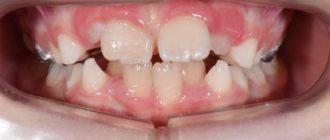
In children aged 5 to 7 years, that is, at the stage of growth of permanent teeth, trauma to the alveolar process can also be diagnosed due to the presence of follicles of permanent teeth in the bone, which subsequently disappear.
Clinical picture of an alveolar bone fracture
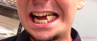
Patients complain of bleeding from the mouth, pain in the upper or lower jaw that increases when closing teeth or chewing food, improper contact of teeth or the inability to close them due to increasing pain. The tissues in the perioral area and cheeks are swollen. There may be abrasions, wounds, and bruises. The patient's mouth is half open, sometimes saliva mixed with blood comes out of it. On the mucous membrane of the lips or cheeks there are lacerated wounds and hemorrhages due to damage to the soft tissues on the teeth at the time of impact.
Fracture of the alveolar process of the upper jaw in the frontal region. Displacement of teeth and hemorrhage along the transitional fold are determined. When a fragment is displaced, a rupture of the mucous membrane of the alveolar process along the fracture gap is possible. In this case, a fracture of the bone tissue can sometimes be seen through the wound on the mucous membrane. The teeth of the broken fragment are displaced into the oral cavity or towards the occlusal plane, which leads to a violation of the configuration of the dental arch. When the jaws close, the teeth of only the displaced portion of the alveolar process come into contact. They are usually mobile, and percussion is painful.
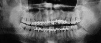
If the fragment is slightly displaced, there may not be a rupture of the mucous membrane. However, hemorrhage along the transitional fold must be determined. Percussion of the teeth between which the fracture gap passes is painful. If during examination the displacement of the fragment is not determined, the fracture gap can be determined by carefully moving the suspected fragment in the anteroposterior or lateral direction and palpating its mobility with the fingers of the other hand. On the radiograph, the fracture gap of the alveolar process of the upper jaw is visible in the form of a strip of clearing with fuzzy and uneven edges. On the lower jaw, the fracture gap is more distinct, which is explained by the difference in its anatomical structure.
Intraoral radiograph. The fracture gap of the alveolar process of the upper jaw is determined
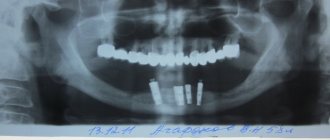
Lateral radiograph of the lower jaw. The fracture of the alveolar part and the displacement of the fragment are determined
Symptoms of alveolar bone fracture
An alveolar ridge fracture can be diagnosed if:
- sharp, spasm-like pain in the jaw when chewing or swallowing saliva,
- swelling of the mucous membrane,
- lacerations of the gums (with or without bleeding),
- painful closing of the jaws,
During palpation, the damaged part may move or a separated area may be palpated. Externally, the fracture is characterized by contusions and bruises on the face in the area of the fracture. External examination and x-ray show a crack in the alveolar process or complete separation of the bone from the main cranial bone.
Depending on the angle at which the blow occurred, the broken part may move in different directions, inward or deep into the mouth. If the blow falls on the lower jaw, in this case the impact occurs with the help of the lower jaw on the upper jaw, and the broken part moves upward.
Classification of alveolar process fractures (K.S. Yadrova).
- Partial - the fracture gap passes through the outer part of the alveolar process. In this case, a fracture of the outer compact plate occurs within the sockets of several teeth and part of the interdental septa. There is no displacement of the fragments. - Incomplete - the fracture gap in the form of a crack passes through the entire thickness of the alveolar process, capturing the outer and inner compact plates and spongy substance. There is no displacement of the fragments. - Complete - two vertical fracture gaps are united by one horizontal one and pass through the thickness of the entire alveolar process. - Comminuted - fracture gaps intersect in several directions. - With a bone defect - the broken part of the alveolar process is torn off. The alveolar process of the upper jaw breaks more often than the lower jaw, which is associated with its anatomical features (it is longer and thinner, its compact plates are thinner and penetrated with a large number of openings for blood vessels and nerve trunks, it is more vulnerable to an impact in the sagittal plane, since the upper jaw in most patients it overlaps the lower one). The alveolar part of the lower jaw is protected by a protruding chin. In case of a blow from the side, the alveolar process of the upper jaw is protected by the zygomatic arch and bone. After an impact, the broken fragment of the alveolar process is displaced, as a rule, in the direction of the acting force: the frontal section - posteriorly into the oral cavity, the lateral - medially, into the oral cavity. Rarely, a fragment can additionally rotate around its longitudinal axis. A fragment of the alveolar process more often retains connection with the periosteum and mucous membrane on at least one side, less often it is completely torn off.
A fracture of the alveolar process is often accompanied by a simultaneous fracture of teeth or dislocation of teeth. The fracture gap often has an arched shape. It starts from the crest of the alveolar process in the interdental space, rises up (on the upper) or down (on the lower) jaw, runs horizontally along several teeth with unequal levels of the apexes of the roots and unequal thickness of the compact plate accordingly, then descends between the teeth to the crest alveolar process. The fracture gap often extends outside the roots of the teeth. Less commonly, it is located within the roots of the teeth, which is combined with their fracture in the apical third. When the lateral portion of the alveolar process of the upper jaw is fractured, the floor of the maxillary sinus may break off.
Diagnostics
The fracture is diagnosed by an oral and maxillofacial surgeon. An x-ray may show different degrees of damage:
- partial damage - damage to part of the bone, not complete detachment;
- non-displaced fracture – damage to all parts of the bone;
- complete fracture - the x-ray shows a gap between the separated part and the skull;
- fracture in different places - damage to the alveolar process in different places, bone fragmentation;
- fracture with deformation - a completely torn off part, displaced at a different angle.
Causes and types of damage
A fracture of the alveolar process of the upper jaw can be caused by:
- various injuries,
- direct blow to the face
- falling even from a small height.
There are additional risk factors. These include diseases that affect the condition of bone tissue and reduce its strength.
Doctors divide jaw fractures into several types:
- partial - only the outer alveolar plate is damaged;
- incomplete – the bone is injured in all layers without displacement of fragments;
- complete – determined by the results of an x-ray;
- comminuted – characterized by the appearance of several fragments;
- with a bone defect - accompanied by a complete separation of the damaged area from the jaw bone.
Non-displaced fractures have the most favorable prognosis.
Treatment
The treatment method is selected depending on the established severity of the fracture. Before any manipulation, the doctor performs anesthesia. Torn tissues are treated with antiseptics to prevent infection. Then reconstruction is carried out (if there are fragments) and fixation of the jaw.
If there is displacement, the doctor opens the gums in the area of the fracture (revision) to eliminate the sharp edges of the fragments, after which he manually puts the displaced part in place. This must be done in such a way that it corresponds to the correct bite. After this, the gum is sutured and an iodoform dressing is applied to the wound.
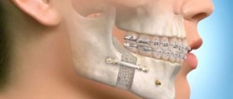
To fix the alveolar process, a smooth splint-bracket is used, which is attached to three healthy teeth on both sides of the chipped part. If this is not possible, that is, if the teeth on one side are not stable or are completely absent, then I use five additional teeth for fixation. For greater immobilization of the dentition, wearing a chin sling may be indicated.
If the injury occurs in the anterior part of the upper molars, then fixation occurs through the installation of a single-jaw bracket, which is fixed on the damaged area (attached with ligatures to healthy teeth).
In case of complete absence of teeth, a splint made of quickly hardening plastic is installed.
After all surgical procedures, the patient is prescribed antibacterial therapy and medications to accelerate healing and prevent the development of inflammation in the injured area.
Alveolar ridge fracture
Treatment of an alveolar bone fracture includes pain relief, antiseptic treatment of damaged tissue, manual reposition of fragments, and immobilization. For pain relief, conduction anesthesia is performed. If the alveolar process is fractured with displacement, the wound is inspected, the sharp edges of the bone are smoothed and the mucous membrane is sutured tightly or the bone wound is covered with an iodoform bandage.
The displaced fragment is installed in the correct position under the control of occlusal relationships. For immobilization, a smooth splint made of aluminum wire is most often used. It is bent from the buccal surface of the teeth. Provided there are no destructive periapical changes in bone tissue and pathological mobility of the teeth in the undamaged area, the splint is fixed to 3 teeth on either side of the alveolar process fracture line. The single-jaw brace is installed using adhesive systems and light-curing composite material or using metal ligatures, which must be changed every week.
If, in case of a fracture of the alveolar process in the molar area, there is only one support for fixing the splint, the number of stable teeth is increased to 5. To achieve more stable immobilization, a chin sling is used. In case of impacted dislocation of the anterior part of the upper jaw, a single-jaw steel bracket is used, which is tied with ligatures to healthy teeth. The displaced fragment is connected to the splint with elastic bands. If, during a fracture of the alveolar process, there are no teeth in the supporting areas, a splint is made from quick-hardening plastic. In the first days, antibiotic therapy and hypothermia are prescribed. Herbal decoctions and preparations based on chlorhexidine digluconate are used as preparations for antiseptic treatment.
Forecast
If the roots of the teeth are not in the fracture line of the alveolar process, the prognosis is favorable. Simultaneous reduction and immobilization can achieve callus formation within 8 weeks. When patients present late, the treatment period is lengthened, the list of drugs for anti-inflammatory and antibacterial therapy is expanded, the options for osteosynthesis are narrowed, and the risks of developing post-traumatic osteomyelitis and pseudarthrosis increase. To reduce stiff fragments, additional devices for extraoral and intraoral traction are used.
If, along with a fracture of the alveolar part, a violation of the integrity of the roots of the teeth is diagnosed, the prognosis is unfavorable. In most cases, consolidation cannot be achieved. As a result of disturbances in innervation and trophism, sequestration and rejection of the broken fragment are observed.
Rehabilitation period
If the trauma of the alveolar process did not damage the roots of the teeth, then with proper reconstruction and fixation, after about 8 weeks, a bone callus will form, and the process will grow back to the jaw. The treatment prognosis is favorable.
If the roots of the teeth are damaged, the broken parts may no longer take root. As a rule, it is not possible to achieve consolidation even with successful treatment.
Rehabilitation after a fracture is aimed at full or maximum restoration of the functions of the injured part of the body. It may consist of a set of procedures, which, depending on the characteristics of the injury, may include:
- physical therapy,
- physiotherapy,
- massage.
In order for further rehabilitation to have a positive effect, each of the components of the complex must be selected by a qualified specialist, taking into account all the individual characteristics of the patient.
Symptoms
A fracture of the alveolar process of the upper jaw has several characteristic features. The most common symptoms:
- sharp pain
- inability to completely close your mouth,
- occurrence of malocclusion,
- dysfunction of chewing and swallowing,
- hematomas,
- dislocations and mobility of one or more teeth,
- severe swelling of soft tissues,
- bleeding.
If you suspect an alveolar bone fracture, you should seek medical help as soon as possible.
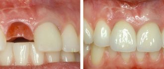
Tooth extraction after a fracture
It is necessary to remove a tooth only if it cannot be restored. If, after a fracture, an element of the dentition becomes loose, it is necessary to seek the help of a specialist as quickly as possible.
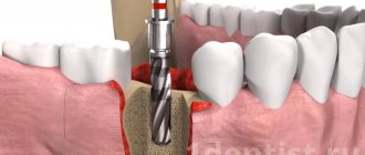
Tooth extraction is performed under anesthesia. Thanks to this, a person can completely relax and also get rid of pain. Usually everything happens very quickly, but in complex cases when a serious fracture has occurred, difficulties may arise. Sometimes the specialist has to cut the gum and pull out the remaining parts of the root, after which sutures are placed in this place. After some time they are removed. Depending on the specifics of a particular case, a specialist may recommend physical therapy and the installation of a dental implant.
Maxillary fracture
During an extraoral examination of patients with type 3 fractures of the upper jaw, a violation of the integrity of the zygomaticalveolar ridges is revealed: tissue swelling, abrasions, an increase in the vertical parameters of the face. At the border of the transition of the immobile mucosa of the alveolar process to the mobile one, as well as on the hard palate, hemorrhages are diagnosed. Displacement of the damaged sections during a fracture of the upper jaw leads to rupture of the mucosa. Downward dislocation of the posterior fragment causes elongation of the soft palate.
During palpation examination, irregularities and recesses are determined on the alveolar process. When pressing on the hooks of the pterygoid processes, the patient feels pain in the area corresponding to the fracture line of the upper jaw. Disocclusion is more often observed in the anterior area, and malocclusion pathologies along the transversal and sagittal planes are less frequently diagnosed. The patient does not feel the tip of the probe touching the mucous membrane of the alveolar process, which indicates a loss of pain sensitivity. A CT scan for a type 3 maxillary fracture reveals areas of integrity disruption in the areas of the pyriform aperture and zygomaticalveolar ridges, and decreased transparency of the maxillary sinuses.
In case of a type 2 fracture of the upper jaw, the symptom of glasses is positive - the periorbital zone immediately after the injury is saturated with blood. Chemosis, exophthalmos, and lacrimation are observed. Pain sensitivity of the skin in areas corresponding to the level of damage is reduced. In the anterior section, as a rule, there is disocclusion. During a palpation examination, the dentist determines the mobility of the maxillary bone at the border with the orbit, in the area of the zygomaticalveolar ridge, as well as in the area of the suture connecting the frontal bone with the upper jaw. These same changes can be diagnosed by radiographic examination.
In type 1 fractures of the upper jaw, diplopia, chemosis, exophthalmos, subconjunctival hemorrhages, and eyelid edema are observed. If the patient is lying down, enophthalmos is detected. In a sitting position, diplopia increases, and when the teeth are closed, it decreases. By palpation, in case of an upper fracture of the upper jaw, it is possible to identify unevenness in the areas of the frontomaxillary, as well as the zygomatic-frontal sutures and the zygomatic arch. The load test is positive. Computed tomography reveals a violation of the integrity of the root of the nose, zygomatic arch, frontozygomatic suture, and sphenoid bone. A diagnostic test to determine the presence of rhinorrhea is the handkerchief test. After drying, the structure of the fabric soaked in liquor remains unchanged. If the scarf has become stiff, it means there is no liquorrhea, serous contents are released from the nasal passages.
It is necessary to differentiate a fracture of the upper jaw from other injuries of the bones of the maxillofacial skeleton. All patients should be examined by an oral and maxillofacial surgeon, as well as a neurologist. If the maxillary sinuses, optic nerve, or skull bones are damaged, treatment is carried out jointly with a neurosurgeon, resuscitator, ophthalmologist, or otolaryngologist.
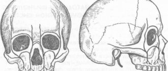
Anatomy
The alveolar process is part of the upper and lower jaw. This is the part of the bone that contains the so-called alveoli - tooth sockets. The structure of the process is such that it consists of two plates (external and internal), the spongy substance of bone tissue located between them, as well as 8 holes for the corresponding teeth.
The outer plate is thinner than the inner one, more susceptible to trauma, and the spongy substance acts as a shock absorber, protecting the bone from a complete fracture of the alveolar process.
However, the type and extent of injury is determined not only by the structure of the alveolar process. An important role is played by such factors as the strength of the mechanical action, its direction, the point of application of the force, and the traction of muscle structures.
With regard to the prognosis, the key factor is the integrity of the root system of the teeth, and if a fracture of the alveolar process is suspected in a child, it is important to resolve the issue of the integrity of the follicles of permanent teeth.
Treatment methods for fractures
Treatment of fractures of the alveolar process of the upper jaw includes several stages:
- pain relief,
- antiseptic treatment,
- repositioning of all fragments,
- immobilization with special structures.
Displaced fractures require complex treatment. In case of serious injuries, the wound is inspected, the sharp edges of bone fragments are smoothed and the mucous membrane is sutured. The bone fragments are returned to the correct position and securely fixed with staples or splints. To prevent complications in the first days after injury, antibacterial therapy and rinsing the mouth with herbal decoctions are prescribed.
With timely provision of qualified assistance, bone callus is formed within 8 weeks. The speed of recovery depends on the individual characteristics of the body.