Who is the study for?
The main indication for ultrasound examination of the salivary glands is the presence of any complaints in the patient. Most often, patients come to the doctor with the following problems:
- sharp pain in the submandibular or parotid area;
- local or general hyperthermia (increased temperature directly in the area of the lesion or in the entire body as a whole);
- increase in the size of the salivary glands;
- dry mouth (xerostomia);
- the presence of compactions (tumors, stones) near the salivary glands.
If the above symptoms are present, as well as after a visual examination, palpation examination and history taking, the doctor, in addition to laboratory tests, refers the patient to an ultrasound scan. The method is used if it is necessary to carry out differential diagnosis for such pathologies as:
- inflammatory diseases of the salivary glands (acute and chronic sialoadenitis, viral and bacterial);
- adenoma;
- salivary stone disease;
- tonsil damage;
- tumor neoplasms (malignant and benign);
- individual anatomical anomalies;
- enlargement of nearby lymph nodes;
- dystrophic lesions of organ tissue.
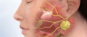
During the study, specialists can assess the condition of the group of large salivary glands: parotid, sublingual, submandibular.
How to prepare for the procedure
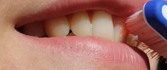
Ultrasound of the salivary glands is an absolutely safe and painless manipulation, for which the patient does not require special preparation. Patients of almost any age tolerate the procedure well, without showing any anxiety before and during its implementation, and therefore it does not require premedication (pre-administration of sedative medications) even in children.
On the eve of the visit to the specialist (about 3-4 hours in advance), the patient is advised to refrain from eating, and immediately before the examination he needs to thoroughly clean the oral cavity.
References
- Malignant tumors of the salivary glands, Clinical guidelines. Association of Oncologists of Russia, 2022. - 55 p.
- Balkanov, A.S., Bychenkova, O.A., Sipkin, A.M. and others. Combined treatment of parotid salivary gland cancer. Almanac of Clinical Medicine, 2022. - No. 4. - P. 309-313.
- Polyakov, V.G., Shishkov, R.V., Ermilova, V.D. and others. Children's Oncology, 2004. - No. 1. - P. 45-47.
- Carlson, E., Schlieve, T. Salivary Gland Malignancies. Oral and maxillofacial surgery clinics of North America, 2022. - Vol. 31(1). — P. 125-144.
How to do an ultrasound of the salivary gland
The examination can be carried out in 2 ways: from the oral cavity and from the external surface. The procedure does not take much time (it usually takes no more than 20-30 minutes to complete). During an ultrasound scan of the salivary glands, the patient does not feel any pain or other discomfort.
During the examination, the patient takes a supine position, places his head on a specially prepared pillow and tilts it slightly back (or turns it to the left or right, depending on the location of the gland being examined).
If the parotid gland is being examined, the device's sensors are placed on the parotid area. When studying the sublingual and submandibular glands - into the oral cavity (sometimes the extraoral method can be used).
Embryology
The parotid gland, like other major salivary glands, develops from the epithelium of the oral cavity. The gland bud appears in the embryo at the 6th week of development in the depths of the groove separating the cheek from the gum, in the form of an epithelial cord, which grows towards the ear. At the 8th week. embryonic development, the distal end of this cord begins to branch and gives rise to excretory ducts and terminal secretory sections of the liver. At the beginning of the 3rd month, gaps appear in the anlage of the excretory ducts, their epithelial lining becomes double-rowed, and in large excretory ducts multilayered. Differentiation of glandular epithelium in the terminal secretory sections of the liver. occurs somewhat later than in other salivary glands.
Ultrasound of the submandibular salivary gland
The norm for an ultrasound examination of the salivary submandibular gland corresponds to the following parameters:
- Fine-grained homogeneous structure.
- The edges of the glands are smooth.
- The excretory duct is well visualized, small in size, without signs of blockage.
Determining the normal size of the submandibular glands is not particularly difficult precisely because of its clear contours. During inflammation (sialoadenitis), the organ increases in size and its edges become blurred.
Research methods
Rice.
1. Pantomosialogram of unchanged submandibular glands: 1 - parenchyma of the gland, 2 - ducts of the gland, 3 - submandibular duct (the contours of the ducts are smooth, clear). To determine the nature of the pathological process, probing of the ducts, sialometry (measurement of the amount of secretion released from the duct per unit of time), cytological examination of the secretion (see Saliva), radiography, pantomography (see) with artificial contrast of the ducts, or pantomosialography (Fig. 1) are used ), thermal imaging (see Thermography), ultrasonic dowsing (see Ultrasound diagnostics) and scanning (see).
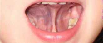
Ultrasound of the sublingual salivary gland
Normally, the sublingual salivary gland has smooth, not very clear contours. They have a homogeneous structure, but echogenicity (the ability to reflect ultrasound) may be slightly increased. In the absence of any diseases, the device will not show the ducts of this organ.
The shape of the sublingual salivary glands displayed on the device screen changes depending on where the sensor is placed. When located in the chin area, the glands have oval contours. If the device is applied parallel to the body of the lower jaw, the glands will be slightly elongated.
Anatomy
Rice.
1. Variants of the parotid gland and parotid duct (but B. G. Ali-Zade and M. K. Artemova, according to S. N. Kasatkin): a - trapezoidal parotid gland and straight parotid duct; b - semilunar-shaped parotid gland and arcuate parotid duct; c - triangular-shaped parotid gland and geniculate parotid duct; d — oval-shaped parotid gland and the ascending direction of the parotid duct; 1 - parotid gland, 2 - parotid duct; 3 - chewing muscle. In the Parotid Gland, there is a superficial part (pars superficialis), adjacent to the masticatory muscle, and a deep part (pars profunda), extending into the mandibular fossa (fossa retromandibularis). Sometimes a pharyngeal process extends from the inner edge of the gland. O.J. most often it has an irregular pyramidal or trapezoidal shape, sometimes crescentic, triangular or oval (Fig. 1).
In a newborn O. has a mass of 1.8 g, contains a lot of loose connective tissue and blood vessels, its secretory function in the first 6 weeks. insignificant. The gland grows most intensively up to 2 years, increasing 5-6 times. At the end of the 2nd year of life, gistol ends. differentiation of the skin, its growth slows down.
In an adult O. weighs 20-30 g; its vertical size is 4-6.5 cm, sagittal 3-5 cm, horizontal 2-3.8 cm. In old age, the size and weight of the body. are decreasing.
Rice. 2. Scheme of the parotid gland bed (horizontal section): 1 - skin; 2 - subcutaneous tissue; 3 - superficial layer of the fascia of the parotid gland; 4 - chewing muscle; 5 - lower jaw; 6 - medial pterygoid muscle; 7 - wall of the pharynx; 8 - deep layer of fascia of the parotid gland; 9 - styloid process; 10 - internal carotid artery; 11 - internal jugular vein; 12 - digastric muscle; 13 - sternocleidomastoid muscle. Rice. 3. Bed of the parotid gland: 1 - temporomandibular joint; 2 - external carotid artery; 3 - chewing muscle; 4 - septum between the parotid gland and the submandibular gland; 5 - submandibular gland; 6 - digastric muscle; 7 - sternocleidomastoid muscle; 8 - styloid process; 9 — wall of the external auditory canal; 10 - external auditory opening.
In front, the parotid gland is adjacent to the masticatory muscle (m. masseter), the branch of the lower jaw (r. mandibulae) and the medial pterygoid muscle (m. pterygoideus med.); at the back it borders with the sternocleidomastoid muscle (m. sternocleidomastoideus), the posterior belly of the digastric muscle (venter post m. digastrici) and the mastoid process (processus mastoideus); medially adjacent to the styloid process (processus styloideus) and the stylohyoid (m. stylohyoideus) and styloglossus (m. styloglossus) muscles, the internal carotid artery (a. carotis int.) and the internal jugular vein (v. jugularis) extending from it int.), hypoglossal nerve (n. hypoglossus) and peripharyngeal tissue; from above it adjoins the zygomatic arch (areus zygomaticus) and the external auditory canal (porus acusticus ext.). These formations limit the bed of the liver. (Fig. 2), which is lined by the fascia of the liver. (fascia parotidea). Fascia O. g. fused with the fascia of the surrounding muscles and attached to the edge of the lower jaw, zygomatic arch, mastoid and styloid processes. Between the angle of the lower jaw and the sternocleidomastoid muscle, the fascia forms a dense septum (Fig. 3), separating the O. from the submandibular gland (submandibular gland, T.; gl. submandibularis).
Rice. 4. Topography of the parotid gland: 1 - auriculotemporal nerve; 2 - superficial temporal arteries and vein; 3 - zygomatic arch; 4 - temporal branch of the facial nerve; 5 - zygomatic branch of the facial nerve; 6 - parotid duct; 7 - buccal branches of the facial nerve; 8 - facial arteries and vein; 9 - chewing muscle; 10 - marginal branch of the facial nerve; 11 - submandibular gland; 12 - cervical branch of the facial nerve; 13 - external carotid artery; 14 - subcutaneous muscle of the neck; 15 - internal jugular vein; 16 - sternocleidomastoid muscle; 17 - external jugular vein; 18 - v. retromandibularis; 19 - parotid gland; 20 - maxillary artery; 21— parotid plexus of the facial nerve; 22 - facial nerve; 23 - auricle; 24 - accessory parotid gland.
Through the thickness of O. zh. large vessels and nerves pass through; external carotid artery (a. carotis ext.) with the maxillary (a. maxillaris) and superficial temporal arteries (a. temporalis superficialis) branching off from it, v. retromandibularis, nerves auriculotemporal (n. auriculotemporalis) and facial (n. facialis). The facial nerve (see) forms the parotid plexus (plexus parotideus) in the thickness of the gland, the branches of which, leaving the gland, fan out to the facial muscles (Fig. 4). This determines the radial direction of the incisions of the gland during operations.
The system of excretory ducts of the gland is represented by intralobular, interlobular and interlobar ducts, which merge into the common parotid duct (ductus parotideus), or Stenon duct, which was first described by the Danish scientist N. Stenon in 1661. Length of the parotid duct 40-70 mm, its dia. 3-5 mm. The parotid duct usually originates from the upper third of the gland, bends around the edge of the masseter muscle and the fatty body of the cheek (corpus adiposum buccae) and opens into the vestibule of the mouth at the level of the upper second molar. In this place on the mucous membrane of the cheek there is a papilla of the liver. (papilla parotidea). According to S. N. Kasatkin (1948), in 44% of cases the parotid duct is ascending, in 23% - descending, less common are straight, cranked, arcuate (Fig. 1), S-shaped and bifurcated parotid duct. In half of the cases, the duct of the accessory parotid gland (glandula parotis accessoria) flows into it. Sometimes a blind canaliculus, the so-called, departs from the parotid duct near its mouth. Shievich's organ is a rudimentary salivary duct. The parotid duct contains valves and terminal siphons that regulate the excretion of saliva.
Blood supply is provided by the branches of the external carotid artery, superficial temporal artery, transverse artery of the face (a. transversa faciei), posterior and deep auricular arteries (aa. auriculares post, et profunda). Intraorgan arteries and veins pass through the interlobular septa. Venous drainage occurs into the pterygoid plexus (plexus pterygoideus) and the mandibular vein.
Lymphatic vessels of the lake. drain into the superficial and deep parotid lymph nodes (nodi lymphatici parotidei superficiales et profundi); their efferent vessels go to the superficial and deep cervical lymph nodes (nodi lymphatici cervicales superficiales et profundi).
Innervation is carried out by sympathetic and parasympathetic nerves. Preganglionic sympathetic fibers originate in the gray matter of the upper thoracic segments of the spinal cord and are interrupted in the superior cervical ganglion (gangl, cervicale sup.). Postganglionic sympathetic fibers go to the lake. as part of the external carotid plexus (plexus caroticus ext.). Sympathetic nerves constrict blood vessels and inhibit the secretion of saliva. The gland receives parasympathetic innervation from the lower salivary nucleus (nucleus salivatorius inf.) of the glossopharyngeus nerve. Preganglionic fibers go as part of this nerve and its branches (n. tympanicus, n. petrosus minor) to the ear ganglion (gangl, oticum). Postganglionic fibers reach the gland along the branches of the auriculotemporal nerve. Parasympathetic fibers stimulate secretion and dilate blood vessels.
X-ray anatomy
Sialography of the Parotid gland (see Sialography) in the anterior direct projection allows you to detect the shadow of the gland. outward from the branch of the lower jaw. In the lateral projection of the lake. projected onto the ramus of the mandible and the fossa retromandibularis area. The parotid duct on the sialogram in the lateral projection runs in an oblique direction from back to front and upward. Its intraglandular part crosses the posterior edge of the ramus of the lower jaw at the border of its middle and lower thirds or lies on the angle of the lower jaw. At the level of the anterior edge of the mandibular ramus, the duct emerges from the gland, and its extraglandular part is located at the base of the coronoid process of the mandible. Interlobar ducts of the lake. They merge into the parotid duct at different angles and have different numbers of branches. Depending on this, S. N. Kasatkin (1948) divides the glands into many branched, moderately branched and few branched.