3485
Not all people have a sparkling, beautiful smile, as there are many factors that can affect aesthetics. One of the conditions for healthy teeth and the entire oral cavity as a whole is correct bite.
In particular, orthognathic occlusion, which is the subject of today’s article, falls into this category.
What is orthognathy
Translated from Greek, “gnathos” means jaw, and “ortho” means straight or regular.
In dentistry, orthognathic is a physiologically correct bite. Its main feature is a slight overlap (up to 1/3 of the crown height) of the lower teeth with the upper ones. This occlusion is considered the most advantageous from an anatomical and physiological point of view (which we will talk about later). Other characteristics of correct occlusion include straight teeth and tight grip of the incisors (there are no gaps between them).
General overview
To begin with, it is worth defining that occlusion in dentistry means the specificity of the occlusal closure of the teeth during clenching of the jaws. The correct ratio is considered to be one in which full functionality is maintained and the aesthetics of the facial contour are not disturbed.
From the point of view of finding the optimal option, the best option is considered to be the orthognathic or normagnothic form of occlusion. This, among other things, is implied in the name itself - in ancient Greek, the word “orthognathia” means “straight upper jaw.” This structure is characterized by the location of the arch and facial bone in the same vertical plane, close contact of the incisors, as well as the impeccable shape of the jaw.
Orthognathic bite: how to determine
The surest way to determine the type of occlusion is to consult a dentist for an examination. Orthognathic bite has the following features, which can be determined independently:
- The upper teeth overlap the lower teeth by 1/3 (or less) of the crown height. This is the basis of a physiological bite. It is important to note that if the closure is direct (when the edges of the upper teeth directly meet the edges of the lower teeth), then this is a type of pathological bite, although such a smile looks very aesthetically pleasing. With such closure, the teeth quickly wear out and are destroyed.
- One upper tooth meets the two lower ones.
- Middle line. Many people mistakenly believe that the middle of the upper jaw should coincide with the middle of the lower jaw. In fact, it is important that the midlines of the upper jaw and face coincide.
- You do not feel any functional impairment. The processes of chewing and swallowing are not accompanied by discomfort and pain. If clicking or crunching is felt, this indicates dysfunction of the temporomandibular joint (TMJ). This symptom is often observed with pathological bites.
- Correct facial proportions. This is an indirect characteristic for physiological occlusion. There are cases when pathological closure of teeth does not affect the symmetry of the face. There are also opposite cases when those with a correct bite have an asymmetrical face. An individual approach and correct diagnosis by a doctor are important here.
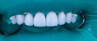
What does an orthognathic bite look like?
Orthognathia: benefits
- Uniform load on teeth . This protects hard tissues from premature destruction.
- No speech impediment . With pathological occlusions, a violation of diction is often observed, since some sounds are difficult for a person to hear.
- No strain on the digestive organs . With an incorrect bite, a person chews food poorly. Relatively large pieces of food enter the stomach, which impairs digestion.
- Effective oral hygiene . The correct shape of teeth is the key to good hygiene. In this case, the risk of developing caries, periodontitis and other dental diseases is significantly reduced.
- Successful dental treatment . People with malocclusion experience difficulties with prosthetics, implantation and other types of dental treatment. Often, before the main treatment, the doctor prescribes bite correction.
- Aesthetic benefits . In most cases, an orthognathic bite provides a beautiful smile and facial symmetry.
What affects the bite of teeth?
The negative impact of defects in the dental system on the condition of the entire organism has been repeatedly proven by scientists. The list of identified health risks is impressive even for ardent optimists.
- Due to improper distribution of the chewing load, tooth enamel wears off faster, teeth begin to react painfully to cold and hot, their necks become exposed, and noticeable gaps form between the teeth.
- Diseases of the temporomandibular joints occur, accompanied by headaches and a characteristic clicking sound when the jaws move.
- The aesthetics of the face suffers: its proportions change, wrinkles form, teeth become crooked, and the smile becomes unattractive.
- It becomes difficult to bite food, chewing functions are disrupted, causing diseases of the gastrointestinal tract.
- Speech defects appear, facial expressions are distorted, and self-esteem decreases.
- Crowding and crooked teeth cause accelerated tooth decay due to frequent chipping and carious lesions.
- The oral mucosa is injured - cheeks, gums, tongue, and the tissues of the oral cavity become inflamed.
- The quality of breathing deteriorates, which leads to diseases of the nasopharynx, trachea, and hearing aid.
How does a correct bite develop?
You need to monitor your bite from early childhood. Pay attention to the position of the tongue, the position of the teeth and the development of the child’s jaw. Let's consider the main stages of bite formation in children of different ages.
- 0-6 months . During this period, the foundation for the closure of the dentition is laid. If the child is fed correctly (so that the masticatory muscles experience the proper level of load), then the dental arches and jaws develop harmoniously (if there are no hereditary or congenital anomalies of the dental system).
- 6 months . The first milk teeth appear, the tongue rests on the roof of the mouth.
- Up to 3 years . Closer to 3 years of age, baby teeth are formed. At this age, the child’s dentofacial arches are fully functioning, he is able to chew food normally and speak.
- 4 years . From this age, the milk bite gradually loses its position.
- 6 years . Permanent teeth appear in place of the lost milk teeth. An overlap of the lower teeth with the upper teeth is formed (no more than 1/3 of the height of the crown). If the child had gaps between the teeth in the primary dentition (tremas or diastemas), then by the time the permanent dentition is formed, these defects disappear.
- 15 years . In adolescents, the formation of dentition ends. The overlap of the upper teeth with the lower ones should remain at 1/3 of the height of the crown.
Malocclusion of teeth
Look at your reflection in the mirror. If you notice an excessively protruding upper or lower lip, teeth that “overlap” each other, or gaps between the dentition when the jaws are closed, this is a clear reason to consult a specialist. Descriptions of dental anomalies and photographs of teeth with malocclusion pathologies are presented in the table below.
Distal (prognathic)
In a distal bite, the upper jaw is more developed than the lower jaw.
Mesial (medial)
With a mesial bite, the lower jaw is pushed forward.
Cross
In a crossbite, the rows of teeth intersect in the manner of scissors when the jaws close.
Deep
In a deep bite, the upper teeth significantly overlap the lower teeth.
Open
The presence of pronounced gaps between the dentition when the jaws are closed indicates an open bite.
The importance of physiological occlusion
Physiological occlusion is important for health for the following reasons:
- the bone tissue of the skull elements develops harmoniously;
- uniform load on teeth and joints;
- no negative effects on the digestive or respiratory system;
- correct facial proportions, lack of psychological discomfort in this regard;
- beautiful smile;
- good oral hygiene.
Anatomy and physiology of the dental system
When it comes to occlusion, not only the location of the teeth is important, but also the functions of the remaining elements of the dental system. These are the upper and lower jaws, jaw joints and muscles that ensure the process of chewing and swallowing.
The greatest load on the teeth occurs when chewing. It is important that at this moment the load is evenly distributed on the surface of the teeth. This way hard tissues will retain their strength and structure longer. Otherwise, the teeth will quickly wear out. This threatens with caries, wedge-shaped defect, gum disease and final tooth destruction.
Under the physiological norm, the position of the jaws should be parallel. The lower jaw resembles a parabolic arch, and the upper jaw is a semi-oval. The coordinated functioning of the jaw joints is also important - without discomfort, clicks and crunches.
Distal occlusion (upper macrognathia, prognathia)
Distal bite therapy can be carried out using the following methods and methods:
- orthodontic treatment,
- hardware-surgical,
- surgical,
- prosthetic
- various combined and combined methods.
During treatment, taking into account certain features depending on the clinical form of the anomaly, the age of the patient, the individual characteristics of the structure of the facial skull and the type of its growth, the following tasks must be solved.
- Regulation during jaw growth using a facebow and extraoral traction or a functional apparatus.
- Restriction of growth and shortening of the dentition of the upper jaw due to the distal movement of the upper molars, canines and elimination of protrusion of the anterior teeth
- When treating distal occlusion, it is advisable to transfer form P2 to form II1, which can be achieved by using arches in the traditional sequence, that is, the primary arch, as a rule, is multi-stranded, flexible, allowing the creation of additional bends when teeth are crowded in order to avoid excessive load on them, then a steel one rectangular.
- Distal movement of the upper anterior teeth without or after extraction of individual teeth (most often premolars).
- Stimulation of growth and anterior movement of the lower jaw
- Expansion of the dentition of the upper and/or lower jaw
- Change in interalveolar height and normalization of the Spee curve.
- Normalization of the function of masticatory and facial muscles
- Retention period
It is not always possible to completely correct the anomaly during these manipulations, but a change in position of approximately 45 mm can be achieved. When planning orthodontic treatment of patients with distal occlusion, teleradiological examination data characterizing the type of growth of the maxillofacial complex, the activity of residual growth and their comparison with orthognathic occlusion are very important.
With a distal occlusion, the proportion of the neutral type of growth decreases (from 71% with orthognathic to 50%) in favor of horizontal, namely to 43% versus 15% with orthognathic. This indicates the predominance of development of the facial skeleton in the anteroposterior direction due to intensive growth of the upper jaw, especially in the period of 7-12 years and somewhat less in 12-15 years. That is why the most optimal for modifying jaw growth are the replacement bite and the earliest permanent bite (for 14-15 years, for 12-13).
In patients with a neutral type of growth of the facial skeleton, the main tasks when correcting a distal bite are, first of all, to restrain the growth of the upper jaw and stimulate the growth of the lower jaw. In such patients, mainly removable equipment of functional or combined action should be used.
With the horizontal type, it is necessary, first of all, to restrain the growth of the upper jaw, with simultaneous distal movement of the lateral teeth, using a face bow with a cervical traction. For adult patients, to reduce the upper dentition, treatment is recommended with the removal of the first premolars, followed by distal displacement of the lateral and anterior teeth. This is indicated for prognathia with a reduced or average size of the base of the upper jaw and for prognathia caused by crowding of the upper front teeth, their sharp protrusion, often together with the alveolar process.
In severe forms of distal occlusion with a pronounced horizontal gap, removal of the first premolars is indicated even in a mixed dentition. After this, when treating II1, you can use a flex arch, then a nitinol one, fixing them first on the teeth of the upper jaw. One of the indications for tooth extraction is a reduction in the retromolar space, which enhances the mesial displacement of the lateral teeth and aggravates the close position of the anterior teeth, with insufficient space for the canines of the upper and lower jaw (Zhulev E.N.).
According to WR Proffit (1986), the indication for serial extraction is a discrepancy between the size of the teeth and the dental arch by 10 mm or more, and Ringenberg (1964) believes that the initial value should be smaller, namely 7 mm. According to the point of view of V.P. Norkunaite, with the length of the segment of the dentition “from the distal surfaces of the crowns of the 12th and 22nd teeth to the mesial points of the sixth teeth” equal to 18.5-21.0 mm, and if the sum of the mesiodistal dimensions of the canines and premolars is 22.5-24, 0 mm, then removal of individual permanent teeth is indicated. It should be noted that non-extraction orthodontic treatment is even relatively simpler, since there is no need to move the teeth a significant distance to close the post-extraction gap.
As a last resort, removal of the second molar (sometimes unilateral) is used and distalization of the dentition is performed using a face bow. It is very difficult to move the first molars distally more than 1.5-2.0 mm even after removing the second molars, since distal tooth displacement is much more difficult than mesial displacement. The latter requires more reliable support and stabilization, as E. Engle wrote about. The extraoral thrust should not be low, otherwise extrusion of the molars will occur.
During the period of replacement teeth, when treating distal deep bite, you can use the forgotten but good method of A. Katz, namely, crowns with spikes on the second milk or first permanent molars (the tooth is not prepared) of the lower jaw. When the mandible is advanced forward, the elongated mesial cusps of the artificial crown should fit into the gap between the first and second primary molars of the upper jaw, widened by preparation. In this case, some separation of the bite occurs, which contributes to the dentoalveolar lengthening of the lateral teeth and a decrease in the incisal overlap. Long-term use of such crowns (8-10 months) leads to the formation of an orthognathic bite.
In children with mixed dentition and a clear tendency to develop distal occlusion, McNamara recommends maxillary expansion with overcorrection, usually a rapid maxilla expander. The subsequent use of a retention plate leads to the movement of the lower jaw to a position more convenient for the patient, pushed forward. This eliminates buccal crossbite and, after some time, improves occlusal relationships in the sagittal direction. Somewhat earlier, this phenomenon was explained by H. Taatz and Reichenbach by the fact that the expansion of the upper jaw contributes to the spontaneous displacement of the lower jaw to the anterior position. If such correction does not occur, then RG Alexander recommends the use of a facebow with extraoral traction until the end of the mixed dentition.
In mixed dentition, removable plate devices and a pre-orthodontic trainer are used in the treatment of prognathia.
But in addition to this, when treating anomalies at the dentoalveolar level, especially when combined with a narrowing of the dentition or crowding of teeth, fixed structures can be used. First of all, this is a “2 x 4” device, that is, rings for the first molars and braces for the 4 upper incisors, or utility arches.
Growth can be stimulated using activators, for example Andresen Haüpl or function regulators R.Fränkel. The activator is a removable two-jaw monoblock plastic, functional apparatus, consisting of upper and lower plates connected to each other; a vestibular arch, springs or a screw may be added to them. In addition to the plates adjacent to the inner surface of the alveolar processes, they have a corresponding bed for the oral surfaces of all upper and lower teeth. It is better to fix all types of plates with arrow-shaped clasps and Adams clasps.
The device holds the lower jaw in an extended anterior position (a constructive bite that must be determined by the doctor before treatment), promoting dentoalveolar lengthening in the lateral areas, while the upper anterior teeth are moved posteriorly due to a reciprocal action. On the upper jaw, the plate touches the mesial edges of the surfaces of the teeth, but lags behind the distal ones. On the lower jaw, on the contrary, it fits tightly to the distal edges and lags behind the mesial ones to move the lower jaw.
The clinical laboratory stages of production are as follows.
First clinical impression taking from both jaws; the first laboratory casting of plaster models and making a wax template for the upper jaw with bite ridges to determine the constructive bite, the boundaries of the wax template: in front are the cutting edges of the incisors, behind is a line passing through the middle of the crowns of the last molars, on the side is the chewing surface of the lateral teeth.
The second clinical stage is the determination of a constructive bite: the patient moves the lower jaw forward to a neutral relationship of the first permanent molars (1 class each), and he is asked to close his teeth until they come into contact with the wax. In this case, the separation of the dentition should exceed the “rest height” and it is necessary to monitor the position of the ridge and the coincidence of the midline. If in the position of the constructive bite the neutral closure of the sixth teeth is not achieved and the discrepancy is 45 mm, then this position is fixed. If the sagittal discrepancy exceeds 6 mm, the first activator is first prepared (at 45 mm), and after 6-8 months the second activator is prepared, but with the movement of the lower jaw to the neutral closure of the sixth teeth.
After fixing the constructive bite, plaster models with a wax template are handed over to the dental technician and the doctor gives him instructions:
- make an apparatus with or without a vestibular arch for retrusion of the upper anterior teeth (the shape is being specified),
- install a screw or other additional elements, springs, levers, lingual arches, etc. F.Ya. Khoroshilkina and WR Proffit proposed installing facebow tubes into the activator (bite blocks in the premolar area) in order to be able, along with the functional action of the device, to create additional distal and vertical force using extraoral traction.
Second laboratory stage: the models are plastered in an occluder, the wax template is removed, a plastic base is made, the listed parts or others (as directed by the doctor), the device is polymerized in a special double cuvette or in a regular one, increasing its vertical size
Third clinical stage: fitting the activator in the oral cavity, first to the upper dentition, and then to the lower one; the activator should fit tightly to the teeth, with lips closed; The rules for using and caring for the device are explained to the patient and the next visit is scheduled. During repeated visits, the device is adjusted in the direction of movement of the upper and lower lateral teeth. During the treatment process, the dental bed is polished according to the direction of movement of the teeth, that is, those that need to be moved in the palatal or lingual direction, and vice versa, the plate should fit tightly to those teeth that need to be moved in the vestibular direction. The device can mainly be used while at home or while sleeping. Treatment is especially successful in the early stages of distal and deep bites.
The device helps restore nasal breathing, since the child is forced to breathe more through the nose due to the closure of the oral fissure by the plate. But it is contraindicated if nasal breathing is completely absent. The activator also helps eliminate the habit of sucking fingers, tongue, lips and various objects. The vestibular deviation of the lower teeth can be prevented by the activator hood, which overlaps them by 1/3 of the height of the crowns, so the plastic in it is polished or the hood is completely removed. Similar actions, depending on the progress of treatment, are taken at each visit. You can also stimulate the advancement of the lower jaw using the Balters bionator.
Treatment of distal occlusion (II2) can be carried out in two stages. First, the upper anterior teeth are deviated, eliminating the blocking of the lower jaw, that is, subclass II2 is transferred to II1 using edgewise therapy, rotating the first molar. The latter should be the first step in the treatment of a class II anomaly if there is a tendency for mesial rotation of the molar around the palatal root. If orthodontic treatment II1 is carried out without tooth extraction, then sometimes it is enough to turn the first upper molar with its buccal surface posteriorly, which allows you to create an additional space of 1.5-3.0-4.0 mm and the subclass will move to II1. This can be done with the help of an extraoral yaga, the Gozhgarin palatal clasp, in which the ends of the clasp, curved in two planes, are fixed into the palatal locks on the molars. The device is activated by unbending the loop.
This treatment method can be used when a class II anomaly is combined with an open bite. To illustrate, we give an example from the clinical practice of Dr. P. Ngan et al.: an 8-year-old patient had closure of the molars on both sides according to E. Engle class II, a 5-millimeter sagittal discrepancy in the frontal area, an anterior open bite and lower retrognathia. The main goal of treatment was to delay the anterior growth of the upper jaw, transfer the molar relationship from class II to class I, reduce concomitant skeletal disorders and open bite.
The treatment apparatus consisted of an activator and an extraoral arch attached to it. Because the base plate of the device did not cover the vault of the palate, a connecting arch (diameter 1.2 mm) was used instead, which increased the space for the tongue. For fixation to the extraoral traction activator, a special tube with a diameter of 1.12 mm (0.045 inches) was mounted in the plastic between the upper and lower dentition. The force of extraoral traction was up to 400 grams on each side. Springs for tilting the front teeth were made of elastic steel wire with a diameter of 0.5-0.6 mm, the lower part of which was fixed with horizontal shanks in plastic. The vertical part of the springs had a point contact in the area of the necks of the teeth.
The mandibular part of the apparatus consisted of an incisal platform for advancing the lower jaw. When determining the constructive bite, the lower jaw moved forward until the incisors made direct contact. In patients with hyperactivity of the muscles of the perioral region, to reduce their effect, lip pads in the form of a “tear” according to R. Fränkel were used, which were located in the vestibule of the oral cavity parallel to the alveolar process. The ratio of molars according to class I. was achieved in about a year and at the same time the size of the open bite decreased, which led to an improvement in the lip relationship. The entire treatment lasted about 14 months.
Sometimes symmetrical or unilateral removal of premolars in the upper jaw is performed. At the second stage of treatment, the lower jaw is set in the correct relationship with the upper jaw. To do this, when there is a sharp narrowing of the lower dentition, it is expanded, and then, based on the clinical picture and X-ray data of the temporomandibular joints, sagittal movement of the lower jaw is carried out using plates with an inclined plane. There are a large number of varieties of plates, including those with an inclined plane. Depending on a particular clinical situation, the doctor selects the appropriate design.
A. Katz's bite block is used to treat prognathia in combination with a deep bite. A special feature of its design is an inclined plane and reversible clasps that bend over the cutting edges of the front teeth onto their vestibular surface. The plate does not adhere to the mucous membrane of the anterior part of the palate and the necks of the anterior teeth. When closing with an inclined plane, the lower teeth slide along its surface, trying to return from a forced (constructive) bite to its original position, and the lower jaw moves forward, and the upper teeth tilt towards the palatine side. In the lateral areas, due to the separation of the bite, a vertical restructuring occurs, that is, dentoalveolar elongation.
The fundamental clinical and laboratory stages of making a plate differ little from those described in the manufacture of the activator: obtaining impressions, making a wax composition of the plate with retaining and reversible clasps, determining the constructive bite, polymerization of plastic, fitting and application of the device.
It should be remembered that when treating distal occlusion in patients 15-20 years old, when using bite blocks before stabilization occurs, a double or “wandering” bite may be established, that is, in a position of physiological rest, the lower jaw is fixed in a neutral position, and during function it moves to the former (distal).
The devices proposed by R. Fränkel are called functional regulators, the main parts of which are side shields and pelota, which relieve the dentition from the pressure of the cheeks and lips. As a result, under the influence of the tongue, the growth of the apical base is stimulated in the transversal and sagittal directions. The parts of the apparatus are held together by metal arcs made of elastic wire. This skeletonization made it possible to increase the strength of the regulators, reduce the size of the plastic shields, lighten the apparatus and make it open in the frontal area for better swallowing and speech. Active elements (screws or springs) can be added to the device to accelerate the movement of individual teeth.
R. Fränkel proposed function regulators of three main types: type I (FR I) is used to eliminate protrusion of the anterior teeth and distal occlusion, combined with narrowing of the dentition, fan-shaped arrangement of the upper frontal teeth and with anomalies of 1 class. E. Engla; type II (FR II) for the treatment of distal occlusion of subclass 2 (II2), that is, in combination with deep overlap and retrusion of the upper anterior teeth; type III (FR III) for the treatment of progenia. The principal clinical and laboratory stages of the regulators have been described previously.
The use of this method is effective in early childhood (the period of primary and mixed dentition), that is, when one can count on the growth of the jaw bones and especially the apical base. Treatment with the regulator, especially during the period of its development, is recommended according to the following scheme: for the first two weeks use the device during the day for 1 hour, for the next 2 weeks every day for 2 hours, then during all free time, removing the device only during meals; in 2-3 months around the clock. After correcting the bite with adjusters, retention devices are not required, since already during the active phase of orthodontic treatment, the conditions that contribute to the occurrence of relapse are eliminated.
Sagittal movement of the lower jaw during prognathism should be considered as the last stage of treatment, based on the considerations that the restructuring of muscles, temporomandibular joints, as well as dentoalveolar lengthening in the lateral areas in the vertical direction are not always successful. When treating severe forms of prognathia with deep overlap, the separation between the lateral teeth should be at least 45 mm. With active protraction of the lower jaw, tissue restructuring occurs in the order of activation (stimulation) of functional hypertrophy, mainly of the lateral pterygoid muscle, which is poorly developed during prognathism.
It is necessary to constantly monitor the separation of the bite and, as contact between the lateral teeth is achieved, to re-create the separation of the bite by correcting the inclined plane. It is also necessary to correct the appliance in the area where its base adheres to the palatal surfaces of the anterior teeth. In most cases, a sagittally moved mandible is secured in its new position due to the close contacts of natural teeth or contacts created by dentures.
The use of devices can be combined with active myogymnastics, but they are incompatible with edgewise therapy, although it would be very desirable to correct the dentoalveolar components of the anomaly simultaneously with the correction of jaw growth. This is possible when using non-removable functional devices or when combining braces with a face bow. There is no point in drawing a sharp boundary between the phases of treatment, expecting, for example, leveling of the dentition, because extraoral appliances also contribute to a certain extent to the correction of the dental components of the anomaly.
RG Alexander is a proponent of the use of extraoral traction both for growing jaws (children, adolescents) and for adults. But in the first, with the help of a facial arch, the growth of the upper jaw is suppressed and at the same time its dentition is leveled, the lower jaw is unblocked, providing the opportunity to achieve its genetic potential. In adults, when growth has stopped, the main purpose of extraoral appliances is to hold the upper molars in place, to avoid their displacement forward.
Intermaxillary traction, in the author's opinion, should be used while the dentition of both jaws is aligned and stabilized, rigid end steel archwires are installed (0.17 x 0.25) and torque control is established to prevent incisor tilting. The arches must completely fill the slots of the braces and be in the mouth for at least a month before installing elastic traction according to class II. Having studied the vector of forces in the traditional position of the above-mentioned thrust, that is, from the upper canines to the lower first molars, RG Alexander determined the presence of an undesirable, very significant vertical component of the force. An increase in the horizontal component of the force can be achieved through a different fixation of the traction, namely, from the second lower molar to the spherical hook of the bracket on the upper lateral incisors. This increases the vector of horizontally acting force and reduces the tendency to “open” the bite, for which, by the way, elastics are not used in the “Vari Simplex Discipline” system.
When treating the first subclass (II1) of prognathia, complicated by a deep or open bite, the incorrect position of the teeth and an anomaly in the shape of the dentition are usually eliminated. If it is necessary to expand the lateral areas of the upper dentition, then a rapid palatal expander (rapid maxilla expander) is used in the early stages of treatment, before the installation of fixed appliances. If the teeth have not erupted, then plastic plates with a screw can be used, and only then the lower jaw is moved forward. When treating a distal bite in combination with protrusion of the upper anterior teeth, their close position and narrowing of the dentition or their asymmetry, one should not rush to eliminate the protrusion, since the upper anterior teeth, which have become palatally inclined as a result of treatment, will interfere with the movement of the lower jaw.
According to many clinicians, with early treatment, distal occlusion can be eliminated in approximately 80% of patients with functional devices. The use of edgewise therapy, in particular the direct arch technique, expands the age-related indications for orthodontic correction, but positive dynamics of treatment are observed only at the dentoalveolar level.
In case of distal occlusion (II2), the method of eliminating crowding of teeth must be planned taking into account the structure of the facial skeleton, the age of the patient and the amount of space in the dentition. The correct choice of method allows you to get an optimal result and avoid complications during and after treatment.
Treatment of adult patients is reduced mainly to leveling the position of the teeth and eliminating deep incisal overlap, if any. The process of “detailing” the molars is either prolonged (as a result of the “distalization” of first the second and then the first molars) or is impossible because adults already have erupted second and third molars. In such patients, extraction is more indicated, and a dilemma arises: which tooth should be removed, the first or second premolar? To resolve this issue it is necessary to take into account:
- the magnitude of the space deficit; if, after straightening the teeth, a residual gap of no more than 2.0 mm is predicted, then the first premolar is removed, and if it is more than 2.0 mm, then the second premolar; this choice can be argued by the fact that during the closure of the spaces between the teeth, the tendency for retrusion of the incisors increases, while the removal of the second premolar has a lesser effect on the position of the incisors;
- dental condition, it is preferable to remove the affected teeth (destroyed crown, endodontic treatment, changes in periapical tissues, large filling or severe abrasion);
- after extraction, it is necessary to retract the canines or “first premolar canines”, for which the full arch technique or the segmental arch technique can be used;
- The full-arch technique in its standard implementation is as follows: at the first stage, braces are fixed on all teeth, using for additional support, for example, a Gozhgarin clasp, when indicated in combination with a face bow; the initial arch, as a rule, is nitinol, with simultaneous “distalization” of the canine or first molar using an eight-shaped ligature (within the dentition this can be done using springs, elastic traction, elastomeric power modules); it should, however, be noted that in this case, a protrusive displacement of the incisors occurs, which is very undesirable in adults, since then it is necessary to retract them to eliminate the sagittal discrepancy, therefore it is better to use the segmental arch technique;
- segmental arch technique: braces are fixed only on the teeth of the lateral segment, with additional stabilization of the supporting teeth, as in the previous version; then a steel edge arch with a diameter of 0.40×0.55 mm is passively inserted into the braces; for “distalization” the usual sliding technique; if the canine has an abnormal position initially, then a nitinol arch should first be introduced, fixing it only in braces on the canines and premolars, with simultaneous “distalization” with an 8-shaped ligature; after normalizing the position of the fang, you can move on to a full arch and fixation of braces on the incisors, the leveling of which is carried out according to the traditional method (nitinol arch, steel arches, TMA); This technique allows for canine retraction without side effects on the incisors.
For surgical treatment, write down clear indications:
- justification for the need for such therapy, for example, the magnitude of the sagittal discrepancy of the jaws is 10 mm or more, the angle of the SNSs (SNA) on the teleroentgenogram is greater than normal, which is ~ 82°
- good state of somatic and mental health, the growth of the patient’s facial skeleton must be complete
- pronounced protrusion of the lower incisors (the angle of inclination is less than 70-80°, while the norm is 90-95°); it is well known that the vestibular movement of the lower anterior teeth is very limited and the limit depends on the value of the initial axial angle; the maximum reasonable limit should not be less than 90-95°, therefore, when using equipment for the vestibular movement of the lower front teeth, you need to be very careful; correction of the sagittal position of the lower anterior teeth can be carried out: by changing their vestibular inclination,
- changes in the length of the lower jaw, but this depends on the age and nature of micrognathia, that is, it is of the condylar type (when the articular process, which is the center of longitudinal growth, is affected) or extracondylar,
- changes in the position of the mandibular head, if the anomaly has developed due to distal displacement of the mandible
An approximate set of myogymnastic exercises for the treatment of distal occlusion. Exercises should be selected according to the child’s age and not be too difficult. L.S. Persia recommends first of all determining the level of development of the child and giving the load not to the point of fatigue, but approximately 75% of it. Exercises to correct a specific anomaly should be dosed and performed against the background of general physical exercise, starting 2-3 weeks before orthodontic treatment.
Muscle contractions must be performed with maximum amplitude, their intensity must be within physiological limits, with a gradual increase in speed and duration; between two successive contractions there should be a pause equal to the duration of the contraction itself.
Exercises to normalize breathing function (performed during morning exercises, physical education lessons or while walking); starting position: the state of correct posture: the head and torso are held straight, the shoulders are slightly pulled back and slightly lowered, the chest is turned out, the shoulder blades are adjacent to the back, the stomach is tucked and the knee joints are straightened.
Exercises to normalize lip closure (can be performed during speech development classes). Starting position: sitting in front of a mirror, head held straight, shoulders slightly pulled back and slightly lowered, chest turned out, knee joints bent, legs together, stomach tucked.
What are the problems with orthognathic occlusion?
Orthognathia does not guarantee the absence of dental problems. Even with a physiologically correct bite, various defects are possible. These include:
- crowded teeth;
- diastema and trema - gaps between teeth;
- edentia (absence of one or more teeth);
- speech defect;
- other violations.
Important. Under no circumstances should you neglect such defects in your primary bite. Many parents believe that baby teeth are not that important because they will soon fall out. This is a big misconception, since the health of permanent teeth largely depends on the condition of baby teeth. Therefore, if you want your orthognathic bite to be preserved in the future, then take care of your teeth from early childhood.
Diagnostics
The correctness of the bite is determined by the orthodontist at the appointment. Such a specialist is available in any dental clinic - both for children and for adults.
He determines the correct position of the teeth and also selects a method for correcting it. Already during the initial examination, large gaps between bone formations, their crowding, extra teeth or knocking them out of the general row are noticeable.
For more accurate diagnosis, X-ray equipment is used. Such an image allows you to clearly determine the distance between the teeth, the location of the jaws relative to each other, the presence or absence of large gaps between bone formations. Also, an x-ray will show how best to correct the bite in the presence of pathologies.
Correct bite: how to maintain
Pathological bites are formed due to hereditary or external factors. If we cannot influence genes, then we can change our lifestyle. To avoid malocclusion, we recommend adhering to the following rules:
- Wean your child off bad habits - thumb sucking, cheek biting, excessive use of the pacifier.
- Provide normal nutrition - introduce solid food in a timely manner so that the jaw experiences normal load.
- Treat respiratory diseases. The child should breathe through his nose. Mouth breathing is one of the risk factors for the formation of a pathological bite.
- Treat dental diseases in a timely manner.
- Visit your dentist regularly for preventative checkups.
- Use an orthopedic pillow while sleeping.
FAQ
Why is correct bite important?
Correct bite means a reduced risk of not only dental, but also other diseases. With pathological occlusion, the load on the teeth is uneven, which leads to accelerated wear of some teeth.
In addition, malocclusion is an additional burden on the digestive system and respiratory organs. Finally, there is psychological discomfort due to facial disproportion.
Can the bite be incorrect if the teeth are straight?
Yes. The straight bite mentioned above can be formed with straight teeth. Therefore, the ideal shape of the teeth can also be observed in pathological occlusions that require orthodontic correction. In addition to straight teeth, straight teeth can also be observed in deep, distal or open bites.
Correct jaw anatomy - what is it?
The main sign of a physiologically correct jaw is its shape. The lower jaw resembles a parabola, while the upper jaw resembles a semi-oval. This is how the teeth are located on the corresponding jaws.
Both jaws should be symmetrical along an imaginary vertical line that runs between the central upper teeth.
What is the correct tongue position?
The entire tongue (both tip and back) should touch the roof of the mouth.
How to recognize an abnormal bite?
Above we looked at the criteria for correct bite. Therefore, deviations from these signs can be considered incorrect occlusion. At the same time, you cannot make diagnoses yourself. Consult a doctor who will accurately determine the type of bite. Let us remind you that among pathological bites the following are distinguished:
- distal – the upper teeth hang excessively over the lower teeth;
- mesial – the lower jaw protrudes strongly forward;
- deep – the upper teeth overlap the lower teeth by more than half;
- open – the presence of a sagittal gap between the teeth;
- cross – displacement of one jaw relative to the other (displacement towards the teeth, molars and premolars);
- straight - the closure of the lower edges of the upper and lower teeth during occlusion.
In what cases may treatment be required?
Typically, treatment by an orthodontist is not for those who have a physiologically correct orthognathic bite. However, the structural features of the dentofacial system in some cases may require a certain correction even with this type of bite:
- Sometimes, with an orthognathic bite, a problem may arise with teeth that are uneven in relation to the rest in the row .
Treatment in this case requires changes in appearance - this situation can greatly spoil the impression of a smile and cause psychological discomfort. - One of the possible problems is that the incisors are too large in relation to the rest of the row . In this case, the closure remains correct in all respects, but a discrepancy occurs in the position of the teeth.
The structure of the dentofacial apparatus is characterized by individual characteristics for each patient, so there may be other cases that require the intervention of an orthodontist.