Inflammation and hypertrophy of gingival papillae - possible causes and treatment methods
The condition of our gums directly affects the health of our teeth. Therefore, when the interdental gingival papillae become inflamed and grow, you need to sound the alarm. It is better to see a specialist as quickly as possible and begin treatment to prevent the pathological process from spreading to the deep layers of the periodontium. Otherwise, you may encounter serious complications, because the dentogingival ligamentous apparatus may also be damaged, and this is a real risk of tooth loss. Read further in the article about what can provoke inflammation and proliferation of gingival papillae and how such diseases are treated today.
The advantage of mesotherapy with Revident
Benefits of hyaluronic acid for gums:
- is able to retain moisture for a long time, due to which it moisturizes the tissue at the injection site;
- helps increase tissue elasticity;
- helps increase resistance to external factors;
- accelerates metabolism in the tissues around the teeth, promotes the rapid elimination of toxins;
- helps cope with gum inflammation and bleeding;
- helps eliminate bad breath and pain.
The main advantages of the method are complete safety and absence of contraindications. Injections are an alternative to surgical treatment of root recession (exposure). Injections of hyaluronic acid help restore the gingival papilla.
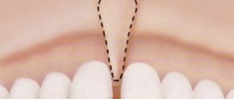
Interdental papillae - structure and functions
Gingival papillae are areas of the marginal gum. They are triangular in shape and are located between the teeth. Experts in the field of periodontology include the following tasks as their main functions:
- protection of the cervical area and roots from carious processes,
- increasing the depreciation of teeth while chewing food,
- retention of teeth in sockets,
- nutrition of hard tissues.
Gingival papillae are areas of the marginal gum.
Gum tissue does not have the ability to regenerate. Therefore, if the integrity of its structure is violated, for example, as a result of too intense cleansing, this process cannot be reversed. To hide the defect, gum grafting will have to be performed.
What diseases lead to gum inflammation
Inflammation of the marginal gums can be caused by mechanical trauma, constant exposure to tobacco smoke, thermal or acid burns, abundant subgingival deposits and many other factors. The following describes gum diseases that occur most often and also lead to inflammation and deformation of the interdental papillae.
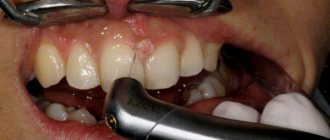
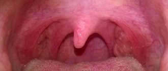
Papillitis – gingival polyp
This disease leads to inflammation of the gingival papillae located near one or several teeth. One of the common reasons for the development of this pathological process is an allergic reaction. In other cases, characteristic symptoms arise against the background of carious processes in the cervical area. According to statistics, in almost 40% of cases, the provoking factors are diseases of the cardiovascular system, as well as malfunctions of the endocrine system and gastrointestinal tract1.
The photo shows a gingival polyp
The clinical picture is as follows: the papillae noticeably turn red or acquire a bluish tint, obvious swelling occurs, and bleeding appears at the time of mechanical irritation. Patients complain of pain while brushing their teeth, eating, and even talking.
Gingivitis – inflammation of the gums
Gingivitis is a disease in which the inflammatory process spreads to the gum tissue and provokes unpleasant symptoms such as redness of the mucous membrane, swelling and bleeding. In this case, inflammation can cover only the interdental papillae, including the marginal gum or its entire surface.
This is what gingivitis looks like
“I had gingivitis just when I was pregnant. The gums were all red and inflamed. I was scared, I remember running to the doctor. There they reassured me, they said that this often happens against the background of hormonal changes, nothing to worry about. We brushed my teeth there, prescribed rinses and some kind of ointment, I don’t remember the name. A week later everything went away. So there is no need to panic ahead of time, but in no case should you delay going to the doctor.”
NikaS, from correspondence on the woman.ru forum
The most common forms are hypertrophic gingivitis with the proliferation of affected tissues and the atrophic, that is, chronic stage of the disease. In the first case, hypertrophy of the mucosa can lead to overlap of the crown by almost half, as well as disruption of the dentogingival bonds. In the International Classification of Diseases, 10th revision (ICD-10), this pathological phenomenon is assigned the number K06.1.
Periodontitis – inflammation of periodontal tissues
Periodontitis is an inflammation of periodontal tissues, in an advanced stage leading to the destruction of the periodontal ligamentous apparatus, loosening and loss of teeth. Most often, this pathology develops against the background of advanced gingivitis. At the same time, the disease is considered incurable, but with a competent comprehensive approach to treatment, it is possible to achieve its stable remission and prevent disastrous consequences. The development of the disease is accompanied by redness and bleeding of the gums and painful sensations.
Symptoms may indicate the presence of periodontitis
Fibromatosis and soft tissue hypertrophy
The pathology is a tumor-like lesion of the mucosa, in which hypertrophy occurs, that is, the growth of the gingival papilla, as well as the marginal and alveolar gums. With this clinical picture, the gingival tissue usually greatly increases in volume, which causes discomfort and bleeding, especially during meals. How it looks from the outside is shown in the photo below.
Fibromatosis is an overgrowth of gum tissue
It should be noted that fibromatosis is often diagnosed in children during the eruption of permanent incisors and molars, as well as in patients against the background of hormonal changes.
Development factors
The cause of the development of pathological inflammation can be a simple failure to comply with the rules of hygienic care. The condition of soft tissues affects the condition of dentin, since all processes are closely interconnected.
If you notice signs such as redness or swelling, it is recommended to contact your dentist immediately. The causes of inflammation can be general and local in nature.
Common ones include:
- lack of minerals in the body;
- hormonal imbalance;
- bad habits;
- diabetes;
- diseases of the gastrointestinal tract;
- infectious diseases;
- disorders of the cardiovascular organs;
- low immunity;
- long-term treatment with certain groups of medications.
There are also local causes that can increase the risk of inflammation of the mucous membranes of the oral cavity, in particular:
- teething;
- chemical or thermal burns;
- mechanical factors affecting delicate gum tissue;
- dental plaque and tartar;
- non-compliance with the rules for caring for the oral cavity, which leads to the accumulation of pathogenic microbes in periodontal pockets;
- improper implementation of prosthetics.
It is noteworthy that in practice there are cases when the cause of the development of pathology is the sharp edge of the prosthesis or an incorrectly cut filling. In this case, inflammation affects exclusively the site of damage to the mucosa.
How to treat inflammation and hypertrophy of gingival papillae
If obvious signs of inflammation appear, you should immediately contact your dentist. The sooner you do this, the easier and faster the doctor will be able to diagnose the problem and solve it. As part of the diagnostic examination, the specialist will conduct a visual examination and will likely send you for x-rays. In some cases, a CT scan, blood and urine tests, and biochemistry may be required.
Having established the exact cause of the symptoms, the specialist will prescribe adequate treatment. Below we briefly describe the main directions of therapy that are relevant in such cases.
Coagulation - cauterization
We are talking about a modern method of surgical treatment, in which wounds and ulcers on the mucous membrane of the mouth are cauterized using an electrode with a force of 2 amperes. Under the influence of current, blood clots and wounds are clogged, which helps stop bleeding. Additionally, coagulation ensures disinfection of damaged tissues.
During coagulation, wounds are cauterized using an electrode
Medication therapy
Therapy using medications is aimed at stopping the inflammatory process and eliminating its characteristic symptoms, including reducing the sensitivity of tissues. For inflammation of the interdental papillae, the following procedures are usually prescribed:
- rinsing with antiseptic solutions such as Chlorhexidine and Miramistin,
- local applications using antibacterial and anti-inflammatory gels and ointments,
- irrigation with lidocaine spray to relieve pain,
- applications using vitamins A and E,
- rinsing with decoctions of medicinal plants, such as chamomile and oak bark.
Ointments and gels promote rapid absorption and action.
Additionally, therapy is carried out aimed at strengthening the immune system and improving metabolic processes in the body. For this, the patient is prescribed multivitamin complexes, for example, “Vitrum” or “Complevit”.
Antibiotics for purulent processes
Powerful drugs related to antibiotics are usually prescribed in cases where the inflammatory process has already led to the release of pus in the soft tissues. Such medications should never be taken without a doctor's prescription. Moreover, the treatment will be effective only if the pathogenic microorganisms are sensitive to the components of the antibacterial agent. For gum inflammation, Amoxiclav, Erythromycin, or, for example, Cephalexin may be prescribed.
For purulent processes, antibiotics are prescribed
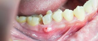
Surgical intervention
Against the background of advanced hypertrophy of gum tissue, the level of the mucous membrane usually changes, and the interdental papillae greatly increase. Subsequently, such an aesthetic defect can be removed surgically. The doctor literally excises the excess overgrown tissue using special instruments or a laser. This operation is usually performed for hypertrophic gingivitis or fibromatosis.
If the situation is the opposite, and the inflammatory process has led to gum recession and exposure of the cervical area, soft tissue is built up by moving one’s own flap of mucous membrane to the place where it is missing, after which the doctor applies sutures. Such an operation is usually required for atrophic gingivitis, periodontitis, to eliminate the consequences of soft tissue necrosis.
The photo shows an operation to eliminate recession
What does traditional medicine offer?
As part of maintenance therapy, doctors often recommend rinses and applications using decoctions and infusions prepared from medicinal herbs. Such products are allowed to be used only after consultation with a specialist. Otherwise, self-medication can lead to worsening of the condition and dangerous complications.
Plants such as sage, oak bark, chamomile, calendula and rosemary have pronounced antibacterial, anti-inflammatory and regenerating properties. You can also prepare a decoction from the herbal collection. To do this, just take a teaspoon of each dried flower, pour boiling water, let it brew, strain and then use the resulting rinse at least 4 times a day.
Herbal decoctions can be used at home
Regeneration of the gingival papilla and mucous membrane in the area of the intermediate part of the bridge
Ramon Gomez Meda Doctor of Dentistry, private practice (periodontics and prosthetic dentistry) (Leon, Spain)
Santiago Mareque Bueno Doctor of Dentistry, private practice (periodontology) (Pontevedra, Spain); Associate Professor at the University of Santiago de Compostela
In order for the restoration to look natural and the restored teeth to perform their function correctly, it is necessary to take into account the structure of the gums, the appearance of the lips and the patient’s face as a whole. Mucogingival surgery is used to treat gum recession [1–5].
The interdental gingival papilla is the area of gum between two adjacent teeth. It not only serves as a biological barrier that protects periodontal structures, but also plays a significant role in the formation of the aesthetic appearance. The absence of interdental gingival papillae can lead to problems with pronunciation, as well as the retention of food debris in the interdental spaces [6].
If the interdental gingival papilla is lost, its regeneration is quite difficult. Only a few such cases are known in dental practice [7–14]. However, none of the reports contain information about methods that allow restoration of the gingival papilla [15]. This report describes a surgical method for restoring mucosa and gingival papilla in the pontic pontic area in the presence of bone deficiency.
Surgical technique
The patient, 45 years old, came to the clinic for treatment of periodontal pathology. She complained about the mobility of the two upper central incisors. The patient wanted to restore her appearance and also eliminate periodontal pathology. The central incisors had grade 3 mobility; the pocket depth during probing was 10 mm [11] and 8 mm [21]. In the area of the right lateral incisor, a periodontal pocket with a depth of 10 mm was also found in combination with a vertical bone defect, which indicated a deficiency of bone tissue under the gingival papilla (Fig. 1 a, b).
Rice. 1a. Recession found on the labial side of teeth 11 and 12
Rice. 1b. Recession found on the labial side of teeth 11 and 12
A 7 mm deep pocket was also found in the area of tooth 22.
When collecting anamnesis, no allergies, concomitant diseases or bad habits were revealed. The patient was classified as ASA class 1. Several weeks before surgery, the patient was taught oral hygiene, in addition, subgingival deposits were removed and root surfaces were cleaned. After removal of granulation tissue in the area of the gingival papilla in the area of the 12th tooth, soft tissue recession to a height of 3 mm was discovered. In accordance with Miller's classification, she was assigned class III. On the vestibular side, in the area of teeth 11 and 12, recession of soft tissues to a height of 2 mm was also detected (Fig. 2).
Rice. 2. Vertical defect and class III mobility of teeth 11 and 21
Due to bone loss around the two central incisors, the decision was made to remove them (Fig. 3).
Rice. 3 a - d. The first large connective tissue graft was used in the area of the intermediate part of the bridge to protect the interincisal gingival papilla. We made sure that the temporary prosthesis does not put undue pressure on the graft
When smiling, the patient's gums were partially exposed (no more than a third of the length of the coronal part). At the same time, the color of the gum mucosa was heterogeneous. Photographs, x-rays were taken, alginate impressions were taken and masticography was performed. Based on digital analysis of photographs, diagnostic models were made, which were then placed in the articulator. The patient was then given treatment options. A tooth-supported bridge represents the most current option for replacing missing teeth, especially as an alternative to complex vertical guided bone regeneration, which would require frequent examinations and strict patient compliance. The use of such a prosthesis is less risky than installing an implant-fixed prosthesis if bone and soft tissue are not present in sufficient quantities. The patient had a high sociocultural level and aesthetic preferences. Taking into account other personal factors, in particular the patient’s place of residence, we were forced to choose the fastest, most effective and reliable solution. During her first three visits to the hygienist, the patient cried. Given her emotional instability, we abandoned a comprehensive therapeutic approach to reduce the risk of psychological trauma and possible failure. After the existing problem was explained to the patient, she agreed to remove two central incisors, correct the gums in the area of the intermediate part of the bridge, as well as the gingival papilla using several connective tissue grafts. On the same day, after appropriate preparation of the canines and lateral incisors, a temporary fixed prosthesis was installed. The neck of tooth 12 was prepared accordingly, taking into account the likely future soft tissue reconstruction. Endodontic treatment of the lateral incisors was required. Silicone impressions were made to create a second, more accurate, long-lasting temporary prosthesis and to re-evaluate the case from a biological, functional, and esthetic perspective. Four weeks later, soft tissue recession was detected due to bone resorption on the vestibular side of the maxillary alveolar process.
First, a large connective tissue graft was used (Fig. 4).
Rice. 4 a - d. After the second stage of surgery, the volume of tissue in the area of the right central incisor and the papilla between it and the lateral incisor was increased
Using several soft tissue incisions, a tunnel was created in the area of the pontic pontic (Fig. 4). A 6-0 nylon suture was used to secure the graft. We ensured that the temporary prosthesis did not put undue pressure on the graft (Fig. 4). Then we took a break for 4 months. At the end of the period, an increase in the volume of soft tissues was revealed, which still remained insufficient (Fig. 5).
Rice. 5 a - d. The connective tissue graft was installed using a tunnel approach after frenectomy
We needed more tissue in the area of the right central incisor and the gingival papilla between teeth 11 and 12. The depth of the pocket during probing is 7 mm (Fig. 5). Given the loss of 3-4 mm of papilla tissue, we can conclude that the probable probing depth was 10 mm with a 5 mm bone defect at the level of the papilla. After this, the second phase of surgical intervention began (Fig. 5). The preoperative status of the interdental gingival papilla was determined using the Norland and Tarnow classification [16]. The interdental gingival papilla, vestibular and palatal gingiva were numbed with local anesthesia using 1 capsule of Ultracaine® (articaine HCl/epinephrine, 40/0.005 mg/ml) and 1:100,000 epinephrine solution. For better visualization of the surgical field, a surgical dissecting loupe was used. First, a semicircular incision was made at the mucogingival junction to reposition the labial frenulum (Fig. 6).
Rice. 6 a - d. A diamond cutter was used to remove part of the transplanted epithelium
The second incision was made with a microscalpel from the lost gingival papilla along the gingival sulcus around the neck of the lateral incisor. The blade was turned towards the bone. The incision was made through the entire thickness of the gum tissue and provided access for a mini-curette. The third incision was made along the apical border of the semicircular incision directly towards the bone (Fig. 6). As a result, a gingival-papillary complex was formed. Its mobility was necessary to create free space under the gingival papilla and install a connective tissue graft. In addition, some mobility of the palate tissue was also ensured. The resulting flap was fixed coronally using a curette directed along the gingival sulcus and a small periotome. The amount of donor tissue required was determined during a preoperative assessment of gingival and incisal height in comparison with the expected new location of the gingival papilla. A section of connective tissue of significant size and thickness with a section of epithelium 2 mm wide was taken from the patient’s palate (Fig. 5). An area of epithelium was taken to obtain denser and more fibrous connective tissue, as well as to better fill the space under the coronally fixed tissue flap. The use of a large volume of tissue increased the chances of successful graft engraftment, since the graft was nourished by blood perfusion from a larger area. An area of epithelium was placed on the buccal side of the coronally fixed tissue flap, but was not covered by it (Fig. 6), since epithelium is denser than connective tissue and therefore better suited as a base for the repositioned flap. The connective tissue portion of the graft was placed in the gingival sulcus of the lost gingival papilla to prevent movement of the tissue flap and retraction of the papilla (Fig. 6). A 6-0 nylon suture (interrupted suture) was used to secure the graft in position and stabilize the wound. This microsurgical approach was made possible by using a Zeiss optical microscope. The wound on the palate is closed with a continuous suture. The patient is prescribed amoxicillin (500 mg, three times a day, 10 days), as well as an alcohol-free mouthwash with chlorhexidine (twice a day, 3 weeks). Keratinizing epithelial cells and food debris could be removed from the wound surface using a cotton swab soaked in chlorhexidine gluconate. After 4 weeks, the stitches were removed. The patient was also prohibited from using mechanical means to clean teeth in the wound area for 4 weeks. An earlier examination of the patient was impossible due to the remoteness of her place of residence. The postoperative period passed without complications. The third stage of surgery took place before installation of the permanent prosthesis. Using a diamond cutter, part of the transplanted epithelium was removed (Fig. 7).
Rice. 7 a - c. Transformation of the intermediate part of the bridge after the first and second operations
The area between the pontic and the lateral incisors was not probed for 6 months. As a result of probing, a gingival pocket with a depth of 5 mm was discovered in the area of the lateral incisor, which was only 1 mm greater than the depth of the gingival pocket in the area of tooth 22.
results
The patient's condition was assessed 3 months after the first surgical procedure. Only horizontal tissue growth was achieved in the area of the intermediate part of the bridge (Fig. 8).
Rice. 8 a, b. After the second stage of surgical intervention, the edge of the soft tissue of the gingival papilla was 3-4 mm closer to the incisors than before the operation, while there was no bleeding, and probing did not give negative results
The depth of probing in the area of the lateral incisor before the second operation was 7 mm. A recession of 3 mm in diameter was found in the area of the right lateral incisor (Miller class III). After the second stage of surgical intervention, the edge of the gingival papilla was 3-4 mm closer to the incisors than before the operation. The depth during probing decreased by 4-5 mm. An examination carried out after 2 years showed that the clinical results recorded 3 months after surgery had improved. In particular, there was no black triangle between the artificial crowns of the lateral and central incisor (Fig. 9 a, b).
Rice. 9 a. When checked after two years, no black triangle was found between the lateral and central incisors
Rice. 9 b. When checked after two years, no black triangle was found between the lateral and central incisors
There was no retraction or compression of the papillary tissue, and the probing depth did not increase. Radiographic examination showed improvement in the condition of the underlying bone (Fig. 10).
Rice. 10 a - d. Radiographic examination showed significant improvement in the condition of the underlying bone, although no bone graft was used
The depth of the gingival groove of the papilla is greater than on the opposite side, there is no bleeding, and probing does not give negative results. The success of the procedure depended on the following factors:
- The space between the bone and the coronally fixed gingival papilla was filled with a connective tissue graft.
- The connective tissue was well stabilized by the suture.
conclusions
In clinical cases that present not only a medical but also an aesthetic problem, reconstructive surgery can mask tissue loss, but the patient rarely achieves an ideal appearance. To improve the results of such intervention, periodontal plastic procedures can be used. The use of optics and microsurgical instruments is recommended. This allows the surgeon to improve visibility, avoid unnecessary incisions, and increase the chances of a favorable treatment outcome.
How to prevent inflammation of the mucous membrane after filling
There are cases when the gums become inflamed after the filling procedure. Of course, the situation is extremely unpleasant, because it would seem that the patient went to the doctor and is now waiting for relief, but in the end a new problem arises. To minimize the risk of encountering such a nuisance, it is better to spend time finding a good clinic and an experienced doctor. And experts in the field of dentistry cite the two most optimal options for preventing such complications.
Use high-quality modern materials to cover proximal cavities
When filling complex cavities, which include approximal ones, it is recommended to use modern materials: photocomposites or glass ionomer cements. Light-curing composites are rightfully considered the best choice for filling procedures. They contain an optimal ratio of large and small particles, which increases their functional characteristics after hardening. Such materials adhere firmly to dental tissues and closely correspond to them in terms of key indicators, including biocompatibility.
When treating teeth, you should choose only high-quality materials
Use restoration tabs
If the cavity is too large or complex, due to which the use of photocomposite or glass ionomer cement cannot guarantee the integrity and reliability of the filling, it is better to resort to the help of a restoration inlay. We are talking about a microprosthesis, which is created from impressions in the laboratory and is used to restore the integrity of a damaged tooth. Such inlays are usually used for various forms of carious lesions, mechanical damage to crowns, pathological abrasion, or after unsuccessful attempts at filling using the classical method.
The photo shows dental inlays
How does the disease manifest itself?
Gum overgrowth affects the outer (buccal) and inner (lingual) areas of the gums, affecting one or both jaws. The general surface of the gums, the gingival margin adjacent to the tooth, as well as the interdental papillae are affected.
Main symptoms of the pathology:
- partial or complete overgrowth of crowns in the area of fibromatosis;
- smooth rounded edge of growths;
- increased density of hypertrophied tissues;
- an even pink tint of the gums and no bleeding.
The intensity of symptoms determines the stage of the disease.
In this case, gum hypertrophy can be generalized or focal. The difference is that if in a focal form only a single lesion is formed, then in a generalized form several pathological areas merge with each other. In severe cases of the disease, fibromatosis can be combined with changes in facial features characteristic of acromegaly.
On a note!
With hereditary pathology, it is impossible to reverse tissue hypertrophy, however, in areas of the gums with extracted teeth, growth usually stops.
Marginal periodontitis – complex treatment
If the inflammation is caused by marginal periodontitis, the most important thing is to eliminate the causative factor as quickly as possible. This could be an incorrectly installed filling or an uncomfortable orthopedic design that creates a constant mechanical impact on the mucous membrane. Next, the patient must undergo appropriate treatment from a periodontist.
Anti-inflammatory therapy using local drugs
To treat marginal periodontitis, doctors usually prescribe a whole range of different procedures aimed at relieving inflammation and restoring affected tissues. As part of the therapy, rinses, applications, gingival dressings, injections and oxygen therapy are prescribed. Additionally, antiseptic and anti-inflammatory solutions, ointments and gels are prescribed for self-use at home.
General drug treatment
If the gums near the tooth become inflamed, more powerful medications, such as antibiotics, may also be prescribed. Medicines of the following pharmacological groups are also usually prescribed: multivitamins, desensitizing drugs, adaptogens, and immunostimulating agents.
Physiotherapeutic procedures
Physiotherapy is also often used to treat gums. This category of procedures includes the following methods: laser or ultraviolet treatment, microwave therapy, heat treatment using paraffin applications, magnetic therapy and electrophoresis.
Physiotherapy is also used to treat gums.
Properties of hyaluronic acid used in dentistry:
- anti-inflammatory – used for gum inflammation, periodontitis, and dental implantation;
- bacteriostatic – suppresses the growth and development of bacteria in the area of inflammation, which is actively used in the treatment of periodontitis;
- regenerative – helps speed up the healing and recovery processes after surgery;
- improves the fixation of bone materials when used for dental implantation (bone grafting), maintaining the stability and volume of the bone graft;
- restores interdental papillae, reconstructing missing tissues;
- helps in the restoration of tissues inside and outside the joint (injections of hyaluronic acid are used for disorders of the temporomandibular joints), reducing pain, eliminating clicks in the joint and restoring mobility to it.
Let's take a closer look at some areas of application of hyaluronic acid in dentistry.
Home care during treatment of inflamed interdental papillae
The main condition for assisted home therapy is proper comprehensive hygiene. It is important to regularly brush your teeth twice a day with the right brush and toothpaste. After each meal, experts recommend using floss and rinsing your mouth. As an additional hygienic tool, it is worth purchasing an irrigator, which, by supplying an air-water jet under high pressure, allows you to efficiently clean even hard-to-reach places. And, of course, it is important to regularly visit the dentist’s office to undergo appropriate therapeutic and preventive procedures.
An irrigator is an additional device for maintaining oral hygiene
Inflammation in the gums spreads very quickly, so at the first suspicious symptoms you should immediately consult a doctor. It is easier to treat any mucosal disease at an early stage than to later try to cope with chronic processes and complications.
1Glebov, K.G. Difficulties in the differential diagnosis of papillostenosis and papillospasm, 2014.