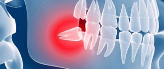
Various diseases of the soft tissues of the oral cavity are quite common cases in medical practice. Gum hyperplasia is one of these pathological diseases, when rapid growth of gum tissue cells occurs, and the swollen tissue significantly covers the surface of the dental crown. The causes of this disease are not fully understood. This may be a consequence of taking any medications, a symptom of blood diseases, or a possible genetic predisposition.
Symptoms of gum hyperplasia
Gum growth in the initial stages may resemble swelling or tumor formation. Specific symptoms gradually appear:
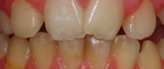
- seals appear on the surface of the mucous membrane or under it, which gradually increase in size and may become more voluminous;
- The volume of the gums increases: they become wider, the gingival margin rises higher. At the same time, the tissues remain dense, there are no signs of edema;
- the interdental spaces are filled with gum tissue;
- the edges of the gums rise, covering the surface of the crowns;
- when chewing or brushing teeth, overgrown areas are injured and can become inflamed, after which their surface becomes compacted;
- the color of the mucous gradually becomes bright pink.
Diagnosis and treatment of gum hyperplasia
Before the dentist selects a treatment method, an in-depth examination will be required - a visual examination of the oral cavity is not enough. As part of the diagnosis, the doctor prescribes:
- collection of material from the pathological focus for histological examination - it is necessary to exclude a malignant process;
- targeted and panoramic photographs of the jaw - make it possible to assess the area of distribution of the pathological process;
- orthopantomogram - lesions of the alveolar region are detected;
- Blood tests for hormones help determine the cause of fibromatosis.
Treatment is prescribed depending on the stage of the disease. Most often, conservative therapy is used based on diagnosis - already prescribed drug treatment is adjusted, hormonal therapy is carried out. Antihistamines or agents that reduce blood supply to pathologically altered tissues often help.
Surgical intervention is used only if the disease is in an advanced stage or has developed due to a genetic predisposition. Surgery is also advisable if drug therapy is ineffective.
It is necessary to treat gum fibromatosis, if only because complications may develop: loosening and loss of healthy teeth, osteoporosis, gingivitis, periodontitis. Over time, overgrown tissues cause bleeding gums and the appearance of wounds on them.
Types of gum hyperplasia
Periodontal tissues can grow in different ways, involving different areas of the gums. There are several types of hyperplasia:
- localized: the growth of new cells occurs in a limited area, next to one tooth or group of teeth (only on one or both sides);
- generalized: the gums grow over the entire surface on one or both jaws;
- papillary: new cells are formed only in the area of the gingival papilla, the pathology does not affect other gingival tissues;
- marginal: the gingival papilla and the area of the gingival margin grow;
- diffuse: growth of both marginal and attached gums occurs (an increase in periodontal tissue to the entire height from the gingival margin to the mucogingival border).
Hyperplasia may be limited. The increase in tissue in this case is similar to a tumor, strictly isolated, and has a wide base or pedicle. It looks like a growth or bump.
Causes of gum tissue overgrowth
Hyperplasia is a fairly rare disease, so it has not yet been fully studied. Specialists in the field of pathological anatomy identify two main reasons for its development:
- genetic predisposition – signs of the disease appear in childhood (up to 10 years);
- long-term use of certain medications - containing estrogen, Cyclosporine and others.
Metabolic disorders and too much accumulation of fibroblasts can also trigger the disease. Sometimes fibromatosis is diagnosed in adolescents and pregnant women - in this case we are talking about a hormonal problem.
Signs of gum tissue overgrowth
Gingival hyperplasia is an excessive formation of connective tissue in the oral cavity. Due to the increase in the number of cells, the gums swell and cover part of the teeth. This phenomenon is characterized by swelling of the mucous membrane. Over time, tumors of varying sizes form and the following symptoms appear:
- change in gum color,
- overgrowth of interdental spaces,
- pain when pressing on the mucous membrane,
- bleeding gums when brushing teeth and eating solid foods.
Overgrown gums cause discomfort and impair the quality of life. It spoils the smile, interferes with hygiene procedures, and contributes to the development of complexes.
The mucous membrane can grow in different ways. Doctors distinguish two types of hyperplasia:
- focal - affects areas near the molars, in the smile zone, near the wisdom teeth;
- generalized - affects almost all areas of the upper or lower jaw.
The clinical picture depends on the type and degree of hyperplasia.
Stages of development of gingival hyperlasia
- At the first stage, inflammation of the gums is observed in the form of a slight increase in the volume of gum tissue, gradually filling the interdental space.
- The second stage is characterized by a voluminous swelling that covers a significant part of the part of the tooth protruding above the gum. This inflamed tissue is prone to bleeding from any external influence.
- In the third stage, the growth becomes significant, covering most of the tooth. Such inflamed gums become homogeneous pink in color. The formation of additional growths is characteristic.
Treatment of gum fibromatosis
Modern medicine provides for the treatment of gum hyperplasia only with surgical methods. A special procedure is performed - gingivectomy, during which the doctor makes an incision into the enlarged gingival margin. The operation lasts 30 minutes and requires local anesthesia. The dentist applies a special periodontal bandage to the damaged area, which protects the wound from infection. In parallel with this, sanitation of the oral cavity is performed, including the elimination of tissue defects and the treatment of dental caries. If the disease affects temporary teeth, the doctor is forced to remove them.
Treatment of gum fibromatosis
It is not uncommon for fibromatosis to return after treatment. In such cases, the doctor is forced to perform another surgical operation. If hyperplasia has a medicinal form, then during treatment the doctor recommends replacing the previously used drug with another. Such a replacement often leads to positive changes, namely to the gum tissue regaining its previous appearance.
With repeated use of drugs that provoked the occurrence of gum hyperplasia, the chances of recurrence of the pathology increase significantly. If replacing the medications does not help, the doctor removes the pathological gum tissue surgically. Regular removal of tartar helps slow down the progression of the disease.
At the dentist
ethnoscience
Folk remedies will not help get rid of hyperplasia, but they can be used as an addition to the main therapy. Due to the antibacterial effect, various infusions of medicinal herbs are used for rinsing the mouth. Below are the most effective recipes for folk remedies used in the treatment of oral diseases.
Table. Folk remedies for gum hyperplasia.
| Product name, photo | Application |
| Horseradish tincture | Pour 500 ml boiling water 3 tbsp. l. chopped horseradish and leave for 50-60 minutes. After straining the finished tincture, use it to rinse your mouth 2 times a day - morning and evening. This will cleanse the blood vessels, relieve inflammation and destroy bacteria in the oral cavity. |
| Propolis chewing gum | Dissolve 60-80 g of propolis in a water bath, then mix it with 5 g of lemon juice, 5 g of lemon balm essential oil and 1 tsp. honey Cut the hardened mass into small pieces. Cooked chewing gum can kill bacteria, which is why it is often used to treat colds. |
| Beetroot compress | Mix 1 tbsp. l. sea buckthorn oil with fresh beets chopped on a fine grater, so that the result is a homogeneous substance. Apply a compress from the prepared composition to the gums 2 times a day. The duration of the procedure is 20 minutes. |
| Oak bark balm | Grind 2 tbsp. l. oak bark and mix it with 1 tbsp. l. linden flowers. Pour 300 ml of boiling water over it and leave for 40 minutes. After straining the prepared balm, use it to rinse your mouth 2-3 times a day. |
| Chamomile decoction | In medicine, chamomile is used for its wound healing and anti-inflammatory properties. To prepare the decoction, pour 200 ml of boiling water into 4 tbsp. l. dried chamomile flowers. Leave for 1-1.5 hours. Rinse your mouth with this decoction 5-6 times a day or every few hours. |
On a note! Refusal to seek medical help, as well as self-medication for gum fibromatosis can lead to serious problems. Eliminating such consequences will be much more difficult than curing hyperplasia.
Treatment of gum disease
Only a doctor can establish an accurate diagnosis and prescribe a course of treatment, taking into account the cause and stage of the disease, the presence of contraindications, age and other individual characteristics of the patient.
Treatment methods for gum disease:
- hygienic teeth cleaning (helps in the initial stages of diseases);
- hardware treatment of gums (allows you to clean periodontal pockets, improve tissue regeneration, remove granulation tissue);
- anti-inflammatory therapy (relieves swelling, reduces inflammation, destroys germs);
- physiotherapy (darsonvalization, massage, vacuum therapy, electrophoresis);
- curettage of periodontal pockets (open and closed, allows you to clean out accumulated deposits from the gum pockets);
- surgical intervention (implantation of a healthy area of the gum on the damaged one, placing a membrane between the bone tissue and the gum that stimulates tissue regeneration, gingivectomy (excision of the edge of the gum to gain access to plaque accumulated under it), flap surgery).
- splinting of teeth (prevents teeth from loosening).
Reasons and forms
A complete list of causes that provoke fibromatosis has not yet been established. However, doctors have identified at least two risk factors. The first, as we noted above, is genetics. If one of the parents suffers from hyperplasia, then with a high degree of probability we can say that the child will inherit this disease. The second risk factor is taking certain medications, including cyclosporine and phenytoin.
There are two forms of gum hyperplasia - localized and generalized.
- Localized hyperplasia. This form is characterized by the formation of one or several separate foci. Most often they are localized on the front teeth, and the overgrown tissue can cover up to 2/3 of the crown part. It is important to note that the formations do not differ in color from normal gums. Moreover, they are painless.
- Generalized hyperplasia. In this case, several foci of growth are formed, which merge into one as the disease develops. In this case, the overgrown tissue can completely cover the teeth. It is also worth noting that the generalized form is observed both in the smile area and on the molars.
Gingival hyperplasia is characterized by pathological growth of soft tissue.
Prevention measures
Among all the possible reasons contributing to the development of gum hyperplasia, insufficient oral hygiene is far from the least important. To prevent the occurrence of an inflammatory process, it is necessary, first of all, to limit the amount of sweets consumed, as they negatively affect the condition of the teeth and oral cavity. After each meal you need to rinse your mouth with a special solution or brush your teeth. It is also advisable to give up bad habits that have a detrimental effect on the condition of the gums and teeth.
Disease prevention
Smoking not only weakens the patient's immune system, but also impairs blood circulation in the gums. Therefore, if you or your relatives have had gum hyperplasia before, try to get rid of this bad habit.
On a note! If you have dentures that do not adhere well to the surface of the gums, replace them with better ones. Otherwise, harmful bacteria will accumulate in the space between the gum and the product.
Dentures
Regular snacking can cause the formation of tartar, which can lead to gum tissue disease. To prevent this phenomenon, you need to use dental floss after each such snack or abandon them altogether. Dental chewing gum can help clean your mouth, but it is not recommended to chew it on an empty stomach.
Whether you have any dental disease or not, it is important to visit your doctor's office regularly for dental checkups. Many oral pathologies do not appear in the initial stages, so only an experienced specialist can identify them in a timely manner. Visit your dentist twice a year and you can avoid many oral diseases.
Teeth cleaning
You can have your teeth professionally cleaned periodically when you visit your dentist's office. This will remove all plaque from the surface of the teeth that cannot be removed with a regular toothbrush. Also, do not forget about the therapeutic diet. Proper nutrition also helps prevent many oral diseases, including hyperplasia. It is better to consult a specialist regarding the preparation of your diet, but in most cases it includes daily consumption of vitamin-containing foods (fresh fruits, vegetables, dairy products, and so on). Following such a diet will not only protect the body, but also lose extra pounds.
Gingival hypertrophy (gingival fibromatosis)
There are two forms of gum fibromatosis: generalized and limited. With generalized fibromatosis , which can be nodular or diffuse, several confluent foci of granular growths of the gums form, which can eventually cover the crowns of the teeth. With more rare limited fibromatosis, single growths are detected in the area of the tubercle of the upper jaw or the lingual surface of the gums of the lower jaw. These growths have a smooth surface, dense consistency and regular round shape, are localized on one side or have a bilateral localization.
Limited fibromatosis is also called focal.
Gingival hypertrophy occurs slowly. Dense formations appear on the gums, do not stand out in color and are painless, mainly on the front teeth. The crowns of the teeth are closed up to two-thirds, less often completely.
It can prevent the loss of temporary (baby) teeth and the germination of molars. Threatens osteoporosis and destruction of interdental septa.
Based on the intensity of the productive process, there are 3 degrees of gum hypertrophy. I degree is characterized by excessive growth of the gum edge, gingival papillae, which are thickened in the form of a cushion throughout the entire affected area and increased by 1/3 of the height of the tooth crown, their shape is changed (round, oval, irregularly shaped). Hypertrophy is more pronounced at the base of the papillae.
Grade II is determined by the progression of hypertrophy of the gingival margin and gingival papillae. The gingival margin is raised in the form of a roller covering the lower part of the crown of the teeth. The shape of the papillae is changed, the growth reaches 1/2 the height of the crown of the teeth.
III degree is characterized by pronounced hyperplasia of the gingival margin and gingival papillae. The increased gum volume covers more than 2/3 of the height of the tooth crowns, often reaching the cutting edge or the surface of the teeth. Enlarged papillae are often covered with multiple small and large bleeding granulations.
Drug-induced gum hypertrophy. Taking certain medications can lead to gum hypertrophy. It is observed in 25-50% of patients taking phenytoin and cyclosporine. Phenytoin is prescribed to prevent epileptic seizures. It stabilizes the excitation threshold and reduces the convulsive readiness of motor neurons. Cyclosporine is prescribed to patients after organ transplantation, as it suppresses the proliferation of T-lymphocytes and prevents the development of a rejection reaction.
Gum hypertrophy is also observed in 1-10% of patients taking calcium channel blockers, for example, nifedipine, diltiazem, verapamil, felodipine, amlodipine. Gum hypertrophy also develops when taking sodium valproate and estrogenic drugs (oral contraceptives and conjugated estrogens), especially in large doses. Although the mechanism of gingival hypertrophy is unclear, it is believed that in many cases it is caused by a disruption of calcium flow through the membranes of gingival fibroblasts, which leads to changes in cellular homeostasis, collagenase activity and local immunity. Gum hypertrophy when taking estrogens may be due to the fact that they increase blood supply to the gums and the formation of inflammatory mediators in them.
Drug-induced gum hyperplasia is possible at any age, regardless of gender. Although gingival hypertrophy is based on a hyperplastic reaction, an inflammatory component caused by bacteria contained in dental plaque may play a role. Drug-induced gingival hypertrophy is usually generalized and begins in the interdental papillae. It is more pronounced on the labial surface of the upper teeth and appears in the form of easily bleeding red nodules. As the gums grow, fibrosis occurs, the interdental papillae enlarge, acquire a densely elastic consistency and a pink color. Over time, they can completely cover the crowns of the teeth, which makes oral care difficult, limits chewing, and causes aesthetic discomfort in patients.
Nadent – Natalia Khvorostinova’s Universal Dentistry in Moscow
general information
Gingival hyperplasia is a pathological growth of gum tissue until it covers a significant surface of the tooth crowns.
As a rule, hypertrophic growth of the gums is not associated with inflammatory processes and does not affect other tissues of the oral cavity (cheeks, tongue, etc.).
This anomaly is often accompanied by hyperemia - increased blood supply to the gum tissue, which entails pain and a high risk of bleeding when brushing teeth or eating rough food.
If gum hyperplasia is not treated in time, it can prevent the replacement of baby teeth with permanent ones, as well as lead to osteoporosis and destruction of interdental septa.
In addition, gum hyperplasia significantly complicates oral hygiene and indirectly provokes the development of various types of oral diseases.
Causes of gum hyperplasia
Despite the fact that gingival hyperplasia is relatively common, the reasons that cause its development have not yet been clearly established.
Very often (in 25-40% of cases), gum hyperplasia develops with the active use of drugs such as phenytoin (prevents epileptic seizures), cyclosporine (taken after organ transplantation, preventing organ rejection).
Up to 10% of patients taking various drugs that block calcium channels are also at risk of this disease: nifedipine, diltiazem, felodipine, verapamil, etc.
Other factors predisposing to this disease include:
- Pregnancy
- Certain blood diseases (such as leukemia)
- Puberty
- Genetic predisposition
- Some malocclusions
- Abundance of tartar
Gum hypertrophy most often occurs in adults. In children, it is observed only in the case of genetic causes of the disease.
Drug-induced hyperplasia is possible at any age.
Types of gum hyperplasia
Experts distinguish two types of hyperplasia:
- Generalized hyperplasia - occurs most often and consists of several foci of granular growths of the gums merging into one, covering a fairly large surface of the teeth
- Limited (focal) hyperplasia is relatively rare and represents single growths of the gums in the area of the tubercle of the upper jaw or in the area of the lingual surface of the gums in the lower jaw. Single growths can be localized either on one side of the gum or have a bilateral localization.
Symptoms of gum hyperplasia
The main manifestations of gum hyperplasia:
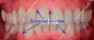
- Swelling of the gums (gingival margin and interdental papillae) and increase in their mass
- Filling the interdental spaces with gum tissue
- Covering of a significant part of the surface of the crown of the teeth with gum tissue
- Thickening of hypertrophied gum tissue
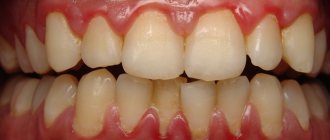
- Homogeneous pink gum color
Development of gingival hyperplasia
Despite the fact that the mechanism of development of gingival hyperplasia has not been fully studied, it has been established that most often this disease is caused by a violation of the movement of calcium through the membranes of gum fibroblasts, which entails a disruption of homeostasis in the cells of the gingival tissue and the active production of collagenase, which promotes gum growth.
Gum hyperplasia develops quite slowly.
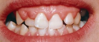
First, small dense formations in the form of a roller appear on the gums (usually in the area of the front teeth), which do not differ in color from the rest of the surface of the gums and do not cause pain when pressed. The crowns of the teeth are covered with gum tissue by about 1/3.
Next, hypertrophy of the gingival margin and gingival papillae begins to progress, the shape of the gingival papillae becomes deformed, and the growing gum covers up to half the height of the tooth crown.
The extreme degree of gum hyperplasia is characterized by maximum growth of the gingival margin, covering more than 2/3 of the height of the crowns and sometimes reaching the cutting edge or chewing surface of the teeth. Swollen gingival papillae are covered with bleeding granules of various sizes.
Diagnosis and treatment of gum hyperplasia
The main task when diagnosing gum hyperplasia is not to confuse it with other gum diseases. Therefore, when diagnosing here, in addition to visual examination, radiographs and histological analysis are often used.
Treatment of gum hyperplasia is currently only possible surgically by excision of excess hypertrophic gum tissue. At the same time, a complete sanitation of the oral cavity is performed.
If gum hyperplasia is caused by taking certain medications, you should replace them with other drugs. In this case, discontinuation of the drug can contribute to the natural return of the gums to their normal state. If this does not happen, the excess gum tissue is removed surgically.
If the patient again starts taking the drugs that caused gum hyperplasia, then the likelihood of a relapse of the disease is high.