When a root canal infection occurs, a formation called a cyst may develop at the top of the tooth root, which appears as a pus-filled cavity with a fibrous lining on the inside.
A cyst, also often called a periodontal abscess, can gradually enlarge over time, causing serious discomfort to a person. The cyst located on the teeth of the upper jaw has the highest growth rate, which is facilitated by more porous bone.
If we look at the x-ray, then a strong darkening in the upper region of the root is a cyst.
Diagnosis of dental cysts
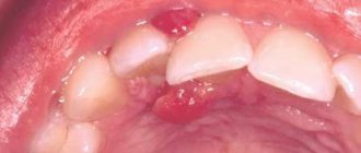
Since at an early stage the disease has practically no symptoms, it can be very difficult to determine. Nevertheless, there are certain diagnostic methods that allow you to identify a cyst:
- external examination, the task of which is to determine whether there is growth of the capsule in the gingival tissue;
- checking the integrity of existing fillings (if there are any violations in the root part, they begin to collapse, which is noticeable to the naked eye);
- X-ray;
- tomography of the jaw.
The surest way to make a diagnosis is an x-ray: the capsule filled with cystic fluid will appear as a bright white area (its shape can be round or oblong) located under the gum pocket.
Based on the location of the formation and the current stage of development of the disease, the doctor will be able to prescribe the most effective treatment.
Hemorrhagic ovarian cysts
A complex hemorrhagic ovarian cyst is formed by bleeding from a Graafian follicle or follicular cyst. On ultrasound, hemorrhagic cysts appear as single-chamber thin-walled cystic structures with the presence of fibrin strands or hypoechoic inclusions, with good permeability to ultrasound. On MRI, hemorrhagic cysts are characterized by high signal intensity on T1 FS scans, while on T2 WI they give a hypointense signal. With Doppler ultrasound, there is no internal blood flow; the component accumulating contrast inside the cyst is not detected on CT or MRI. The wall of a hemorrhagic cyst has variable thickness, often with the presence of vessels located in a circular pattern. Despite the fact that hemorrhagic cysts usually manifest with acute pain symptoms, they can be an incidental finding in a patient who does not present any complaints.
Sonograms reveal a hemorrhagic cyst with a blood clot simulating a neoplasm. However, Doppler ultrasound did not reveal internal blood flow in the cyst, and its permeability to ultrasound was not reduced.
MRI picture of a hemorrhagic ovarian cyst: in the T1 VI mode without fat suppression, a complex cyst is determined, characterized by a hyperintense signal, which can be caused by both the fatty component and the blood. On T1 fat-suppressed imaging, the signal remains hyperintense, allowing confirmation of the presence of blood. After administration of gadolinium-based contrast, no contrast enhancement is observed, which allows us to confirm the hemorrhagic nature of the ovarian cyst. In addition, it is necessary to include endometrioma in the differential diagnostic range.
Ultrasound reveals a soft tissue (solid) component in both ovaries. However, ultrasound permeability on both sides is intact, suggesting the presence of hemorrhagic cysts. Dopplerography (not shown) shows no blood flow in the formations.
How to distinguish a hemorrhagic cyst on MRI? In T1 mode, a component with high signal characteristics (fat, blood or protein-rich fluid) is detected in both formations. With fat suppression, the signal intensity does not decrease, which generally makes it possible to exclude a teratoma containing adipose tissue and confirm the presence of hemorrhagic fluid.
Types of cysts
To prescribe the most effective treatment, the dentist needs to determine the type of cyst and identify why it formed.
Cystic formations are:
- radicular (occur at the root of the tooth or in close proximity to it);
- residual (formed after tooth extraction);
- retromolar (formed as a result of problematic eruption of wisdom teeth).
Based on their origin, cysts are divided into:
- odontogenic (they are caused by various dental diseases);
- non-odontogenic (their formation is not associated with dental diseases).
Separately, it is worth highlighting follicular cysts, which contain the tooth germ: they are formed as a result of improper care of baby teeth.
Corpus luteum cyst
The corpus luteum can become obliterated and fill with fluid, including blood, resulting in the formation of a corpus luteum cyst.
Ultrasound: corpus luteum cyst. Small complex ovarian cysts are visible with blood flow in the wall, which is detected by Doppler ultrasound. Typical circular blood flow during Doppler examination is called the “ring of fire.” Note the good permeability of the cyst to ultrasound and the absence of internal blood flow, which correlates with the changes characteristic of a partially involuted cyst of the corpus luteum
It should be noted that women taking hormonal oral contraceptives that suppress ovulation usually do not develop a corpus luteum. Conversely, the use of drugs that induce ovulation increases the chance of developing corpus luteum cysts.
Pelvic ultrasound: corpus luteum cyst. On the left, the sonogram shows changes (“ring of fire”), typical of a corpus luteum cyst. On the right, in the photo of the ovarian specimen, a hemorrhagic cyst with collapsed walls is clearly visible.
Corpus luteum cyst on MRI. An axial T2-weighted tomogram reveals a cyst of the involuted corpus luteum (arrow), which is a normal finding. The right ovary is unchanged.
Main symptoms of an abscess
A dental cyst may not reveal itself for a long time: a person may not realize that there is something wrong with his teeth and gums. Sometimes an abscess causes mild pain when biting or pressing on the gum. But barely noticeable and far from constant pain can force few people to go to the doctor: a cyst at this stage of development is discovered by chance when a person goes to the dentist about completely different teeth.
An infection in the abscess cavity can worsen if immunity decreases: the process of pus formation accelerates, severe pain appears, the cheek swells, the person feels weak and lacks strength, and the temperature rises.
Inflammation of the appendages (salpingoophoritis) and tubo-ovarian abscess
Tubo-ovarian abscess usually occurs as a complication of an ascending (from the vagina to the cervix and fallopian tubes) chlamydial or gonorrheal infection. CT and MRI reveal a complex cystic formation of the ovary with a thick wall and lack of vascularization. Thickening of the endometrium or hydrosalpinx makes the diagnosis of tubo-ovarian abscess more likely.
An axial contrast-enhanced CT scan reveals a complex cystic formation on the left, resembling an abscess, with a thick wall accumulating contrast and gas inclusions inside.
On a CT scan in the sagittal plane (left), it can be seen that the ovarian vein approaches the mass, confirming its nature (arrow). On the coronal tomogram (right), the anatomical relationships of the mass and the uterus can be assessed. A gas bubble is visualized in the uterine cavity, which suggests an infectious onset here, with subsequent spread of infection through the fallopian tube to the ovary.
Why does a cyst occur?
The main reason for the development of an abscess is the appearance of infection in the root canals. But there can be several reasons for infection:
- Advanced caries that has developed into pulpitis due to ignoring the need for its treatment. In the tissues on which the carious formation has appeared, pathogenic microorganisms settle, first entering the pulp, as a result of which its inflammation begins, then, if the pulpitis is not treated, penetrating beyond the boundaries of the tooth, resulting in a periodontal abscess.
- Unsatisfactorily filled root canals. Canal filling is a fairly common procedure that dentists have to perform during treatment. However, despite the routine nature of the operation for a dentist, not all specialists, due to lack of experience or due to inattention, carelessness and the desire to complete the work as quickly as possible, do it efficiently. The canal must be sealed along its entire length, since in the unsealed part there is a high probability of infection, which, just as in the case described above, will sooner or later go beyond the boundaries of the tooth and lead to the appearance of an abscess. Official statistics on poorly filled canals are depressing: in more than half of the cases, dentists make mistakes during the procedure, which subsequently result in the appearance of a cyst. In the picture, the unsealed part of the canal is always clearly visible. Sometimes it happens that the root canal is completely filled, but the material used for filling is loose, which is also not good.
Mature teratoma (dermoid cyst) of the ovary
A mature cystic teratoma, also called a dermoid cyst, is an extremely common ovarian mass that can be cystic in nature. "Mature" in this context means a benign formation as opposed to an "immature", malignant teratoma. Benign cystic teratomas usually occur in young women of childbearing age. On CT, MRI and ultrasound they appear unilocular in (up to) 90% of cases, but can be multilocular or bilateral in approximately 15% of cases. Up to 60% of teratomas may contain calcium inclusions in their structure. The cystic component is represented by a fatty fluid produced by the sebaceous glands located in the tissue lining the cyst. The presence of fat is a diagnostic sign of teratoma. On ultrasound, it has a characteristic cystic appearance with the presence of a hyperechoic solid nodule in the wall, called Rokitansky's node or dermoid plug.
Ultrasound visualizes Rokitansky's node or dermoid plug (arrow).
Liquid-fat levels may also be detected due to density differences (fat, as a lighter, less dense substance, floats on the surface of the water). You can also visualize thin echogenic lines (“stripes”), the presence of which is caused by “hair” in the cyst cavity. Mature cystic teratomas, even benign ones, are most often removed surgically, as they pose an increased risk of ovarian torsion.
Complications of ovarian dermoid cyst:
- Ovarian torsion
- Infection
- Rupture (spontaneous or as a result of trauma)
- Hemolytic anemia (a rare complication that resolves after resection)
- Malignant transformation (rare)
What does an ovarian dermoid cyst look like on an MRI? A cystic formation with hyperintense signal is visible, within which there are septations (found in approximately 10% of such cysts). In the fat suppression mode, the suppression of signal intensity is determined, which makes it possible to confirm the presence of a fatty component and make a conclusion about a teratoma.
Tooth cyst: treatment
Treatment can be divided into therapeutic (use of drugs) and surgical (gum incision and root resection).
Conservative treatment is justified if:
- no unsealing required;
- if the canal is unsatisfactorily sealed along its entire length;
- if the diameter of the cyst is large (over 1 cm), which is accompanied by pain and swelling noticeable even to a non-specialist.
Surgery is recommended when:
- there is a pin in the channel;
- there is a crown on the causative tooth;
- the root canals are unsealed at the upper part of the root for 1/3 of their length;
- The gums swell, which is accompanied by pain.
Description of therapeutic method of treatment
This method takes a lot of time, requiring a lot of patience from the patient (you will have to visit the dentist more than once), a lot of free time and significant financial expenses.
The process consists of a number of sequential stages:
- First, the doctor works with the canals. The dentist looks to see if they have been filled: if not, the pulp is removed, and the canals are processed using the instrumental method; if they were, then they need to be unsealed before proceeding with subsequent actions.
- The cyst contains pus, which is why after unsealing and getting rid of the pulp, repeated thorough rinsing of the canal with an antiseptic solution will be required.
- A potent medicine is removed from the apex of the root.
- The canal is temporarily filled with a paste that has an antiseptic effect.
- Since the medicine needs to be changed once every certain period of time, the patient will have to come to the dentist every time for this over the next couple of months. After the next change of medication, the canal is again filled with medicinal paste.
- From time to time, an x-ray is taken to monitor the progress of treatment and evaluate its progress. If the cyst in the picture becomes smaller over time, then this indicates positive dynamics.
- When the cyst shrinks, the doctor fills the canal with gutta-percha - this filling is permanent and periodic refilling is not required.
- Installation of a seal.
But even after placing a permanent filling, the patient still must visit the doctor for an image for another two to three months. It should show a reduction in the size of the existing abscess and restoration of natural tissue.
Cystadenoma and cystadenofibroma of the ovary
These formations are also common cystic ovarian tumors (cystomas), which can be either serous or mucinous (mucous). On ultrasound, mucinous cystadenoma often appears as an anechoic, unilocular mass that may resemble a simple cyst. Mucinous cystadenomas often consist of several chambers, which may contain complex fluid with inclusions of protein debris or blood. “Papillary” protrusions on the walls suggest a possible malignancy (cystadenocarcinoma).
Ovarian cystoma on ultrasound. Transvaginal examination (top left) reveals a left ovarian cyst measuring 5.1x5.2 cm (anechoic and without septa). However, a nodule is found on the posterior wall of the cyst with no evidence of internal blood flow on Doppler examination (top right); The differential diagnostic range includes a follicular cyst, an accumulation of debris, and a cystic neoplasm. On MRI (below), thin septa accumulating contrast are detected in the formation. No tumor nodes, lymphadenopathy, or peritoneal metastases were detected. The minimum amount of ascitic fluid is determined. The formation was verified as a cystadenoma by biopsy.
Ovarian cystoma: MRI. On MRI scans performed on the same patient five years later, the mass had grown. On T2 WI a complex cyst is visualized in the left ovary with a solid node on the posterior wall. After contrast administration, a slight increase in signal intensity from thin septa and a node in the wall is detected on T1 FS. MRI data did not allow differentiating between benign (eg, cystadenoma) and malignant ovarian neoplasms. Histological examination of the resectate confirmed cystadenofibroma.
Drug treatment of dental cysts
If the size of the mass is less than 1 centimeter, your doctor may recommend trying medication.
The range of prescribed drugs usually includes:
- antibiotics (for example, azithromycin, amoxicillin, etc.);
- immunomodulatory agents (arbidol, dibazon);
- antihistamines (ketotifen, acrivastine, etc.).
Depending on the individual characteristics of the development of the disease, multivitamin complexes and antifungal drugs may be prescribed. On average, treatment takes about 2 weeks minimum; every 4 days it is advisable to visit a dentist, who, after an examination, will adjust the medication intake.
The main types of cystic formations
Ovarian
This is a fluid-filled bladder that causes the ovary to enlarge, leading to pain and sometimes infertility. The main reason for its appearance is hormonal imbalance. The disease often occurs without symptoms; when the formation is large, the cycle is disrupted, pain is felt in the lower abdomen, a false urge to urinate occurs, and bleeding occurs outside of menstruation. Methods of surgical treatment: oophorectomy (complete removal of the ovary), laparoscopic cystectomy (excision of the cyst), adenxectomy (surgery to remove the uterine appendages). In most cases, the choice is not classical surgery, but gentle endoscopic removal of the cyst while preserving the patient’s reproductive function.
Read more about ovarian cyst removal
Coccygeal
Most often occurs in men aged 15-30 years. It is an opening in the area of the gluteal fold, approximately 10 cm from the anus. Externally it looks like a fistula. It can be congenital or acquired - due to too much hair in this area. It manifests itself as pain when walking, sitting, redness of the tailbone, a feeling of discomfort and the presence of a foreign body in the area where it is located. At a later stage, pus is released from the hole.
Read more about the treatment of coccygeal cyst
Bartholin gland
Appears due to blockage of the gland duct due to infection or chronic inflammation. The formation has a capsule and is filled with secretion, which gradually gels. If it is large, it interferes with walking and sitting, makes intimate intimacy inaccessible, and can become infected and lead to an abscess. Usually reaches 2 cm, but there are formations up to 9 cm. The main causes are chronic bartholinitis, candidiasis, bacterial vaginosis, decreased local immunity.
Where and how to remove a Bartholin gland cyst
Eye
A hollow formation filled with non-inflammatory fluid - products of the activity of the cornea or conjunctiva. May originate from the cornea, iris, conjunctiva and other eye membranes. Causes: inflammation and trauma, congenital anomalies. The disease is manifested by pain, a feeling of fullness, blurred vision, and the presence of translucent dots in the field of vision.
Maxillary sinus
A cavity containing fluid and having a membrane attached to the wall (usually the lower) of the maxillary sinus. A cystic formation can be true (its walls consist of mucous membrane) or pseudocyst (the mucous membrane is split and fluid accumulates in it). It manifests itself as headache, difficulty breathing through the nose, a feeling of fullness and heaviness in the eye and cheek area, mucus discharge from the nose and its flow down the wall of the pharynx, discharge of yellow transparent fluid from the nose, frequent sinusitis with suppuration. It is formed due to the peculiarities of the anatomical structure of the nose, blockage of the excretory duct of the glands of the maxillary sinus, inflammation of the teeth, spreading to the roots.
Read more about the treatment of abscesses and cysts of the ENT organs
Mammary gland
A cavity bounded by a capsule of connective tissue and filled with fluid is formed in the ducts and can be single or multiple. It is formed due to an increase in the duct of the mammary gland, the accumulation of secretions in it. The lesion may be round, oval, or irregular in shape. The disease is asymptomatic for a long time; over time, pain and burning appear in the mammary gland, and may be accompanied by suppuration and inflammation. One of the varieties is a fatty cystic formation that occurs when the sebaceous gland of the skin is blocked and filled with secretions. The main provoking factors are mastitis, thyroid disease, inflammation of the genital organs, and ovarian dysfunction.
Learn more about symptoms and treatments
Uterus and cervix
These are dilated and clogged glands, inside of which secretions (mucus) accumulate. The disease occurs against the background of endocervitis, cervitis. Provoking factors: abortion, childbirth, infections, menopause, hormonal imbalance, use of an intrauterine device, infections. Nabothian cystic formations are localized in the vaginal area of the uterus and are not removed until they reach a certain size. Retention occurs due to an excessive amount of secretion in the gland duct. The disease is often asymptomatic; its indirect signs are frequent inflammation.
Read more about surgical treatment of uterine and cervical cysts
Brain
A cystic formation is an accumulation of fluid in the substance or membranes of the brain. When large in size, it entails intracranial hypertension and puts pressure on the surrounding brain structures. Can form at any age. Depending on the location, cerebral (intracerebral) and arachnoid formations are distinguished. The first are found in the internal structures of the brain, in areas of necrosis. The second ones are in the meninges. Provoking factors: inflammatory diseases, trauma, including birth, cerebrovascular accident, parasites, complications after surgery. They manifest themselves as nausea, a feeling of pressure on the eyes, decreased performance, sleep disturbances, pulsation or noise in the head, and visual impairment.
Thyroid gland
Small formations (up to 5 mm) may not have pronounced symptoms. If the thyroid cyst has dense inclusions and a complicated structure, then special studies (for example, ultrasound), tests and biopsy are needed, because such a condition may be a sign of malignant degeneration.
Read more about surgical treatment of thyroid cysts
Larynx
Cysts of the larynx can be located in any part of the larynx. They do not grow into the mucous membrane, but grow towards the lumen of the larynx and thereby narrow it. The cause of retention cysts is blockage of the excretory ducts of the laryngeal glands. There are cysts of the vocal cords that arise due to constant irritation.
More information about surgical treatment of laryngeal cyst (laryngocele)
You can find out the prices for cyst removal endoscopically or by another method, as well as the cost of preoperative diagnostics on our website or by calling honey. +7 (812) 435 55 55.
Removal of a tooth cyst
The operation is called root resection. It takes no more than an hour: the procedure time depends on the location of the damaged tooth (back teeth are easier and faster to treat, front teeth take much longer).
The operation consists of the following steps:
- A couple of days before the procedure, the canals are sealed (days, not weeks, otherwise early filling can cause purulent inflammation).
- Immediately before the operation, the doctor gives the patient a pain-relieving injection - local anesthesia (it does not hurt, the only thing is that it may hurt a little when the injection begins to “go away”).
- The gum is cut, exposing the bone. A hole is made in the bone tissue near the tooth in which the cyst has formed, for which a drill is used (due to the anesthesia, the patient will not feel anything).
- The hole will allow the doctor to see part of the root and the cyst attached to it. This area of the root is cut off with a drill and removed from the resulting wound using tweezers.
- Since the cyst occupied a certain space in the tissue, after removal a cavity appears in its place. If the cyst is large, the doctor fills it with synthetic bone tissue, which stimulates the growth of normal bone.
- The dissected mucosa is sutured, and for a more effective outflow of the ichor, a drain is installed, which must be removed after a few days.
Resection in no way affects the lifespan of the tooth. The doctor’s task is to completely get rid of the cyst, since even leaving the slightest remnant of it can lead to its re-formation.
Endometrioid ovarian cyst (endometrioma)
Cystic endometriosis (endometrioma) is a type of cyst formed by endometrial tissue growing into the ovary. Endometriomas are found in women of reproductive age and can cause long-term bothersome pain in the pelvic area associated with menstruation. Approximately 75% of patients suffering from endometriosis have ovarian damage. On ultrasound, signs of endometrioma can vary, but in most cases (95%) endometrioma appears as a “classic” homogeneous, hypoechoic cystic formation with the presence of diffuse low-level echogenic areas. Rarely, endometrioma is anechoic, resembling a functional ovarian cyst. In addition, endometriomas can be multilocular and contain septa of varying thickness. In approximately one third of patients, careful examination reveals small echogenic lesions adjacent to the wall, which may be due to the presence of cholesterol accumulations, but may also represent blood clots or debris. It is important to distinguish these lesions from true wall nodules; if they are present, the diagnosis of endometrioma becomes extremely likely.
A transvaginal sonogram visualizes a typical endometrioma with hyperechoic foci in the wall. Doppler ultrasound (not shown) failed to detect blood vessels in these lesions.
Endometrioid ovarian cyst: MRI (right) and CT (left). Computed tomography is used primarily to confirm the cystic nature of the formation. MRI can usually be used to better visualize cysts that are poorly differentiated by ultrasound.
On MRI, hemorrhagic contents within the endometrioma lead to increased signal intensity on T1 WI. On T1WI with fat suppression, endometrioma remains hyperintense in contrast to teratomas, which are also hyperintense on T1WI but hypointense on T1FS. This sequence (T1 FS) should always complement MR imaging because it detects small lesions that are T1 hyperintense.
Polycystic ovary syndrome
Radiation diagnostic methods suggest polycystic ovary syndrome (PCOS), also called Stein-Leventhal syndrome, or are used to confirm the diagnosis.
Radiation criteria for PCOS:
- Presence of 10 (or more) simple peripheral cysts
- The characteristic appearance of a “string of pearls”
- Enlarged ovaries (at the same time, in 30% of patients they are not changed in size)
Clinical signs of polycystic ovary syndrome:
- Hirsutism (increased hair growth)
- Obesity
- Fertility disorders
- Acne
- Male pattern hair growth (baldness)
- Or increased androgen levels
What does PCOS look like? On the left, the MRI scan shows a typical “string of pearls” pattern. On the right, in a patient with an increased level of androgens in the blood, an enlarged ovary is visualized, as well as multiple small simple cysts located along the periphery. Obvious is the accompanying obesity. In this patient, MRI can confirm the diagnosis of PCOS.
Removal or treatment of a dental cyst?
The question is of great importance for people who are afraid of dental operations.
Before deciding on a treatment method, you need to consider a number of nuances:
- if you treat the cyst with medication, it will take at least 3 months; in case of surgical intervention, you will have to visit the doctor only 3-4 times;
- Nowadays, in most surgical operations, a laser has replaced the scalpel, but in some cases it is still impossible to do without traditional incisions;
- if there are deep cracks on the tooth, you cannot do without surgical intervention - no modern medications will help in this case;
- Regardless of the chosen treatment method, the probability of cyst recurrence is about 10%.
In any case, the final decision regarding the method of treatment remains with the doctor, who, based on the analysis of the x-ray and other measures, can make the right choice about the development of the disease.
Ovarian hyperstimulation syndrome: theca-luteal cysts
Ovarian hyperstimulation syndrome is a relatively rare condition caused by excessive hormonal stimulation of hCG (human chorionic gonadotropin) and usually manifests as bilateral ovarian damage. Excessive hormonal stimulation can occur with gestational trophoblastic disease, PCOS, as well as during treatment with hormones or during pregnancy (rarely in a normal pregnancy with a single fetus) with independent resolution after the birth of the child (according to research results). Excessive hormonal stimulation occurs more often with gestational trophoblastic disease, erythroblastosis fetalosis, or multiple pregnancies. Radiation studies usually reveal bilateral enlargement of the ovaries with the presence of multiple cysts, which can completely replace the ovary. The main differential criterion for ovarian hyperstimulation syndrome is characteristic clinical and anamnestic data.
A sonogram performed on a young pregnant woman reveals multiple cysts in both ovaries. On the right, an invasive formation in the uterus is determined, comparable to gestational trophoblastic disease. The conclusion about this disease was made on the basis of characteristic clinical and anamnestic data (the fact of pregnancy in a young woman) and a sonogram, which revealed signs of an invasive form of gestational trophoblastic disease.
Complications of a cyst on the root of a tooth
If the cyst was detected too late, complications cannot be ruled out. The following scenarios are possible:
- deformation of the dentition, which is fraught with the installation of removable orthodontic appliances to correct the situation;
- purulent inflammation that develops into abscesses and fistulas;
- pulp destruction;
- damage to healthy teeth;
- blood poisoning and further transfusion.
The only way to avoid complications is timely detection of the disease, so it is extremely important to visit the dentist at least once a year, even if there are no obvious reasons for this.
Is it possible to get rid of a cyst without visiting a doctor?
Dental treatment has long ceased to cause severe pain, which has become possible thanks to the development of medical technology, but many people still do not like the hassle of going to the dentist, delaying the visit until the last minute.
A person who notices a cyst often tries to get rid of it with the help of strong antibiotics, hoping that the drugs will eliminate pathogens and stop the inflammatory process. However, none of the most modern medications will change the situation for the better if you do not get rid of the infected tissue, treat the root canals with an antiseptic and do not seal them thoroughly. Yes, antibiotics are also needed, but they play the role of an auxiliary, not a primary remedy.
All other traditional methods of treatment (applying lotions, rinsing with various solutions, using homeopathy) bring temporary relief and do not solve the problem. The pain may subside, and the cyst may even decrease slightly for a while, but the inflammatory process will continue to progress.
An abscess is quite insidious because it can develop over a long period of time without any symptoms at all. Dentistry knows many examples when a cyst grew to enormous sizes, turning into a pus-filled sac 6 cm in diameter. In such advanced cases, saving the tooth becomes extremely problematic.
Functional ovarian cysts
Much more common are benign functional ovarian cysts, which are Graafian follicles or corpus luteum, which have reached significant sizes, but otherwise remain benign. In the early postmenopausal period (1–5 years after the last menstrual period), ovulatory cycles may occur, and ovarian cysts may also be detected. And even in late menopause (more than five years after the end of the menstrual period), when ovulation no longer occurs, small simple cysts can be found in 20% of women.
What is a functional ovarian cyst? If ovulation has not occurred and the wall of the follicle has not ruptured, it does not undergo reverse development and turns into a follicular cyst. Another variant of a functional cyst is an enlargement of the corpus luteum with the formation of a corpus luteum cyst. Both formations are benign and do not require drastic measures. An expert second opinion helps distinguish them from malignant variants.
Prevention measures
To avoid cyst formation, you need to:
- brush your teeth twice a day, or better after every meal. If this is not possible, then it is worth purchasing an irrigator that will allow you to rinse the mouth;
- once a year take a photo of the jaws and maxillary sinuses;
- correct any dental disorders at an early stage, preventing their development;
- try not to injure the jaw;
- Visit the dentist twice a year to examine the condition of your teeth and gums.
Reasons for appearance
The exact cause of a cyst on the cervix, the so-called. "Ovuli Naboti", still not installed. The Nabothian glands, located in the body of the cervix, secrete a special secretion that protects a woman’s internal genitalia from infections. If an inflammation process begins in the cervical canal or vagina, it can seize the excretory ducts of the glands, leading over time to their blockage and, as a consequence, to disruption of the excretion of the mucous secretion produced. Constantly filled in the absence of outflow, the glands begin to increase in volume and protrude above the mucous membrane of the cervix in the form of tubercles and balls. This is the mechanism of occurrence of Nabothian cyst.
Provoking factors:
- colpitis, cervicitis,
- salpingoophoritis, endocervicitis,
- trauma to the cervical canal during childbirth, abortion,
- wearing an IUD,
- diagnostic curettage,
- erosion or ectopia,
- hormonal imbalance.
Why are Ovuli Naboti dangerous, why is it better to get rid of them, even if they don’t bother you? During the next examination, the doctor made this diagnosis and the question arose before you - what should be done? It is best to remove cervical cysts, especially multiple ones, by opening them up and performing a “cauterization” operation, preferably using the radio wave method, since very often a purulent infiltrate can form in large and multiple retention formations.
Photo of cervical cyst
Nabothian gland cysts on the cervix, photographs of which can be seen below, were found in young girls and women who have already given birth. The findings were made by a gynecologist during an initial gynecological examination of patients who came to the clinic for completely different problems. This confirms the statistical data on the predominantly random discovery of ovuli naboti, a common pathology of the internal genital organs.
| The photo shows a healthy neck | Small cysts on the cervix, in the photo they look like a ball | Large Nabothian cysts |
| Many endocervical cysts | Large cysts of the cervix, in the form of tubercles | Large Nabothian cysts |
Choosing a clinic for cyst treatment
A large number of hospitals and dental offices have now opened, but not all of them provide truly high-quality services: situations where people, having parted with money for treatment, not only did not get rid of the disease, but ended up with an even bigger problem, are, unfortunately, not uncommon .
To avoid mistakes, you should try to find out the following information before your visit:
- how long has the dentist been practicing, does he have enough experience to perform surgical interventions;
- The clinic also has dental microscopes in its arsenal, which allow us to examine the affected area in as much detail as possible and efficiently fill the canal.
Choosing a professional doctor is the key to quality treatment the first time.
Methods for diagnosing the disease
Today, ovarian cysts are quite well diagnosed using a number of tools:
- An examination by a gynecologist, during which the patient’s complaints are clarified, and it is also determined whether the appendages are enlarged and whether there is pain in the lower abdomen.
- Pregnancy test. It is necessary not only to exclude ectopic pregnancy, but also to determine the possibility of performing a computed tomography scan.
- Ultrasound examination, which allows you to quickly and accurately determine the presence of a cyst and monitor the dynamics of its development.
- Laparoscopic examination. Its advantage is that it gives absolutely accurate results and, if necessary, precise and minimally invasive surgery can be performed during the procedure.
- Computed and magnetic resonance tomography.