Visually, a fistula is a hole in the gum, directly connected to the source of inflammation located near the upper part of the root of any of the teeth. Typically, the fistula opening is located just above the diseased tooth, which is either filled or has a carious formation. Periodontitis is a disease characterized by the appearance of a fistula.
Development of a fistula on the gum
The process of fistula formation takes a fairly long period of time: for this to happen, inflammatory processes must occur in the tissues of the damaged tooth.
The stages that a tooth usually goes through before a fistula appears are as follows:
- caries is formed;
- the disease goes into a deep form;
- the development of caries leads to the formation of pulpitis;
- the last stage is periodontitis.
If you ignore periodontitis and do not take measures to treat it, the disease becomes chronic. At the moment of exacerbation, purulent masses come out through the fistulous tract formed in the gum.
Symptoms accompanying the appearance of a fistula:
- pain, which, however, may not be present;
- mobility of the tooth next to which there is a hole;
- swelling and redness of the affected gum;
- purulent discharge from the hole.
It is difficult and excruciating for children to endure pain, so if these signs are detected, you should seek medical help immediately. Doctors at Family Dentistry know how to work with young patients, and many years of experience guarantee a correct and painless solution to the problem.
At the clinic, the doctor will first identify the cause of the disease. They may be:
- Insufficient oral care. The consequences are caries, and in the absence of proper treatment - pulpitis on one or more teeth. Later, the inflammation descends into the ligamentous tissue of the tooth, where a cavity with pus is formed;
- inflammation of the gums;
Main symptoms of fistula
Before the formation of a fistula, the tooth begins to hurt: when pressing on it and the surrounding area, painful sensations arise, it seems that the tooth is bursting from the inside.
Body temperature may rise and the gums may swell. When the pus comes out, the patient feels relief, thinking that the attack is over. However, this is a misconception: the disease continues to develop, although at this time the person feels much better. The tooth stops hurting, and pus no longer forms.
If no action is taken, the damage will affect the bone tissue, which will require more complex and lengthy treatment.
The main risks of gum fistula and the risk of complications
Without proper timely treatment, fistulas often cause complications. They can affect not only the oral cavity, but also the internal organs of the baby.
There are several common risks:
- Severe intoxication of the body. Occurs because pus is constantly secreted from the wound. The pathogenic microflora that develops poses a great danger.
- Tooth loss. Sometimes it is necessary to remove only the one whose root has been affected. But cases with gradual spread of inflammation are also common, when several teeth need to be removed.
- Damage to the periosteum. It has far-reaching consequences associated with inflammation of tissues and bones, and the need for surgical intervention.
- Sinusitis. The infection quickly spreads to the human maxillary sinuses and begins to rapidly progress, covering ever larger areas.
Other dangerous potential complications include the need for perforation of the cheek, flux, removal of tonsils, and gastrointestinal disorders.
As with any other type of inflammation, the hours begin to count. Therefore, if a problem occurs, you need to seek dental help as quickly as possible.
How to treat a fistula on the gum: diagnosis of the disease
Any treatment is preceded by a diagnosis: the doctor needs to understand what to treat the patient for, as well as decide on the methods.
How to diagnose a fistula? First of all, the patient complains of pain. Often a person may find a small lump in the mouth from which fluid periodically leaks.
A more effective way to establish a fistula is a tomogram or x-ray. Gutta-percha is injected into the fistula tract under anesthesia. An image is taken to determine the location of the inflammatory process.
X-rays performed using modern equipment allow us to establish a complete picture of the disease, which makes it possible to use the necessary treatment methods.
Diagnosis of an abscess
The dental clinic conducts a comprehensive examination of the abscess, which includes:
- Collection of medical history. Complaints of persistent localized pain or swelling. Increased discomfort when biting. The cause of decreased immunity and the presence of common diseases are determined.
- Visual inspection. The doctor determines how swollen and reddened the gums are, the tissue at the root of the tooth, and the gingival margin. Periodontal pockets are probed.
- Pulp vitality test (PVT). With an apical abscess, the nerve fibers of the dental pulp are damaged, but with a periodontal abscess, the pulp is viable.
- Physical diagnostics. On palpation, the lump is soft and fluctuates when pressed (fluctuation). Severe sensitivity and sharp pain when percussing (tapping on the tooth). The mobility of units is determined.
- Radiography. X-ray images show the source of infection - a dark cavity in the periodontal pockets and swelling of the soft tissue. The affected pulp looks like an empty tooth socket.
- Other clinical tests. A bacterial culture is taken to identify the type of pathogen (infection), and the analysis determines the body’s immune response. The clinic selects material and checks: - Complete blood count. With an abscess, an increased level of leukocytes is observed. - Analysis of urine. The presence of protein and red blood cells indicates an inflammatory process.
Fistula on the gum with untreated caries
A carious formation requires treatment as quickly as possible: if you delay contacting a doctor, the caries will develop into pulpitis, the infection from which, going beyond the boundaries of the tooth, will cause the formation of a purulent abscess.
When a fistula appears, there is no pain or swelling, since pus can easily come out through the hole. Pain and swelling occur just before the fistula appears. Basically, the tooth in the projection of the fistula will either be affected by caries, or it will have a crown or filling. Quite often, the patient experiences severe pain when biting on the tooth that caused the inflammation.
With inflammation, accompanied by the formation of pus, at the apex of the root, the infection accumulates in the root canals. To eliminate it, it will be necessary to remove the pulp and high-quality filling of the canals. It is extremely important that the dentist seals the canal along its entire length, without leaving a single free millimeter, otherwise the inflammatory process may develop again after some time.
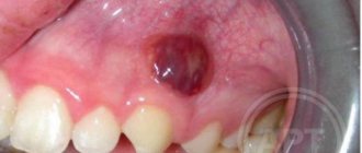
What is a fistula, what does it look like, symptoms
Externally, the fistula is a convex formation with a noticeable white spot on it. This means that the pathological process is accompanied by the formation of pus in large quantities.
There are two types of fistula:
- External, when the gum fistula comes to the surface;
- Internal, when the process remains hidden inside the gum, due to the fact that the fistula channel does not reach its surface. The inflammatory process can only be detected by x-ray examination.
Symptoms of a dental fistula are detected primarily by visual examination. There is a noticeable bulge on the gum, from where pus comes to the surface. There are other signs:
- The pain is cutting, stabbing, spasmodic. Becomes stronger with impact (for example, when chewing), weakens slightly after some of the purulent contents come to the surface;
- Putrid odor;
- Foreign tastes in the mouth;
- Tooth mobility with pathology;
- Swelling of the gums in the area of inflammation;
- Discharge of mucus with pus;
- Increased temperature, symptoms of intoxication of the body;
- Inflammation of the lymph nodes.
The appearance of a fistula due to unsatisfactory canal filling
Canal filling is a fairly common dental procedure indicated in the treatment of diseases such as periodontitis, pulpitis and in preparing teeth for subsequent prosthetics. However, in more than half of the cases, the canals are not filled as well as required by the rules. The most common mistake is filling not to the very top of the root.
Due to a mistake made by the doctor, an infection will form in the unsealed area of the canal over time, which sooner or later will go beyond the boundaries of the tooth and lead to inflammation. In addition to filling the entire length of the canal, the substance used to fill the canal during the procedure must be packed tightly: the appearance of voids and the formation of pores is unacceptable.
The main symptom indicating problems with the tooth is pain when biting. If you look carefully, in the immediate vicinity of the diseased tooth you can easily find a small hole through which pus will come out.
The appearance of a fistula on the gum, the treatment of which is not recommended to be delayed, indicates that the tooth will not stop hurting on its own. To treat it, the tooth is unfilled, an anti-inflammatory medicine is placed in the canals, which is removed after a certain period of time, and the canals are then filled again.
If a tooth has a crown or a pin, unfilling the canals becomes a real problem for the doctor. It is not always possible to remove the pin, and the crown will have to be re-installed after the procedure is completed, which is fraught with additional financial costs for the patient. Sometimes, if the canal is not sealed at its very end, the doctor decides to resort to a procedure for resection of the upper part of the root.
Resection is far from the most difficult operation: its duration is no more than an hour. The operation consists of several main stages:
- Preparation, during which the canals are carefully sealed a couple of days before the main procedure.
- Anesthesia. The operation is performed under local anesthesia: the patient does not feel pain during the doctor’s manipulations, but discomfort may occur later when the effect of the injection wears off.
- Gaining free access to the upper region of the root. To do this, the dentist cuts the gum, exposing the bone tissue, in which a hole is then cut using a drill.
- Through the resulting hole, the doctor cuts off the tip of the root, to which, as a rule, the cyst is attached, which causes inflammation.
- After removing part of the root, an empty cavity remains in the bone. The doctor can fill the space with synthetic tissue, which speeds up the growth process of natural bone.
- The final stage is suturing the gums. For intensive outflow of ichor, drainage can be installed for several days.
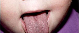
Features of fistula of a milk tooth
Many parents wonder whether there is a risk of developing a fistula when the child’s baby teeth have not yet been replaced by molars. There is a common myth that in this case there will be no chance of suppuration.
But in practice, even with baby teeth, a fistula can develop. Its main source is caries. If you do not deal with timely treatment, caries can greatly destroy the tooth, a dangerous infection will be introduced into the gums, and the gradual development of suppuration will be stimulated.
The more damaged the tooth, the higher the risk. Therefore, our dentists recommend paying close attention to preventive examinations of baby teeth in children. It is necessary to visit the dentist's office regularly to prevent an increase in the risk of the disease.
Fistula due to root perforation
Working with canals requires the dentist to be careful, attentive, and precise, controlled movements. The slightest carelessness can lead to the formation of a non-physiological hole (perforation), which will subsequently cause inflammation and, as a consequence, the appearance of a fistula.
A common mistake that leads to perforation and inflammation is the fixation of the pin when it is installed not along the canal, but its removal into the bone tissue.
With perforation, treatment of the fistula is the most problematic of all cases of its formation. Moreover, the main difficulty is not in medical procedures, but in the behavior of dentists who do not inform patients about the complication that has arisen, so as not to question the professionalism of themselves and their colleagues. As a result, a person who goes to the hospital for dental treatment returns again due to pain and inflammation: often the process becomes so advanced that it is no longer possible to do anything other than remove the tooth.
But still, perforation is not a death sentence, and the tooth can be saved. The sooner the patient applies, the greater the chance that the treatment will be completed successfully. True, this requires expensive materials (Pro Root MTA is often used), with the help of which the perforation area is sealed. Sometimes filling is performed directly through the canals, sometimes a surgical incision is required to gain visual access to the damaged area.
Features of treatment of fistula with concomitant diseases
Are there any particularities in the treatment of fistula if the process is accompanied by other diseases in the oral cavity, such as periodontitis? No, in fact there is no particular connection. Periodontitis is an inflammation of the periodontium of a tooth, while periodontitis is an inflammation of the apical tissues surrounding the tooth. Of course, in some very severe forms, periodontitis can lead to the formation of fistulas, but often these are all unrelated, and one should be treated separately. First, cure periodontitis, if the tooth is healthy, if it does not wobble and can be preserved, and then treat periodontitis.
How long does it take for a patient to recover after fistula treatment?
The treatment of chronic periodontitis itself is not quick, since the patient must first open the tooth canal, clean it, disinfect it with special antimicrobial drugs, and only after that the tooth canal is sealed. If we talk about rehabilitation, when the results are visible, the fistula “goes away” almost immediately, and the bone takes a long time to recover.
If we talk about the surgical method
treatment of periodontitis, when the apical part of the tooth is opened according to the type of resection, but the apical part of the root is polished, the mucous membrane is restored quite quickly - this is
3-5 days
.
Fistula on the gum: what to do?
The most important thing that a person who has pain in the gums should understand is that they need to see a doctor as soon as possible, and not wait for the symptoms to disappear and not use various gels and other means. Yes, they can bring relief, but it will be a temporary relief of symptoms, not healing.
There are several treatment options for fistula.
Endodontic method for treating gum fistula
The method is applicable if the following conditions are met:
- the dental canal and its branches are visible;
- it is clearly visible in the tooth canal whether there are cracks or not.
Treatment is carried out under a microscope. The apical part is sealed with modern high-quality materials that are biocompatible with natural tissues. Canal therapy is performed, which allows the fistula to be eliminated.
Mixed treatment
If the lesion is quite severe, the therapist will need the help of a surgeon.
The therapist cleans the canals and seals the apical part of the root. The surgeon's job is to polish this area of the root. Polishing is carried out using ultrasonic nozzles, which allows you to achieve an ideal result.
Surgery
If the inflammatory process has gone too far, the tooth will have to be removed. The source of inflammation is eliminated along with the tooth and root.
In the future, an implant can be installed in this place at the request of the patient: this is a rather expensive procedure, so it is not recommended to delay the treatment of the fistula. The earlier the patient consults a doctor, the easier and cheaper it will be to cure the disease.
Autotransplantation
This is a type of surgical treatment. In this case, the implant is another tooth of the patient (as a rule, it is a wisdom tooth, which is a kind of reserve, since it is not involved in chewing food).
The advantage of the method is that the person’s own tooth is transplanted, which negates the risk of tissue rejection.
Diagnosis and treatment of fistula
To carry out a diagnosis, you must contact your dentist. The doctor orders an x-ray and conducts a visual examination. Treatment is carried out in a clinical setting. Therapy depends on the cause of the fistula. For example, fillings are used to treat caries, and when an infection is detected, appropriate medications are used. The dentist can also perform treatment with ultrasonic waves or laser.
In some cases, a fistula appears due to poorly done filling of the canal. Then the patient is given an anesthetic, the filling is removed, the dental canal is cleaned and then a new high-quality filling is performed.
If the cause is a cyst, it is removed, which also requires the intervention of a dentist. For recovery, the use of antimicrobial agents, antibiotics, and antihistamines is indicated. If swelling occurs, the doctor may prescribe rinsing with a salt solution.
Cause of primary fistula
Sinusitis is one of the causes of primary fistula. After removing a diseased tooth in the upper jaw, there is a risk of the formation of a fistula tract (ostium) between the maxillary sinus and the oral cavity through the tooth socket. Bacteria penetrate through it from the oral cavity, which provokes inflammation of the sinuses. Usually the hole lasts from 7 to 10 days, then it epithelializes and takes the form of a fistula tract. This is the so-called primary fistula. A secondary fistula forms after an unsuccessful operation.