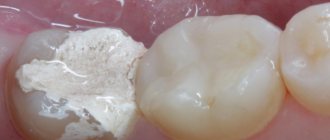
Caries is a local lesion of dental tissues, implying a long process of demineralization, during which a carious cavity is formed. In dentistry, there is a clear classification of dental caries.
The basic principle of caries classification is determined taking into account the depth of damage to dental tissues. The causes of tooth decay are bacteria that need to be removed by brushing your teeth daily.
Caries: its types and classification
Uncomplicated caries:
This is the name of the initial stage of damage to tooth tissue. Caries manifests itself as a small focus of demineralization on the surface of the tooth enamel in the form of a small, light or dark spot. The primary stage of the second degree is characterized by the formation of a carious cavity that does not reach the border between enamel and dentin. Deep tissue damage is manifested by the fact that caries penetrates deeper into the tooth, but without yet affecting the pulp, which is protected by a thin layer of dentin.
Complicated caries:
When caries becomes more severe, it manifests itself as:
- pulpitis – acute inflammation of the neurovascular bundle of the tooth;
- periodontitis is an inflammation of the oral tissues between the alveolar process and the tooth.
Histological classification consists of several points:
- primary damage to tooth enamel;
- damage to the enamel and thin layer of dentin;
- damage to cement:
- dental caries in remission.
K02.0 Enamels
Initial caries or chalk stain is the primary form of the disease. At this stage, there is still no damage to hard tissues, but demineralization and high susceptibility of the enamel to irritation are already diagnosed.
In dentistry, 2 forms of initial caries are defined:
- Active (white spot);
- Stable (brown spot).
During treatment, caries in an active form can either become stable or disappear completely.
The brown spot is irreversible; the only way to get rid of the problem is by preparation and filling.
Symptoms:
- Pain – toothache is not typical for the initial stage. However, due to the fact that demineralization of the enamel occurs (its protective function is reduced), the affected area may experience strong susceptibility to influences.
- External disorders - visible when caries is located on one of the teeth of the outer row. It looks like an inconspicuous white or brown spot.
Treatment directly depends on the specific stage of the disease.
When the stain is chalky, remineralizing treatment and fluoridation are prescribed. When the caries is pigmented, preparation and filling are performed. With timely treatment and oral hygiene, a positive prognosis is expected.
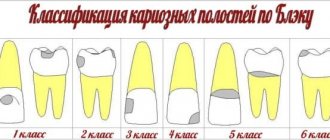
Classification of caries according to Black
This classification of caries intensity was proposed in 1896 by Dr. Black. The essence of the classification is to standardize the methods of filling and removing affected dental tissues. Each class of cavity should be treated using its appropriate filling technique. Levels of carious lesions:
- lesions in the area of natural fissures and in the pits of all teeth in the mouth;
- damage to all surfaces of premolars and molars;
- lesions in the area of the incisors and, rarely, the canines, which do not violate the integrity of the cutting edge;
- damage to all canines and incisors with deformation of the cutting edge;
- damage to all teeth located on the vestibular surface.
- extensive carious lesion.
K02.4 Odontoclasia
Odontoclasia is a severe form of dental tissue damage. The disease affects the enamel, thinning it and leading to the formation of caries. No one is immune from odontoclasia.
The appearance and development of damage is influenced by a huge number of factors. Such prerequisites even include poor heredity, regular oral hygiene, chronic disease, metabolic rate, and bad habits.
The main visible symptom of odontoclasia is toothache. In some cases, due to a non-standard clinical form or an increased pain threshold, the patient does not feel this.
Then only the dentist will be able to make the correct diagnosis during the examination. The main visual sign indicating problems with enamel is tooth damage.
This form of the disease, like other forms of caries, is treatable. The doctor first cleans the affected area, then fills the painful area.
Only high-quality oral cavity prevention and regular dental examinations will help to avoid the development of odontoclasia.
How to treat caries?
It is worth noting that in order to prevent the development of caries, it is recommended to visit the dentist once every six months. When the first stage of caries is detected, a high-quality filling is installed, and the carious area is removed with a drill. If caries is not treated, it progresses to more complex stages, with a large affected area, and then surgical intervention cannot be avoided. If you notice dark spots on your tooth enamel, contact your dentist immediately.
Medium caries is treated in one visit to the doctor, deep caries in 2 visits, and caries in particularly complex forms requires 3 or more visits to the clinic.
Today, caries can be treated without pain, effectively and comfortably for patients. So there is no need to delay going to the doctor. In addition to the drill, caries treatment methods include the microinvasive method, ozone therapy, air abrasive treatment and laser treatment.
K02.1 Dentine
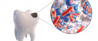
A huge number of bacteria live in the mouth. As a result of their vital activity, organic acids are released. They are the ones responsible for the destruction of the basic mineral components that make up the crystal lattice of the enamel.
Dentin caries is the second stage of the disease. It is accompanied by a violation of the structure of the tooth with the appearance of a cavity.
However, the hole is not always noticeable. It is often possible to notice irregularities only at a dentist’s appointment when a diagnostic probe is inserted. Sometimes it is possible to notice caries on your own.
Symptoms:
- the patient is uncomfortable chewing;
- pain from temperatures (cold or hot food, sweet foods);
- external disturbances, which are especially visible on the front teeth.
Painful sensations can be provoked by one or several foci of the disease, but quickly disappear after the problem is eliminated.
There are only a few types of dentin diagnostics - instrumental, subjective, objective. Sometimes it is difficult to detect a disease solely based on the symptoms described by the patient.
At this stage, you can no longer do without a drill. The doctor drills the diseased teeth and installs a filling. During the treatment process, the specialist not only tries to preserve the tissue, but also the nerve.
Basics of Black caries classification and detailed description.
Read here about the advantages and disadvantages of the Icon method for treating caries in a pediatric clinic.
At this address https://www.vash-dentist.ru/lechenie/zubyi/na-desne-poyavilas-belaya-shishka-foto.html we will tell you why a white bump may appear on the gum.
Stages of treatment of deep caries
Treatment methods for deep caries depend on how much the disease has damaged the tooth tissue and whether it has affected the pulp.
Treatment of deep dental caries without damage to the pulp (deep dentin caries)
- Initial consultation, x-ray, electroodontodiagnosis (EDD for deep caries) to assess the condition of the pulp.
- Anesthesia and preparation of affected tissues.
- In case of deep caries, a therapeutic pad is placed on the bottom of the treated cavity. It contains calcium and has pronounced antimicrobial properties.
- Installation of an insulating lining for deep caries for better fixation of the treatment and isolation of the filling material.
When treating deep caries, pads are necessary before installing a temporary or permanent filling. If the indications are positive (the required dentin thickness and no risk of infection entering the pulp chamber), the doctor can place a permanent filling, and then the entire treatment will take one visit. If a temporary filling is placed, the patient comes to the appointment one more time.
WHEN SHOULD OLD FILLINGS BE CHANGED?
Replacing old fillings is a method of treating and preventing secondary caries. This makes it easier to prevent the disease from getting worse and treat it conservatively in one visit to a specialist. Therefore, old fillings that have stood for more than 5 years should be changed if:
- the tight fit of the filling to the tooth is broken, i.e. pieces of food get stuck between the material and the walls of the cavity, there is a high risk of damage to hard tissues;
- there is excess material and it “hangs” over the neighboring unit or gum, which causes a local inflammatory reaction on the gum, bleeding and the risk of destruction of the root system of the unit due to the accumulation of food and pathological microorganisms;
- abrasion of the filling has occurred, which over time leads to bite pathology and problems with the TMJ;
- The filling material did not restore the anatomy of the chewing surface, i.e. has changed the natural shape, and thus increases the risks of joint diseases;
- the aesthetics are impaired, especially the frontal group of teeth, which are visible when smiling;
It turns out that replacing fillings is important because of maintaining the health of teeth and gum tissue. If the attending physician considers it necessary to replace an outdated filling, you should agree for the sake of continued oral health.
Deep caries after treatment
Considering the serious therapeutic intervention, it is quite normal for a tooth to hurt for 1-2 days after treatment of deep caries (of course, we are not talking about unbearable pain). If the pain continues for a long time, then we are talking about complications, especially when the pulp was initially preserved. In the most difficult cases, therapeutic treatment is not possible, so the only option is to remove the diseased tooth. To avoid such an end, we recommend that you do not delay visits to the doctor: the sooner you come to the dentist’s office, the greater the chance of maintaining your health and money.
Contraindications
Caries is a disease that has no absolute contraindications to treatment. But if certain factors are present, treatment may be delayed or interrupted until they are eliminated. These factors include: the presence of intolerance to drugs and materials that are planned for use in treatment; concomitant diseases aggravating treatment; inadequate psycho-emotional state of the patient before treatment; acute lesions of the oral mucosa and red border of the lips; acute inflammatory diseases of organs and tissues of the mouth; a life-threatening acute condition/disease or exacerbation of a chronic disease (including myocardial infarction, acute cerebrovascular accident), which developed less than 6 months before seeking this dental care; periodontal tissue diseases in the acute stage; unsatisfactory hygienic condition of the oral cavity.
Treatment of deep dental caries with pulp damage
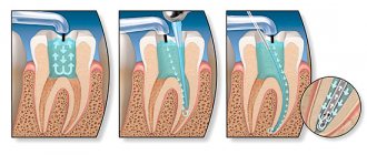
When infection penetrates into the pulp chamber, in the vast majority of cases, tooth depulpation and root canal cleaning are performed. There are methods for complete or partial preservation of the pulp (biological and vital methods of treating pulpitis), however, conservative treatment is possible only at the very early stages of the disease and does not always guarantee a 100% result. After depulpation of the canals, depending on the extent of the damage, the doctor chooses the method of treatment.
- Classic filling of deep caries.
- Installation of filling and stump inlay.
- Crown installation (orthopedic treatment).
Complications
Galloping caries is the most unfavorable form of the disease, since extensive lesions quickly lead to the involvement of neighboring, healthy teeth in the pathological process.
In the absence of timely treatment, the progressive destruction of hard tissues leads to chronic reactive inflammation of the pulp - pulpitis. As a result of inflammation of the pulp, the infection spreads to the periodontium (the space between the cementum of the tooth root and the alveolar bone in which the tooth is located) and causes its inflammation.
If the pathological process is not eliminated in time, the infection can spread to the bones of the facial skull and the soft tissues of the head, causing dangerous complications such as periostitis, abscess and phlegmon.
