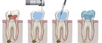
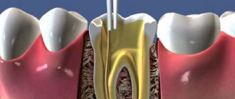
Endomotor
A small electronic device with a tip - an endomotor
— allows:
- treat even severely curved dental canals with the highest quality possible,
- reduce processing time,
- ideally prepare the canals for subsequent filling - completely remove infected tissue from the canals.
The dentist works with an endomotor quickly, efficiently, safely and as comfortably as possible, since modern models of endomotors are so advanced that the doctor’s task comes down to the simplest thing - choosing the desired mode of operation of the instrument. The rest, as they say, is “a matter of technique.”
And the “technique” often depends on the tools that the dentist uses in his work.
Innovative dental instruments Reciproc
- made of an alloy of titanium and nickel - indispensable when working with complicated forms of caries.
Reciprocal instruments in dentistry have long proven themselves to be the best, as they are both flexible and super-strength, helping to create the canal shape that is necessary for high-quality filling.
In addition, a tool that works in reciprocal mode has the superpower of first moving clockwise and then vice versa. This feature allows you to easily separate the instrument from the canal walls.
Reciprocal files reduce the risk of tool breakage from cyclic and torsional fatigue due to the fact that the reciprocal movement settings preset in the endomotor ensure that the tools rotate clockwise and counterclockwise at angles that do not exceed their elastic limit. In addition, as the study showed, reciprocal movement significantly reduces tool fatigue compared to tool operation with continuous rotation.
If you decide to buy reciprocal instruments, the price will pleasantly surprise you. Comfort at work will be an excellent motivation to solve complex professional problems. And instructions for working with machine reciprocity files will bring clarity to the workflow.
A good solution would be to purchase a SILVER RECIPROC endomotor, which is specifically designed to work with nickel-titanium systems in rotary and reciprocal modes. Silver allows you to form a root canal using one tool, without first forming a carpet.
We will consider other models of endomotors with reciprocity below.
Root canal technique using the Mtwo rotating nickel-titanium instrument system
Article provided
Abstract: The authors reviewed the design features of the new Mtwo rotating nickel-titanium tools. A sequence for using files in various clinical situations and depending on the anatomy of the root canal is proposed. The principles of using the Mtwo A and Mtwo R instruments are also discussed. Using computed tomography, the consequences of processing the apical third of the Mtwo A with files at different distances from the apical foramen were assessed.
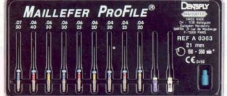
Mtwo endodontic instruments (VDW, Munich, Germany) are a new generation of nickel titanium instruments recently introduced to the market. The standard set of this system includes four basic tools with tip sizes ranging from #10 to #25 and tapers from .04 to .06 (#10 is .04 taper, #15 is .05 taper, #20 is .06 taper and # 25 - taper .06).
Upon completion of the canal preparation with instruments in this sequence, its size will be 25/.06, after which the doctor is offered three different options for completing the root canal treatment. The first sequence allows you to increase the apical diameter using instruments 30.05, 35.04 or 40.04. The second treatment option results in a final taper of .07, facilitating vertical condensation of the gutta-percha, while leaving the apical preparation diameter at #25. The third sequence includes processing with Mtwo apical files, the use of which is the subject of this article (Fig. 1).
Rice. 1. Mtwo tools, basic set and additional files for apex processing.
Design features of Mtwo tools
A colored ring on the shank identifies the tool size according to ISO standards. The number of divisions on the handle indicates the taper of the tool: one division is .04 taper, two divisions is .05, three divisions is .06 and four divisions is .07. There are tools with lengths of 21 mm, 25 mm, and 31 mm. These instruments are produced with both an extended cutting part of 21 mm and a traditional cutting part of 16 mm in length, which makes it possible to process the orifice of the root canal, as well as the wall of the access cavity, where overhanging areas of dentin are often located (Fig. 2).
Rice. 2. Basic set of Mtwo instruments with an extended working part of 21 mm, designed to remove overhanging edges of dentin in the orifice of the canal and on the wall of the access cavity.
Methodology
In cross section, the Mtwo looks like the Latin letter S with two cutting edges. (Fig. 3).
Rice. 3. Cross section of the Mtwo tool (SEM). The surfaces of the two cutting blades form the Latin letter S.
By definition, the major rake angle is the angle formed by the cutting edge and the cross section taken perpendicular to the long axis of the tool (Arens, 1996). The Mtwo's main rake angle is moderately negative. It should be noted that there are practically no nickel titanium tools on the market that have a positive rake angle (Chow et al, 2005). This may be due to the metallurgical properties of the nickel-titanium alloy. Determining the rake angle is one of the most effective ways to measure the cutting efficiency of nickel-titanium tools (Figure 4). In addition, Mtwo tools have a non-cutting tip (Fig. 5).
Rice. 4. Microphotograph of the Mtwo tool 25.06. It can be seen that the cutting edges of the tool have an effective rake angle (magnification x200).
Rice. 5. Microphotograph of the non-cutting tip of the Mtwo tool (magnification x200).
The helical angle (or the angle of inclination of the helical groove to the longitudinal axis. Ed.) is the angle formed by the cutting surface of the instrument and the dentinal wall in a longitudinal section (Buchanan, 1996, 1998). The helical angle is determined by the pitch of the helical cutting of the tool: the larger the pitch, the more open the helical angle will be. And vice versa, the smaller the spiral pitch, the sharper the angle. The helical angle is a very important parameter that determines not only the cutting efficiency of the tool, but also the mechanical stability and its dynamic properties.
The helical angle in Mtwo instruments is specific to each file (Fig. 6). A more developed angle corresponds to larger file sizes (fewer spiral grooves per tool length), and a sharper angle corresponds to smaller file sizes (more spiral grooves). This determines cutting efficiency for larger file sizes and better mechanical stability for smaller file sizes. The spiral grooves become deeper from the apex to the handle, effectively trapping and removing dentin chips. In addition, in large files (20/.06, 25/.06) the helical angle changes along the tool: it increases in the direction from the tip to the handle in the same way as the helix pitch, while it remains unchanged in small files, especially in 10/.04, from which root canal treatment begins. Changes in the helical angle of the instrument reduce the likelihood of it being “sucked” into the root canal.
Rice. 6. Microphotograph of the Mtwo #25/.06 tool, side view: the helical angle increases from the tip towards the shank (magnification x50).
The ability to move freely within the root canal is necessary for small instruments to successfully navigate the canal in the initial phase of treatment. The operator should treat the canal with a “sweeping” motion, holding the instrument while rotating, in order to more effectively prepare the canal wall, removing dentinal filings. Since the patency of the root canal is pre-established with a steel file with a 0.10 mm apical diameter, the Mtwo file 10/.04 can rotate freely in the canal over its entire working length.
Mtwo A and Mtwo R
The Mtwo system includes three files specifically designed for apex treatment - Mtwo A, as well as two types of Mtwo R files for endodontic re-treatment.
The three files for apical processing - Mtwo A1, A2 and A3 differ in tip diameter and taper. The innovative feature of these instruments is the large taper in the last apical millimeter and the .02 ISO taper on the remaining working surface (Fig. 7). The tip diameter (D0) in tool A1 is 0.20 mm with a taper of .15 in the first millimeter, respectively, the diameter of D1 is 0.35 mm. The apical diameter (D0) of A2 instruments is 0.25 mm with a .15 taper in the first millimeter, so D1 is 0.40 mm. In A3 tools, the diameter D0 is 0.25 mm and the taper in the first millimeter is .20, which means D1 is 0.45 mm. The remaining length of the working part of these tools from D1 to D16 has a 2% taper. When designing the instrument, the spiral cutting in the apical millimeter was replaced by two straight blades (Fig. 8). The instrument was designed to widen the apical portion of the canal while maintaining the anatomy of the apical foramen. This is because the diameters of the apical part of the root canals are larger than the average reaming commonly used by practitioners (Orstavik et al, 1991; Wu et al, 2000; Card et al, 2002). The increased taper of the apex also provides a canal shape that is resistant to pressure from gutta-percha condensation during obturation and prevents extrusion of the filling material beyond the apex (Serote et al, 2003).
Rice. 7. Tool Mtwo A1, side view. The cutting edges and unique apical design with increased taper in the last apical millimeter are visible (SEM, ×50 magnification).
Rice. 8. Mtwo A1 instrument tip: innovative tip design with two straight blades in the last apical millimeter (SEM, magnification x200).
Mtwo R instruments have been specially developed for endodontic re-treatment. For this purpose, Mtwo R 15/.05 and Mtwo R 25/.05 files are used (Fig. 9), which have an active tip, with which the tool can easily penetrate the filling material (Fig. 10). The helical angle in these files is constant (Fig. 11), which improves the passage of obstructed canals. The Mtwo R 15/.05 should be used at 200-300 rpm, the same as the standard Mtwo; while the Mtwo R 25/.05 can be used at speeds up to 600 rpm when passing through the mouth of straight channels for increased efficiency.
Rice. 9. Mtwo R tools: 15/.05 (left) and 25/.05 (right).
Rice. 10. Mtwo R Tool Top: Note the presence of cutting blades on the top (SEM).
Rice. 11. The Mtwo R instrument has a constant helical angle throughout the entire working part (SEM, magnification x200).
Sequence of use of tools
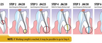
The traditional Mtwo system sequence includes four tools, which are applied from smallest diameter and taper to largest in the following order: 10/.04, 15/.05, 20/.06, 25/.06.
Mtwo tools are used at speeds of 250-350 rpm. Mtwo R 25/.05 should be used at 600 rpm to remove dentin chips from the ostial and middle part of the root canal during endodontic re-treatment. When using Mtwo tools, the torque must be higher than when working with less efficient tools. Typically a torque in excess of 200 g/cm is applied. When using endodontic motors designed to work with Mtwo, torques are set separately for each instrument.
When using Mtwo instruments in a simultaneous technique, there is no need to pre-expand the orifice (Foschi et al, 2004). After establishing the carpet with a #10 K-file, the dentist inserts each instrument to working length, applying light pressure to the apex. When a sensation of screwing occurs, the instrument should be pulled out 1-2 mm so that it can work passively, sweeping movements, removing overhanging dentin and then moving again to the apex.
Instruments are applied with lateral pressure to treat all canal walls one at a time (Plotino et al, 2007a, Grande et al, 2007) (Figs. 12 and 13).
Rice. 12. Cross-section of the root of the upper premolar with oval anatomy of the root canal (left) and after (right) instrumentation. Microcomputed tomography.
Rice. 13. Three-dimensional reconstruction based on microcomputed tomography of the second premolar of the mandible with oval and curved root canal anatomy. Double exposure of the root canal state before (yellow) and after (red) instrumentation. Both in the mesio-distal and in the bucco-lingual projection it is clear that the anatomy of the root canal is preserved (together with R. Bedini and R. Pecci, Italian National Institute of Health, Department of Technology and Health, Rome, Italy).
It is recommended to determine the working length before starting canal treatment, since already at the initial phase of the surgical procedure the files are inserted to their full working length. To do this, you can use an apex locator and then check the resulting measurements radiographically. The latest generation of apex locators allow you to obtain accurate information about the working length in 95% of cases. Although working length can be accurately measured using an apex locator, it is also important to verify it using x-rays, the clinician's touch and the paper point test (Gordon and Chandler, 2004; Plotino et al, 2006). Attaching the apex locator directly to the file or using endodontic motors combined with the apex locator (Fig. 14) is very convenient when using a simultaneous technique, since it takes into account the variation in length during processing and eliminates the need to measure the length of files (Schroeder et al, 2002; Davis et al, 2002; al, 2002).
Rice. 14. Endodontic motor combined with an apex locator and equipped with special settings for the Mtwo instrument system.
Apex treatment
After processing the canal with instruments in the described sequence, the apical diameter will be equal to 0.25 mm. This expansion is considered insufficient to remove debris and clean the apical part of the root canal (Card et al, 2002; Rollison et al, 2002; Iqal and Ku, 2007; Mickel et al, 2007). The diameter of the apical zone should be expanded to 0.40 mm for anterior teeth, single-rooted premolars and for the roots of molars having oval canals (lower distal and upper palatal). The first upper premolars and roots of molars with narrow canals (lower mesial and upper buccal) can be widened to 0.30 or 0.35 mm. From a study of root canal diameters in the mesio-distal and buccolingual directions at a distance of 1 mm from the apical foramen (Wu et al, 2000), it follows that the apical diameter is wider in canals that connect at the same root than in canals that open independent apical foramina.
When determining the final size of the apical foramen, all of the above factors should be taken into account. Traditional rules for determining the apical diameter by wedging a file of a given canal size have been found to be inconsistent (Wu et al, 2002; Bauch and Wallace, 2005; Kfir et al, 2006), and the dentist should determine the appropriate apical preparation diameter on a case-by-case basis using by all available methods.
Treatment of the apical part of the canal with the Mtwo system
After applying the traditional algorithm, the apex can be processed using an additional file sequence that contains tools with sizes 30.05, 35/.04, and 40/.04. In the simultaneous technique, each of these instruments should be introduced to working length passively and without sweeping movements. Reduced file taper with widened apical diameters allows for better passage of even the most curved canals (case report no. 1).
Another option for apical treatment after the traditional Mtwo sequence is the use of one of the apical files, allowing treatment of canals up to a diameter of 0.35 mm (A1), 0.40 mm (A2), and 0.45 mm (A3) 1 millimeter from the apical foramen, using only one instrument: A1 for narrow root canals, A2 and A3 for wide root canals. The .02 taper in the coronal and middle portions of the file allows Mtwo A files to be processed only in the last 3 millimeters of the apical portion. During treatment, the dentist will feel resistance 2-3 millimeters from the apex. This means that the instrument can be inserted to its full working length using 2-3 pecking movements with light apical pressure. When passing the apical part of the root canal with Mtwo files, the apical foramen remains narrow, and the apical diameter 1 mm from the foramen becomes quite widened. Leoni et al., in a recent study (2007), examined the results of apical canal preparation of Mtwo A molars with files at different distances from the apical foramen after preliminary preparation of the canal with a 25/.06 file using micro-computed tomography (Fig. 15).
Rice. 15. Transverse sections of the distal root of the first molar of the upper jaw at a distance of 1, 2 and 3 mm, respectively. from the apical foramen before processing, after processing with the traditional Mtwo instrument sequence up to size 25/.06 and after using Mtwo A3.
Mtwo apical files made it possible to effectively expand the apical 3 mm in the canals of the maxillary molars after pre-treatment with a 25/.06 file and form a circular canal, thereby creating better conditions for medicinal treatment and obturation. It should be noted that all studies concerning measurements of the apical diameter of root canals describe values corresponding to the size of the canal at a distance of 1 mm from the apex (Wu et al, 2000; Kerekes & Tronstad, 1977). Only one research study has been conducted to examine the apical foramen on the external surface of molar roots (Marroquin et al, 2004). In this study, it was shown that the diameter of the hole was smaller than the internal diameter of the canal measured after cutting the roots. The average values of the diameters of narrow and wide physiological foramina in molars are as follows:
- 0.20-0.26 mm in mandibular molars
- 0.18-0.25 mm in the anterior-buccal and posterior-buccal roots of the upper molars
- 0.22-0.29 mm in the palatal roots of the upper molars.
The above studies confirm the rationality of the following approach: the root canals should be expanded at a distance of 1 mm from the apex, while leaving the apical foramen diameter equal to 0.25 mm, which is easily achieved using Mtwo A files.
Discussion
The above described sequence of application of Mtwo instruments refers to the crown-down technique, according to which at each stage each nickel-titanium instrument reaches the apex.
Rice. 16. Three-dimensional reconstruction using microcomputed tomography of the first molar of the mandible in the bucco-lingual projection before and after treatment with the Mtwo system (with R. Bedini and R. Pecci, Italian National Institute of Health, Department of Technology and Health, Rome, Italy).
According to this technique, root canal treatment is carried out in the direction from the mouth to the apex, using smaller and then larger instruments, as in the step-back technique. The simultaneous technique for treating canals is so called because the instrument is immediately inserted to the entire working length. Instruments should not be inserted into the canal with pressure. As soon as the dentist feels a screwing sensation, it is necessary to withdraw the instrument 1-2 mm so that it can work passively, creating space for passage to the apex (Fig. 16a, 16b). When using the instrument, applying lateral pressure (Grande et al, 2007) and gently moving through the canal (until a screwing sensation) makes the process more effective. The flexibility and fatigue resistance (Grande et al, 2006) of Mtwo instruments allow even the most curved canals to be treated effectively and safely (Veltri et al, 2005; Schafer et al, 2006a, 2006b) (Case reports no. 2, 3 and 4). The use of sweeping movements with nickel-titanium instruments is a recent innovation in dentistry (Clauder and Bauman, 2004, Ruddle, 2005). Older generation nickel titanium files could only be used in a passive technique, keeping them centered in the root canal and avoiding lateral pressure (Ruddle, 2002).
Rice. 16A. Three-dimensional reconstruction using microcomputed tomography of the first molar of the mandible in the mesio-distal projection before and after treatment with the Mtwo system.
Rice. 16B. Three-dimensional reconstruction using microcomputed tomography of the mandibular first molar in the bucco-lingual projection before and after treatment with the Mtwo system.
A recent study was conducted on the effect of lateral movement on the fatigue resistance of NiTi Mtwo files (Plotino et al, 2007). Each instrument was used to treat 10 molar root canals that were classified as complex, with and without brushing. The authors observed no instrument failures and only a slight decrease in the fatigue performance of instruments used with lateral sweeping movements, demonstrating that Mtwo NiTi instruments can be used safely in clinical practice. Other research work has focused on controlled clinical fatigue testing of Mtwo instruments (Plotino et al, 2006b). The study results showed that 10 molar root canals were safely treated with each Mtwo nickel titanium file.
The proposal to use a simultaneous technique when working with nickel-titanium instruments has led to a divergence of opinions about the crown-down method. As follows from the definition, during the treatment of canals using the crown-down method, the mouth of the canal first expands, and then the apical part. In the era of nickel-titanium tooling, the crown-down method first used larger diameter and tapered tools to work the mouth, followed by smaller tools to move toward the apex (Ruddle, 2002). The crown-down technique was first described in literature by ES Talbot in 1880. In his work, he wrote the following: “If the canal is curved, you should first go through it with as large an instrument as possible, then replacing it with a small one to pass the curvature. A thin tool can be bent more and travel a greater distance than a larger diameter tool.”
This was the first attempt to describe the crown-down technique, which was difficult to implement at that time due to the rigid steel files used by hand (Fig. 17).
Rice. 17. Article by ES Talbot in 1880, which first describes the crown-down technique. The article also demonstrates the tools used at that time.
With the advent of the latest generation of extremely flexible nickel-titanium files, various ways of applying the crown-down technique have emerged, involving initial processing either with large tools or with more flexible small ones. In fact, in the simultaneous technique, the orifice of the canal is processed before the apical part using small instruments. The use of small instruments first does not contradict the crown-down technique, since in it the canal is also processed from the orifice to the apical part, even if all instruments reach the apex. In other systems, such as ProTaper (Maillefer, Baillagues, Switzerland), each file is produced with different taper values varying within the same tool. The first files that are inserted to full working length, S1 and S2, have a large taper at the mouth and a thin tip with less taper. This instrument design was intended for coronal expansion, facilitating advancement into the canal to its full working length. This leads to the fact that the amount of dentin removed from the canal is quite significant; the file is in close contact with the dentin walls in the apical part and a certain pressure must be applied to advance the instrument apically. In the simultaneous technique, small files are not in close contact with the canal wall, and coronal expansion is achieved only through lateral brushing movements with better tactile control, since apical pressure is not required. When working with other techniques, the resulting diameter of the root canal after treatment corresponds to the diameter of the file inserted into it, while in the simultaneous technique the diameter of the Mtwo file itself is smaller than the diameter of the canal being treated.
Clinical case No. 1.
a) The second premolar of the upper jaw on the right with signs of acute inflammation in the pulp. b) Determination of working length using a steel file. Note that the root of the tooth is strongly curved and has a thin dentinal wall in the apical third. c) Tool Mtwo 20.06 is inserted to working length. d) Filling the root canal system. The Mtwo 40.04 tool was used as a master file. Preservation of canal anatomy and sparing treatment of root tissues was achieved even with relatively large apical expansion.
Clinical case No. 2.
a) The second premolar of the upper jaw on the right with signs of acute inflammation in the pulp. b) Tool Mtwo 20.06 is inserted to working length. c, d) Filling the root canal system after preparation with Mtwo instruments in the standard sequence and Mtwo A1 (in different projections).
Clinical case No. 3.
a) The first premolar of the upper jaw with indications for endodontic treatment. Determining the working length using a steel file. b) Filling the root canal system after preparing the canal with Mtwo instruments in the standard sequence and Mtwo A1.
Clinical case No. 4.
a) The second molar of the upper jaw on the left with indications for endodontic treatment. b) Filling the root canal system after preparing the canal with Mtwo instruments in the standard sequence and Mtwo A2. c) Re-examination after a year.
Clinical case No. 5.
a, b) The first molar of the lower jaw on the left with indications for endodontic treatment. c, d) Determination of working length. The filling material was removed from the canals using Mtwo R instruments. Mtwo master file 35.04. e, g) Root canal filling (view in different projections).
Clinical case No. 6.
a) The second premolar of the upper jaw with indications for endodontic treatment. b) After removal of the filling material using Mtwo R files, apical preparation was carried out with two additional Mtwo instruments to size #40. Filling the root canal system demonstrates the complex anatomy of the apical part of the root.
Clinical case No. 7.
a) The third molar of the lower jaw with signs of acute apical periodontitis. b) Filling the root canal system after treatment with a hybrid technique using steel files and Mtwo system instruments. An apical size of #25 was achieved using a steel file. c, d) When re-examined after 6 months (left) and 1 year (right), healing of the pathological focus is noted.
Clinical case No. 8.
a) The first molar of the upper jaw with signs of acute inflammation in the pulp. b) Filling the root canal system after preparation with Mtwo instruments in a standard sequence. The mesial and distal buccal canals in the apical region were additionally prepared with an Mtwo A2 file, and the palatal canal with an Mtwo A3 file.
Rice. 18. Microcomputed tomography of the first molar of the mandible. A series of five sections was made at the level of the coronal (No. 1, 2 and 3), middle (No. 4) and apical (No. 5) part of the roots. Top row A illustrates the condition before treatment. Middle row B illustrates the appearance of the canals after preparation with two different NiTi instrument systems: the mesial-buccal canal (top left) - with Mtwo 10.04 and 15.05 to the full working length and the mesial-lingual canal (bottom right) - with Orifice Shapers #2, # 3 and #4 using the crown-down technique to a depth of 5 mm. It can be seen that in the simultaneous technique, already with the use of the first two instruments from the basic Mtwo sequence, expansion of the coronal and middle part of the canal was achieved. Row C shows the result of processing channels with the Mtwo system up to size 25.06 and ProFiles up to size 25.06. Please note that both mesial canals are the same size and taper, and have a circular cross-section throughout. The distal canal was processed with a system of instruments not included in the experiment (together with R. Bedini and R. Pecci, Italian National Institute of Health, Department of Technology and Health, Rome, Italy).
A recent study showed that after treatment of the root canal with two initial Mtwo files, the diameter of the canal at the orifice was greater than when treated with high-taper orifice shapers files, specifically designed to expand the coronal part of the canal (Fig. 18). This approach facilitates the treatment of the canal even in the most difficult cases, reducing the likelihood of errors that can be made in the initial phase of treatment, when the canal must be processed along its entire length with a steel file with a diameter of at least 0.20 mm. It should also be noted that the simultaneous technique is based on enlargement of the orifice, which removes the amount of dentin necessary to advance the file to the apex and no more, in contrast to the standard technique, which involves non-selective removal of dentin from the coronal and middle third of the canal (Plotino et al, 2007b). Reducing the diameter of the root canal in the ostial and middle parts increases the mechanical strength of endodontically treated teeth (Rundquist BD and Versluis A, 2006).
Conclusion
The new instrument design, which took full advantage of the unique properties of nickel-titanium alloy, allowed a step forward in the mechanical treatment of root canals, especially in difficult cases. Working with flexible and efficient nickel-titanium instruments at full working length is the future direction for improving root canal cleaning and shaping (Cases 5-8).
Types of endomotors
Today there are two options for endomotors - wired and wireless.
Each model has its own advantages.
Endomotors with an apex locator allow the dentist to accurately determine the size of the apical constriction using a sound or light signal, and see where the instrument is, even while working in a humid environment.
If the endomotor is not equipped with an apex locator, but it is possible to connect one, the apex locator can be purchased separately.
Considering the fact that an apex locator is simply irreplaceable in dentistry, approach your choice seriously and judiciously.
If you want to buy the best new generation apex locator, don't hesitate to choose the Raypex apex locator. The price of the device is absolutely acceptable, given its European quality and a whole list of advantages.
If you need a portable apex locator, the Propex portable apex locator is at your service. Compact dimensions, color display, battery operation, sound indicator with four volume modes and, of course, price are the main advantages of the device.
Which endomotors are better and how to choose an endomotor - we will look in detail using the example of branded models of endodontic miracle technology.
Review of the best endomotors
E-Cube Saeshin (South Korea)
E-Cube Saeshin, an ultra-modern endomotor designed for high-quality treatment of dental canals with rotating “Ni-Ti” instruments, significantly reduces the dentist’s work time and, accordingly, significantly reduces the patient’s time in the chair.
The E-Cube endomotor is equipped with:
- control unit
, which can be powered either from the built-in rechargeable battery or from the mains. The battery charges in no more than 2 hours, while the device holds a charge for 8 hours, that is, it can work continuously for a whole working day; - a large LCD display
with an intuitive interface that displays basic information as concisely and clearly as possible, which helps the doctor concentrate on the work process; - intuitive membrane keyboard
, with which you can easily set torque (torque), rotation speed, direction of rotation, gear ratio; - “M” memory button
, which allows you to quickly select the desired program (out of 9 available); - an “auto-reverse” button, which is responsible for protecting the tool from overload. So, for example, if the maximum permissible torque (specific for each specific file) is exceeded during operation of the endomotor, then the file will automatically rotate in the opposite direction and immediately after removing the excess load, the tip will either stop working, or after stopping it will begin to spin as expected, clockwise;
- branded handle-micromotor
, made of plastic, with a pleasant design, brushless micromotor, which operates almost silently and ensures the smoothest running of “Ni-Ti” tools; - lowering contra-angle handpiece (16:1)
.
The E-Cube endomotor is successfully used by dentists to treat tooth canals with rotating Ni-Ti instruments. The kit includes a foot pedal, 9 programs and auto reverse.
E-Cube is compatible with all NSK end heads.
Advantages of the E-Cube endomotor:
- 9 memory programs,
- auto reverse function,
- micromotor torque from 0.6 to 5.2 N/cm,
- maintaining constant torque,
- transmission ratio: 16:1,
- foot pedal with on/off function,
- compact,
- convenient, functional design,
- operation from a rechargeable battery and directly from the network,
- The battery holds a charge for up to 8 hours.
The E-Cube endomotor is No. 1 for the dentist. If the doctor works with the E-Cube, then the canal filling will be 100% successful.
E-Cube is an analogue of X-Smart, an endomotor that is highly valued by all endodontists, including because of the ideal price/quality ratio.
X-SMART Plus, Dentsply-Maillefer (Switzerland)
The X-Smart Plus endomotor is compact and ergonomic, easy to operate, has 8 programs and a large display, and does not have a foot pedal.
X-Smart Plus operates in sequential and reciprocal modes, is equipped with an auto-reverse function, can last 2 working hours without recharging, and is fully charged within 5 hours.
Advantages of X-Smart Plus:
- Doesn't block the view.
- Clear interface.
- Color display.
- Small size and weight.
- Angle handpiece rotation speed from 100 to 800 rpm.
X-Smart Motor, Dentsply-Maillefer (Switzerland)
X-Smart Motor is an endomotor complete with a handpiece and a 16:1 reduction head.
X-Smart Motor has 9 programs, each of which is very easy to adjust both in terms of rotation speed (120-800 rpm) and torque (0.6-5.2 Ncm). Switching programs is also easy.
The device is equipped with auto reverse. The motor is turned on and off using a button on the handpiece. There is no pedal, but it is possible to connect one.
Advantages of X-Smart Motor:
- A light weight.
- Torque table included.
- Two power sources - built-in battery and power cord.
X-Smart Plus Protape, Dentsply-Maillefer (Switzerland)
The X-Smart Plus Protaper Kit endomotor is easy to operate, reliable and ergonomic.
Main advantages:
- Compact size.
- Miniature head.
- 12 programs.
- Lightweight tip handle.
- Reciprocal mode.
- Torque control function.
X-Smart Plus Protaper does not have a foot pedal, but this does not deprive the dentist of comfort.
Y1001-027 Endo-Mate TC 2, NSK (Japan)
Endo-Mate is a cordless dental handpiece with min. head, equipped with a torque control function and auto reverse. Endo-Mate has only 5 programs, but the quality of work does not suffer from this.
Benefits of Endo-Mate:
- Ease of control.
- Liquid crystal display.
- Auto reverse function.
- Sound alarm function.
- Compatible with many tools from major manufacturers.
Endo-Mate TC 2 MPA Pack, NSK (Japan)
This is a portable, wireless endodontic micromotor with an MPA head, auto reverse and the ability to connect an apex locator.
Benefits of Endo-Mate TC 2 MPA Pack:
- Miniature head and thin neck.
- Wide LCD display
- Intuitive interface
- 5 programs for files
- Memory function for used settings.
- Contactless charging in 1.5 hours.
- It is possible to connect an apex locator.
- Compatible with many brand tools.
You can always buy endomotors profitably in the Dental Stom online store. Call by phone 8
.
Our specialists are always happy to answer any of your questions and are ready to help you choose a product and place an order.
A good endomotor will do the job
An endomotor is an electronic device with a tip - the main instrument of an endodontist.
A high-quality modern endomotor will provide real help even when the root canals are “unrealistically” curved. At the same time, it will significantly reduce the processing time and help to perfectly clean the affected tissues.
Speed, efficiency and safety - these are the main advantages of the best modern models of endomotors!
The key to such success is the ideal tools.
Reciproc tools guarantee your craftsmanship 100%.
Made from an alloy of titanium and nickel, Reciproc is ideal for working with complicated forms of caries.
Flexible and durable Reciproc prepares the tooth canals for further ideal filling.
But the main feature is the movement! Working in a reciprocal mode - moving first clockwise and then vice versa - Reciproc facilitates easy separation of the instrument from the walls of the dental canal.
At the same time, the price of Reciproc is more than acceptable. So when you decide to buy Reciproc, know that you are taking the first giant step towards solving your most difficult professional problems!
Follow the instructions for working with machine reciprocity files - and success is guaranteed.
Step two - SILVER RECIPROC endomotor.
The ultra-modern SILVER RECIPROC is designed to work with nickel-titanium systems in rotary and reciprocal modes.
When you have Silver in your hands, you will form a channel without first forming a carpet.
But this is not the limit. Other models of endomotors with reciprocity definitely deserve attention.
